
Abstract
Prenatal and early postnatal are the most sensitive and high-risk periods to expose to electromagnetic fields (EMFs). This study aimed to investigate the effect of prenatal and early postnatal exposure to 900 MHz radiofrequency waves (RFWs) emitted from a base transceiver station antenna on passive avoidance learning and memory (PALM) and hippocampus histomorphology. Female Sprague Dawley rats (190–230 g) were paired with males. The mated rats, confirmed by observing a vaginal plug, were divided into two groups; control and exposed. The control group (n = 7) was not exposed to RFW. The exposed group was divided into three subgroups (n = 8); exposed Ⅰ, exposed during the gestational period (fetal life), and exposed Ⅱ and Ⅲ (postnatal exposure), exposed to RFW during the first 21 days of life, for 2 h/d and 4 h/d, respectively. PALM was evaluated by a shuttle box in 45-day-old pups. Learning and memory of animals were demonstrated as the duration of remaining within the light area, which is called the lighting time. Histological sections were prepared from brain tissues and stained with hematoxylin and eosin. An impairment in the PALM performance was noticed in all exposed subgroups (Ⅰ, Ⅱ, and Ⅲ) (p < 0.05). Learning (short-term memory) and retention (long-term memory) behaviors were more affected in exposed subgroup Ⅰ (prenatal exposed) compared to other postnatal exposed subgroups (Ⅱ and Ⅲ). Also, a mild decrease in the density of pyramidal cells was observed in the hippocampus of exposed subgroups (Ⅰ and Ⅲ). Prenatal and early postnatal exposure to 900 MHz RFW adversely affected PALM performance and hippocampus tissue in rat pups with more impact for prenatal period exposure.
Introduction
Today, exposure to electromagnetic waves has become one of the main environmental health concerns. Base transceiver stations’ (BTSs) man-made sources used for mobile phone communication are important sources of radiofrequency waves (RFWs). Although Non-Ionizing Radiation Protection (ICNIRP) and Institute of Electrical and Electronics Engineers (IEEE) established the highest level of RFW exposure, however, they do not cover pregnant women and their infants (Guler et al., 2010).
Generally, neurogenesis in the brain begins during fetal life and is completed before birth. However, this process in some parts of the brain areas such as the hippocampus continues during the early postnatal period in all species (Guidi et al., 2005; Lima and Gomes-Leal, 2019). Hippocampus, a curved, paired structure located in the temporal lobes near the amygdala, is one of the most important parts, which is involved in learning and memory (Liu et al., 2018). Exposure to detrimental factors during fetal life may lead to hippocampal development retardation or neurobiological and behavioral defects (Lemaire et al., 2000).
It has been reported that electromagnetic fields (EMFs) may affect neuronal functions, comprising regulation of synaptic plasticity, neurotransmitter release, neuronal survival, learning, and memory (Manikonda et al., 2007; Sakatani et al., 2002). A deleterious effect of exposure to 900 MHz RFW on learning and memory in adult rats has already been reported (Azimzadeh et al., 2018; Azimzadeh and Jelodar, 2020; Jelodar et al., 2018). Moreover, similar evidence has been reported with different frequencies (835, 900, 1800, and 2100 MHz) in addition to devastating effects on hippocampal morphology (Gokcek-Sarac et al., 2017; Krishna Kishore et al., 2019; Kim et al., 2018; Kivrak et al., 2017). However, exposure to 900 MHz EMF has not adversely affected spatial nor nonspatial memory (Dubreuil et al., 2003; Sienkiewicz et al., 2000; Tan et al., 2017).
There is controversy about the effects of EMF emitted by mobile phones on brain morphology. While some studies have reported alteration in brain morphology (Bas et al., 2009; Odaci et al., 2008), others have failed to observe such effects (Azimzadeh et al., 2018; Ragbetli et al., 2009). For example, defects in the dentate gyrus granule cell development in the rat hippocampus in addition to the decrease in the number of granule cells in the dentate gyrus and pyramidal cell have been reported in prenatal (Bas et al., 2009; Odaci et al., 2008) and postnatal (Bas et al., 2009; Sonmez et al., 2010) exposure to 900 MHz RFW. Impaired memory in mice exposed in utrus to 800-1900 MHz following use of light/dark box, object recognition, and step-down assays has been reported (Aldad et al., 2012). Our previous study revealed an impaired passive avoidance learning and memory (PALM) without brain histomorphological alteration in adult male and female rats exposed to 900 MHz RFW (Azimzadeh et al., 2018).
The prenatal and early postnatal periods are considered as the most sensitive period of brain development; however, the majority of investigation in the field of biological health effects of EMF have focused on adults. Therefore, the present study aimed to evaluate the potential effects of prenatal and early postnatal exposure to 900 MHz RFW on passive avoidance learning and memory (PALM) and hippocampus histomorphology in rat pups.
Materials and methods
Experimental design
Female Sprague Dawley rats (190–230 g) at the proestrus stage of the reproductive cycle were paired with male rats (two female/male). The following day, the presence of a vaginal plug confirmed the mating and was considered as day first of pregnancy. Then, pregnant rats were divided into two major groups; control and exposed. The control group was not exposed to RFW.
The exposed group was further divided into three subgroups (n = 8/ea); exposed Ⅰ (prenatal), exposed during the gestational period (fetal; 21 days), and exposed Ⅱ and Ⅲ (postnatal exposure), exposed to RFW for the first 21 days of life, for 2 h/d and 4 h/d, respectively. PALM was evaluated using a shuttle box in 45-day-old pups.
Signal generator
The 900 MHz RFW exposure apparatus was made in the Department of Telecommunication and Electronics Engineering, Shiraz University. A simulator with a 12 cm antenna emitting 900 MHz RFWs circularly with an average power density of 0.6789 mW/cm2 was used. The output of the device was monitored by a spectrum analyzer (FSH6, Rohde and Schwarz, Germany). The simulator was placed centrally 1 m away from the cages of the exposed subgroups (Ⅰ, Ⅱ, and Ⅲ) on the floor of the experimental room. The whole-body average specific absorption rate was 0.035 W/kg and was measured 1 m away from the signal generator by a SPM2 field meter (WPF18 Field Probe 300 kHz to 18 GHz, Wavecontrol, Spain).
PALM apparatus and evaluation
A two-way shuttle box (Aryoazma Co., Iran), which contained light and dark compartments was used. When animals moved from the light-safe compartment into the dark compartment, they received a foot shock of 0.6 mA (for 1 s) with a latent period of 1 s (Azimzadeh et al., 2018; Jelodar et al., 2018). Evaluation of PALM was according to the method of Bures et al. with some modifications as was described in detail in our previous investigation (Azimzadeh et al., 2018; Bures et al., 2016). Briefly, the evaluation was performed over 5 consecutive days as introduction, initial latency, learning, memory consolidation, and memory retention. Foot shock was applied to animals on the initial latency and learning day. If the animals did not move to the dark compartment after 120 s in the course of learning, memory consolidation, and memory retention, they were considered as completely learned.
Histological examination
On the last day of the experiment, the brain was dissected out and fixed in 10% neutral buffered formalin, embedded in paraffin, sectioned at 5 μm, and stained with hematoxylin and eosin (H&E) for histomorphological evaluation of the hippocampus by light microscope.
Statistical analysis
All data are presented as mean ± standard error and were analyzed by the Statistical Package for Social Sciences (SPSS, version 21.0). The nonparametric Kruskal–Wallis test was used to compare results separately for each group. Then, if results were significant, pairwise comparisons were made using the Mann–Whitney U test. A value of p < 0.05 was considered statistically significant.
Results
The PALM test was assessed under the parameter of the latency for the animal stay in the light compartment (lighting time). On the learning day, there was a significant decrease in lighting time in the exposed Ⅰ (p = 0.008), Ⅱ (p = 0.044), and Ⅲ (p = 0.013) subgroups compared with the control group. Also, a significant decrease was observed in lighting time in the exposed Ⅰ subgroup compared to the exposed Ⅱ (p = 0.016) and Ⅲ (p = 0.044) subgroups (p < 0.05) (Figure 1).
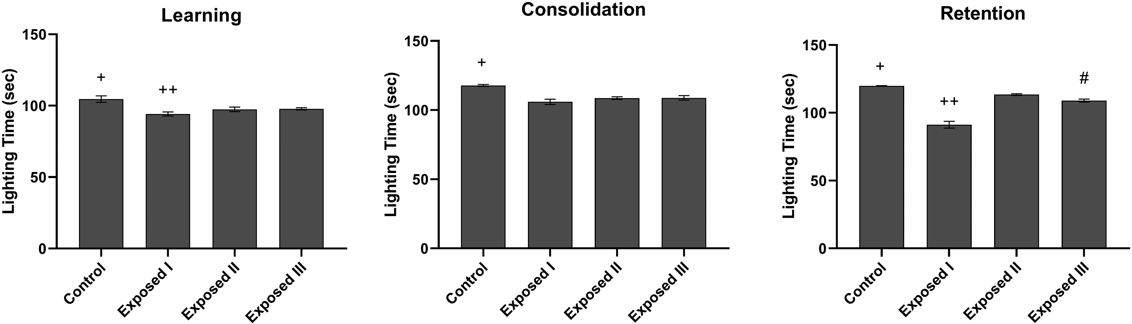
Passive avoidance performance (learning, memory consolidation, and retention) evaluation in the animals were exposed to 900 MHz radiofrequency wave emitted from BTS. Columns represent mean ± SEM (p < 0.05). Exposed Ⅰ: exposed during the gestational period (fetal). Exposed Ⅱ and Ⅲ: (postnatal exposure), exposed to RFW for the first 21 days of life, for 2 h/d and 4 h/d, respectively. Passive avoidance learning and memory evaluation was done on 45-day-old pups. +Significant differences with all groups. ++Significant differences with exposed Ⅱ and Ⅲ. #Significant differences with exposed Ⅱ. BTS: Base transceiver station; SEM: Standard error of mean.
On the consolidation day, the exposed Ⅰ, Ⅲ (p = 0.009), and Ⅱ (p = 0.008) subgroups showed a significant decrease in lighting time compared with the control group (p < 0.05) (Figure 1).
On the retention day, a significant decrease in lighting time was observed in the exposed Ⅰ, Ⅱ, and Ⅲ subgroups compared with the control group (p = 0.007). The exposed Ⅰ subgroup showed a significant decrease in lighting time compared to exposed Ⅱ and Ⅲ (p = 0.009). Moreover, there was a significant decrease in lighting time in the exposed Ⅲ (p = 0.008) compared with the exposed Ⅱ subgroup (Figure 1).
Learning (short-term memory) and retention (long-term memory) behaviors were more affected in the exposed Ⅰ subgroup (prenatal exposed) compared to other exposed subgroups (Ⅱ and Ⅲ), and this indicated the high sensitivity of pregnancy period to exposure to RFW compared to other periods of life. A mild decrease was observed in the density of pyramidal cells in the hippocampus tissue of 37% of animals in subgroup Ⅰ that were exposed to RFW during fetal life 4 h/d and in 50% of rats in subgroup Ⅲ that wereexposed after birth 4 h/d for 21 days compared to the nonexposed group (Figure 2).There was no remarkable histological change in the hippocampus of subgroup Ⅱ rats.
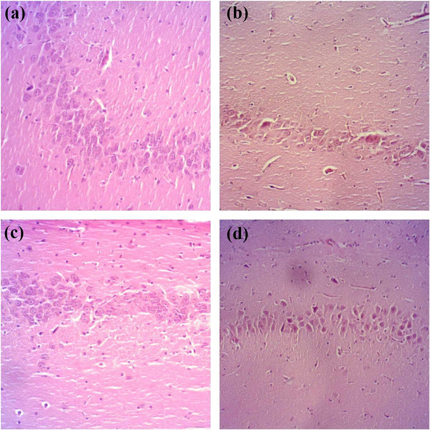
Comparison of histological sections of hippocampus of nonexposed and exposed groups to RFW. A decrease in the density of pyramidal cells in the hippocampus tissue was observed in the exposed subgroups Ⅰ and Ⅲ (a) control group, (b) exposed I, (c) exposed Ⅱ, and (d) exposed Ⅲ. Exposed I, exposed during the gestational period (fetal). Exposed Ⅱ and Ⅲ (postnatal exposure), exposed to RFW for the first 21 days of life, for 2 h/d and 4 h/d, respectively. H&E. ×100. RFW: Radiofrequency wave; H&E: hematoxylin and eosin.
Discussion
Impaired PALM performance in 45-day-old rats exposed to 900 MHz RFW during the prenatal (fetal) and early postnatal (2 and 4 h/d for 21 consecutive days) periods was noticed in this study. Moreover, a decrease in the density of pyramidal cells in the hippocampus tissue was observed in the two exposed subgroups Ⅰ and Ⅲ.
The impact of exposure to EMF and cognitive behaviors has been studied, and various results have been reported. For example, one study showed that the fetus of mice exposed to microwaves (800–1900 MHz) had impaired memory function and behavior and also neurophysiological alterations that persisted up to adulthood (Aldad et al., 2012). The effect of exposure to 900 MHz EMF during pregnancy (from day 13 to day 21, 1 h/d) has been examined and was reported to compromise passive avoidance learning and histopathological changes in the hippocampus (İkinci et al., 2013). Exposure to 900 MHz RFW during the fetal period was reported to induce cell death and also inhibit the differentiation of neuronal stem cells to neurons (Salford et al., 2003). Moreover, impairments in the development of dentate gyrus granule cells and pyramidal cell loss due to inhibition of neurogenesis following prenatal exposure to 900 MHz RFW have been reported (Bas et al., 2009; Odaci et al., 2008). However, no deleterious effects were reported in the pre- and postnatal development of the pups exposed during the prenatal period to a 2450 MHz Wi-Fi signal (Poulletier de Gannes et al., 2012). Another study showed that exposure to low-level EMF (17.5–75 mW/kg) during pregnancy did not affect cognitive performance (Bornhausen and Scheingraber, 2000). Several investigations provided no evidence of exposure-related changes in the offspring’s hippocampus (Ragbetli et al., 2009), cerebellum (Haghani et al., 2013), learning skills, and behavior (Klose et al., 2014) in rats exposed to 900–1800 MHz EMF during gestation. Differences in experimental designs and applied methods such as radiation devices, power density, frequency, orientation, distance to the radiation source, and duration of exposure might be the cause of the discrepancies in the results.
The majority of published papers considered oxidative stress as the main mediator of EMF side effects (Ertilav et al., 2018; Kerman and Senol, 2012; Othman et al., 2017). Microwave radiation can induce oxidative stress, which leads to neuronal apoptosis via the oxidative damage of cellular constituents (Shahin et al., 2015). For example, one study reported that prenatal exposure to the mobile phone (900–1800 MHz) induced oxidative stress in tissues of dams and their offspring (Bahreyni Toossi et al., 2018). Other studies have also reported that prenatal exposure to EMF (900 and 1800 MHz) caused oxidative stress and histopathological changes in the heart (Turedi et al., 2015), brain, and liver in growing rat pups (Cetin et al., 2014). Moreover, increases in uterine oxidative stress in pregnant rats and their offspring as a result of long-term exposure to electromagnetic radiation from mobile phones (900 and 1800 MHz) and Wi-Fi (2450 MHz) devices was reported (Yuksel et al., 2016).
Since dielectric properties and conductivity of organs are the main factors in the rate of EMF absorption, increased water uptake during pregnancy and subsequently increased whole-body electrical conductivity make this period more sensitive to EMF (Kismali et al., 2012). On the other hand, considering that children start using a mobile phone at an early age and lack of morphological development of the skull in childhood may cause greater penetration of EMF into the brain (Christ et al., 2010; Lenhart et al., 2010). The prenatal development of the rat hippocampus can be compared with the development of the hippocampus in the third trimester in humans (Jacobson, 1991; Rodier, 1980). Therefore based on our results and the available evidence from previous investigations, exposure to RFW during the pregnancy and early postnatal periods can adversely affect the development and function of the central nervous system and lead to impaired behavioral and cognitive functions involving short-term memory and learning in postnatal life.
Conclusions
Prenatal and early postnatal exposure to 900 MHz RFW affected both PALM and the hippocampus tissue in rat pups. Alteration of learning (short-term memory) and retention (long-term memory) behaviors was more obvious following prenatal exposure than early postnatal exposure and indicated a greater sensitivity during the fetal period compared with other periods of life. A remarkable decrease in the density of pyramidal cells in the hippocampus was noticed in exposed subgroups I and Ⅲ.
Footnotes
Acknowledgments
The authors acknowledge the Department of Telecommunication and Electronics Engineering, Shiraz University for providing the signal generator.
Declaration of conflicting interests
The author(s) declared no potential conflicts of interest with respect to the research, authorship, and/or publication of this article.
Ethical approval
All investigations were conducted in accordance with the “Guiding Principles for the Care and Use of Research Animals” approved by Shiraz University.
Funding
The author(s) received no financial support for the research, authorship, and/or publication of this article.