
Abstract
Synthetic amorphous silica (SAS) nanomaterial – consisting of aggregates and agglomerates of primary silicon dioxide (SiO2) particles in the nanorange (<100 nm) – is commonly used as excipient in pharmaceuticals, in cosmetics and as food additive (E551). The available data suggest that SAS nanoparticles (NP) after intravenous (IV) exposure persist in liver and spleen; however, insufficient data exist to verify whether SAS may also induce adverse effects. The aim of the present study was to verify the potential long-term effects of SAS NP (NM-203) on spleen and liver as target organs following short-term exposure. Adult male and female Sprague-Dawley rats were treated by IV injection in the tail vein with a single (1-day) dose (SD) and repeated (5-day) doses (RD) of 20 mg/kg bw per day of SAS dispersed in sterile saline solution as vehicle. Histopathological examinations of target organs were performed after 90 days. Tissue biodistribution and full characterization of NM-203, primary particle size 13–45 nm, was performed within the framework of the Nanogenotox project. No mortality or general toxicity occurred; histopathological analysis showed splenomegaly in the RD group accompanied by inflammatory granulomas in both sexes. Granulomas were also present in liver parenchyma in the RD (both sexes) and SD groups (male only). The histopathological results indicated that SAS NP have the potential to persist and induce sex-specific chronic inflammatory lesions in spleen and liver upon short-term treatment. Overall, the data showed that the widespread use of silica in drugs might elicit chronic reactions in spleen and liver prompting to the need of further investigations on the safety of SAS NP.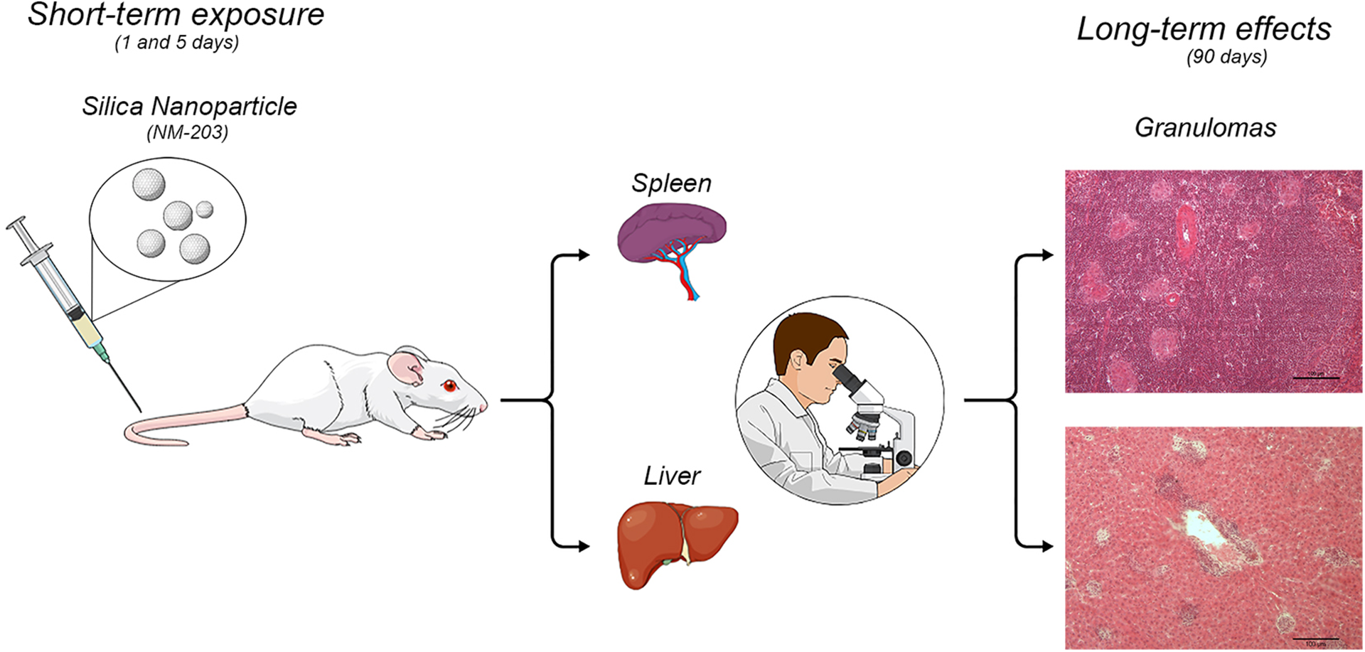
Introduction
Synthetic amorphous silica (SAS) is a material composed of primary nanoparticles (NP < 100 nm) variably agglomerated and aggregated according to the conditions of production and use (Tassinari et al., 2020; van Kesteren et al., 2015). SAS has been widely used for its biocompatibility and easy functionalization with targeting molecules. In fact, it is utilized as nanocarrier for drug delivery, excipient in vitamin preparations and cosmetics, without any limitation of particle size (Alvarez-Berrios and Vivero-Escoto, 2016; Murugadoss et al., 2017), and it is extremely promising as haemostatic agents for preventing the blood loss after damage (Gryshchuk and Galagan, 2016). Engineered SAS is among the most widespread nanomaterial (NM) in the world, but few studies on workplace airborne exposure have been carried out and, currently, no official occupational exposure limits have been established, as for several NMs (Boccuni et al., 2020; Di Cristo et al., 2020). As food additive ‘silicon dioxide (SiO2) or ‘E551’, it has been used for decades in the EU (as stabilizer, carrier, clearing and anticaking agent). The most recent and reliable data on SAS - described in the European Food Safety Authority (EFSA) report (EFSA, 2018) - revealed that in the available in vivo studies after the administration of fumed or precipitated SAS, the silicon content of the liver and kidney, and occasionally of the spleen, was slightly increased; moreover, studies in rats after repeated SAS oral applications indicated no accumulation of silicon. In humans, there was little indication on absorption of SAS after ingestion although SiO2 (of unknown origin) was found in liver and spleen. In general, SAS showed low acute toxicity after repeated oral administration, and no adverse effects were detected even at the highest dose levels. SAS did not raise concern regarding genotoxicity (EFSA, 2018). After intravenous (IV) exposure, the liver and spleen remain the main target organs (De Jong et al., 2013; Waegeneers et al., 2018), and measurable concentrations of silica still persist in such tissues after the 90-day long recovery period. Because of the increasing human exposure to SAS (Fruijtier-Polloth, 2016), it appears necessary to expand knowledge about the possible persistence and long-term effects after acute administration by routes of exposure different from oral intake. This study has been performed within the framework of the Nanogenotox (Nanogenotox: The project, 2013) Joint Action, and the present article reports the complete and detailed elaboration of the data concerning potential long-term toxicological effects on spleen and liver following short-term IV exposure in male and female rats. Indeed, the specific aims of the present elaboration were to ascertain whether (i) the Si exposure may be responsible for the granuloma formation in spleen as in liver and (ii) if present, the granuloma may be reversible or may persist over a long-term time period.
Material and method
Nanomaterial
NM-203 pyrogenic (NM repository of the European Commission Joint Research Centre, Institute for Health and Consumer Protection, Ispra, Italy). NM-203 was fully characterized (Rasmussek et al., 2013).
Animal experiment
The study has been conducted according to the European Community Council Directive 2010/63/UE, the Italian Law 4 marzo 2014, n. 26 and the OECD Principles on Good Laboratory Practice. The study plan has been evaluated and approved by the Italian Ministry of Health according to the D.lgs n. 116/1992 art. 9 (Decree n. 218/2010 B). Moreover, the quality criteria for in vivo toxicity studies performed with NMs, according to Fernández-Cruz et al.(2018) were met.
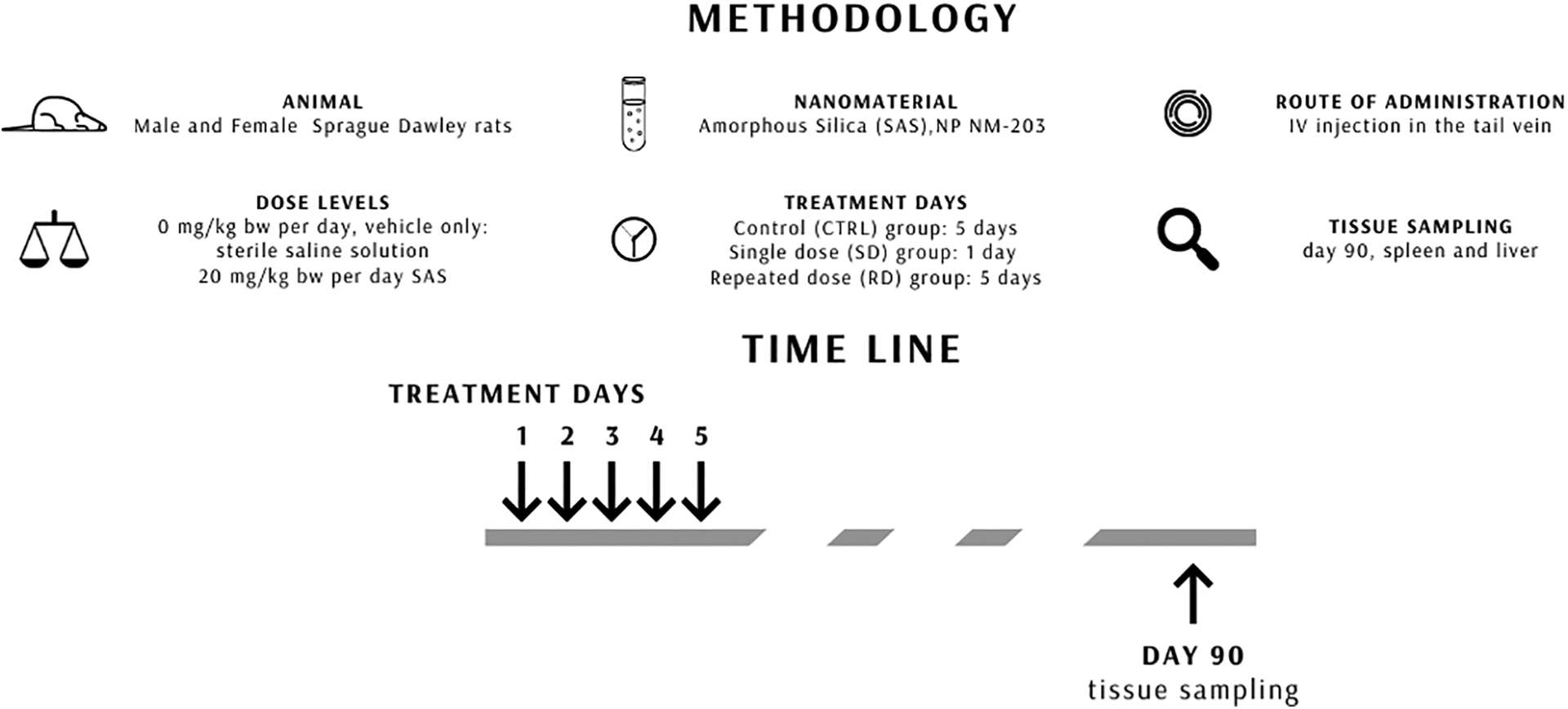
The scheme of experiment is shown in Figure 1. Eighteen young adult outbred Sprague-Dawley rats of both sexes (150–200 g body weight (bw)) were purchased from Charles River (Italy). They were kept under standard laboratory conditions (22 ± 0.5°C room temperature, 50–60% relative humidity, 12-h dark–light alternation with 12–14 air changes per hour), with water and food available ad libitum.
Experimental design.
After approximately 5 days of acclimatization, they were divided into three treatment groups as follows: Control (CTRL): Three male and three female rats received vehicle only (sterile saline solution) for 5 days. Single dose (SD) group: Three male and three female rats received 20 mg/kg bw per day of SAS NP NM-203 for 1 day. Repeated dose (RD) group: Three male and three female rats received 20 mg/kg bw per day of SAS NP NM-203 for 5 days.
The sample size of three rats/sex/group was calculated with STATA 14.2 software (level: 0.05 and power: 80%) considering previous data on rats treated with same dose of NM 203, in the same window of exposure, showing significantly increased SAS biodistribution in liver (De Jong et al., 2013).
Treatments were performed by IV injection in the tail vein. The SD group received a dose of 20 mg/kg bw and the RD group received a total cumulative dose of 100 mg/kg bw. Dose level was selected on the basis of preliminary studies, and on the information concerning maximum dispersibility of SAS NP, stability of dispersions and considering the maximum volume that can be administered orally to each rat (2–2.5 ml/kg bw) (Jensen et al., 2011).
The suspensions to be administered to rats were prepared according to the Nanogenotox protocol (Jensen et al., 2011), using a Bandelin Sonopuls Ultrasonic Homogenizer HD3200 series (Merck, Italy) equipped with an SH 213 G booster horn and a sterile KE 76 tapered tip. The quality of the NM-203 dispersions was evaluated using asymmetric flow field flow fractionation with optical (MALS and UV) and ICP-MS detection (De Jong et al., 2013). Food consumption and bw of each rat were recorded daily during the treatment and twice a week until euthanization. After a 90-day follow-up period, rats were euthanized by CO2 inhalation. Spleen and liver were collected, weighed and stored: a portion for histopathological examination and a portion for tissue distribution of SAS. SAS biodistribution was evaluated according to the method described in the Deliverable # 7 of Nanogenotox project (De Jong et al., 2013) using Agilent 8800 triple quadrupole inductively coupled plasma mass spectrometer (ICP-QQQ-MS; Agilent Technologies Inc., Tokyo, Japan).
Histopathological analysis
Immediately after the euthanization, to avoid any possible post-mortem artefacts, portions of liver and spleen were fixed in 10% buffered formalin and stored in 80% ethyl alcohol. The specimens were embedded in paraffin, cut into 5-µm sections and stained with haematoxylin and eosin for the examination under a light microscopy (Nikon Microphot FX, Italy) with different lenses. Tissues were blindly assessed on coded slides.
Quantitative analysis in tissues was performed by means of an image analysis system (Nis-Elements D) applied to the optical microscope (Nikon Microphot FX), as follows:
Spleen: Red pulp and white pulp areas were measured using a 2× objective; ratio between red and white pulp areas was calculated (Maranghi et al., 2013).
Liver: Using a 20× objective, granulomas size (µm2) and granulomas density, as ratio between number of granulomas and a predetermined liver area, were measured (Yu et al., 2017).
Statistical analysis
Statistical analysis was performed with JMP 10 (SAS Institute Inc., Cary, North Carolina, USA). The GraphPad Prism 6.0 software was used to perform all graphics. Body weight gain, food consumption, absolute and relative organ weight and data of histomorphometrical analysis are presented as means ± standard deviation (sd). Non-parametric Kruskal–Wallis test was applied for all the continuous variables to test the statistical significance of the differences among groups, followed by the post hoc Dunn’s test comparison, where appropriate. For categorical variables (histological end points), data were expressed as proportions of quantal data; pairwise comparisons of treated groups with CTRL group were analysed by means of the two-tailed Fisher exact test. To identify dose–response trends, the Mantel–Haenszel χ2 trend test was used. Significance level was set at p < 0.05 for all the analyses.
Results
NM-203 characterization data are shown in Table 1.
Pyrogenic synthetic amorphous silica nanomaterial NM-203: main physico-chemical characteristics.
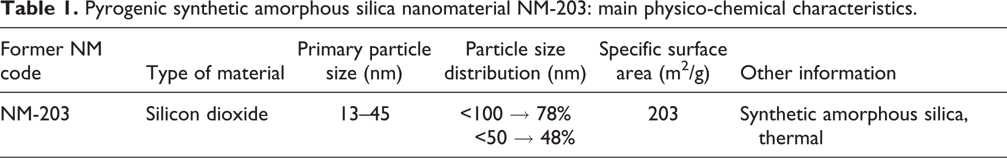
NM: nanomaterial.
The quality evaluation of NM-203 dispersion showed a bimodal size distribution with a first peak indicating the presence of the primary NP (about 20 nm diameter) and a second peak indicating the presence of agglomerates (about 200–300 nm diameter) (De Jong et al., 2013).
Toxicokinetic and biodistribution data of NM-203 were reported in the Deliverable # 7 (De Jong et al., 2013); briefly, the data on spleen and liver distribution of SAS – measured as total Si content – after 90-day IV administration of NM 203 are summarized in Table 2 (De Jong et al., 2013)
Tissue distribution of Si (mg Si/kg fresh weight) in male and female rats IV treated with 0 mg/kg bw per day (CTRL – vehicle only), SD, RD (5 days) of 20 mg/kg bw per day of NM-203 and euthanized after 90-day recovery period.a
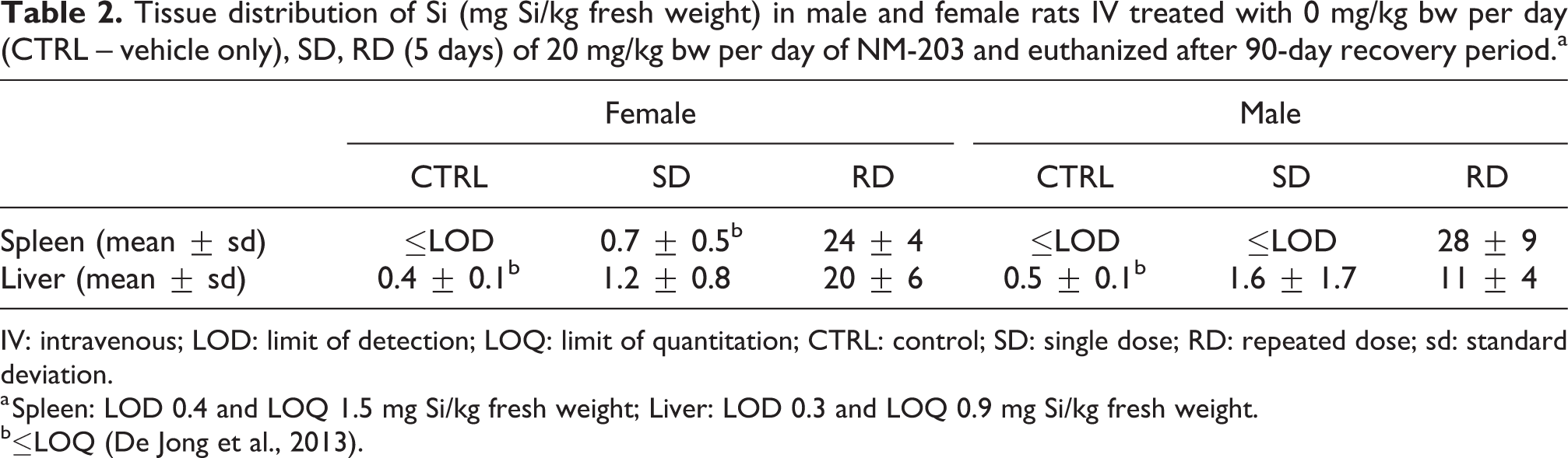
IV: intravenous; LOD: limit of detection; LOQ: limit of quantitation; CTRL: control; SD: single dose; RD: repeated dose; sd: standard deviation.
a Spleen: LOD 0.4 and LOQ 1.5 mg Si/kg fresh weight; Liver: LOD 0.3 and LOQ 0.9 mg Si/kg fresh weight.
b≤LOQ (De Jong et al., 2013).
None of the animals died or showed clinical signs or general toxic effects during the treatment and recovery periods. The bw gain and food consumption were unaffected in both sexes.
Spleen
Macroscopic observation of all RD-treated rats of both sexes showed evident splenomegaly. Absolute and relative weight was significantly increased in male and female rats of RD group in comparison to CTRL (Figure 2). The increase was greater in male (factor 5) than in female rats (factor 3) in comparison to the CTRL group.
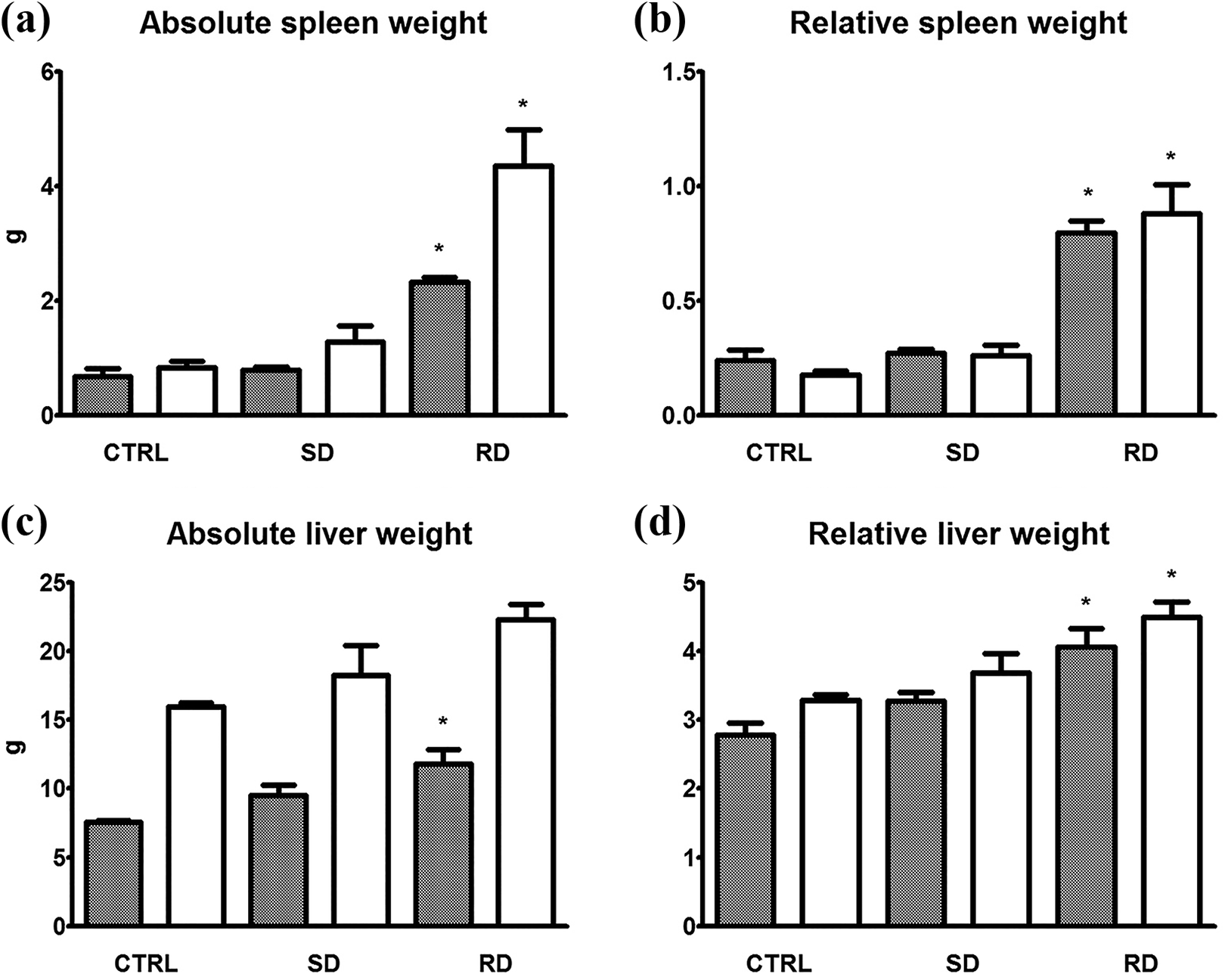
Spleen [(a) (b)] and liver [(c) (d)] absolute and relative weight of male (white column) and female (black column) rats IV treated with: 0 mg/kg bw per day (CTRL – vehicle only) and 20 mg/kg bw per day in SD or RD (5 days) of NM-203 and euthanized after 90-day recovery period; *p < 0.05. IV: intravenous; CTRL: control; SD: single dose; RD: repeated dose.
No treatment-related alterations were present in male and female SD rats.
The presence of inflammatory infiltrates consisting of both mononuclear cells and granulomas (Suttie, 2006) was also observed, significantly greater in both sexes of the RD group in comparison to CTRL. Granulomas were also present in one female rat and of two male rats of the SD group, and a dose-dependent increase of borderline significance (p = 0.0528) was observed for both sexes.
No granuloma was observed in CTRL animals (Table 3 and Figure 3).
General toxicological and histopathological data of male and female rats IV treated with 0 mg/kg bw per day (CTRL – vehicle only), SD, RD of 20 mg/kg bw per day of NM-203 for 5 days and euthanized after 90-day recovery.a
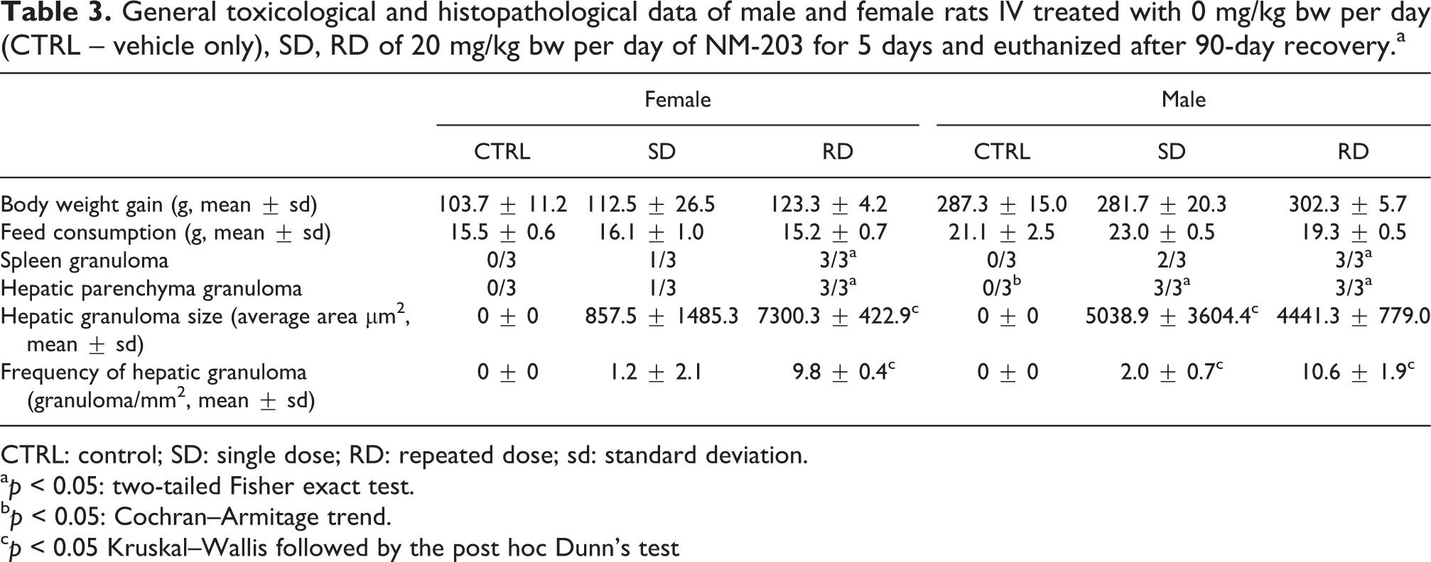
CTRL: control; SD: single dose; RD: repeated dose; sd: standard deviation.
a p < 0.05: two-tailed Fisher exact test.
b p < 0.05: Cochran–Armitage trend.
c p < 0.05 Kruskal–Wallis followed by the post hoc Dunn’s test
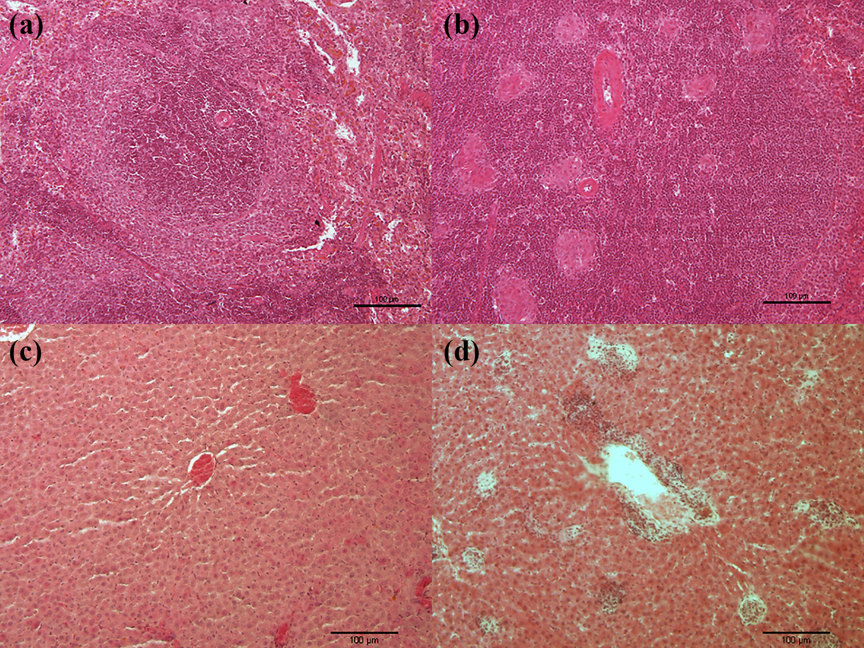
(a) Spleen of control male rat treated with 0 mg/kg bw per day (vehicle only). Germinal centre. (b) Spleen of male rat IV treated with repeated dose (5 days) of 20 mg/kg bw per day of NM-203 for 5 days. Inflammatory infiltrates and granulomas. (c) Liver of control male rat treated with 0 mg/kg bw per day (vehicle only). (d) Liver of male rat IV treated with repeated dose (5 days) of 20 mg/kg bw per day of NM-203 for 5 days. Parenchyma granuloma with lymphocytic infiltration. Bar = 100 mm (original magnification. ×10; H&E stain). H&E: haematoxylin and eosin.
The evident tissue disorganization due to granuloma formation did not allow us to perform quantitative analyses in one SD group female rat and in two female and three male rats in RD group.
Liver
Absolute and relative liver weights were significantly increased in female rats of the RD group in comparison to CTRL. In RD group male rats, an increase of borderline significance (p = 0.0507) was observed in the absolute weight, whereas relative weight was significantly increased in comparison to CTRL (Figure 2).
Both absolute and relative weight were unaffected in male and female rats of SD group.
In male rats, a dose-dependent increase of granulomas with lymphocytic infiltration, significantly greater both in the SD and RD groups in comparison to CTRL, was recorded. In female rats of the RD group, in comparison to CTRL a dose-dependent increase of borderline significance (p = 0.0528) was observed.
Granulomas were located mainly within the parenchyma, close to the centrilobular veins.
Significant differences were found also in the frequency of granulomas and in granuloma size in RD female rats in comparison to CTRL. In RD male rats, group-related significant differences were found in frequency of granulomas (Table 3 and Figure 3).
Discussion
The present article showed, for the first time, that the short-term IV exposure of male and female rats to SAS NP NM-203 induced severe damage in spleen with splenomegaly and granulomas with inflammatory infiltrates, still evident after a long-term recovery period (90 days). The granuloma structure appeared as a disordered but rather dense array of large macrophages intermixed with sparse granulocytes and lymphocytes. Inflammatory cell granulomas were also present in liver parenchyma and around the centrilobular veins. Tissue biodistribution data (Table 2) confirmed the persistence in both sexes of measurable Si concentrations even after 90-day post-treatment. IV administration of the same SAS (NM-203) to male rats at 5, 10, or 20 mg/kg at 48, 24 and 3 h prior to tissue collection induced micro-granuloma formation in liver, and histopathological alterations in spleen at the higher dose tested (cumulative dose = 60 mg/kg bw), not described as granulomas (Guichard et al., 2015). Single 7-mg/kg IV infusion of 13-nm SAS NP in rats evaluated at 7, 30 and 60 days post-treatment caused extensive liver remodelling, with the formation of multiple body-type granulomas starting from day 7 post-treatment, and subsequent development of fibrosis; no effects were described in spleen (Zhuravskii et al., 2016). The longer recovery period of the present study in comparison to other approaches contributed to highlight the presence of spleen granulomas and – at the same time – to confirm the presence and persistence of liver granulomas. Indeed, the granuloma formation in liver appeared to be a common effect of silica NP, independently from dimensions, time and dose levels (Guichard et al., 2015; Zhuravskii et al., 2016). Although toxicokinetic data showed that Si concentration in liver and spleen clearly decreased (De Jong et al., 2013; EFSA, 2018), it was still detectable and higher than CTRL after the 90-day recovery period. This suggests an elimination time longer than 90 days, potentially due to alteration of excretion/elimination processes. According to this, Si concentration/excretion can account for the granuloma formation in both tissues, and a longer period of Si persistence in spleen in comparison to liver. The present approach did not allow to evaluate the process of granuloma formation since the tissues were not sampled at different time points (e.g. 30, 60 days) after treatment, but the persistence over a long-term period is clearly evident. Moreover, the present data suggested that the recorded effects were significantly sex-related, with male rats being more susceptible than females: This was evident both in general toxicity data and in granuloma formation in both spleen and liver. This enhanced male susceptibility to SAS effects was not accompanied by increased Si bioaccumulation in target tissues, suggesting that different and sex-specific mechanisms of biodistribution and toxicity do exist. Overall, such data suggest that a body burden of Si might be established upon the widespread use in drugs, eliciting chronic reactions in target tissues and prompting to the need of further and more in-depth investigations on the safety of SAS NP.
Footnotes
Declaration of conflicting interests
The author(s) declared no potential conflicts of interest with respect to the research, authorship, and/or publication of this article.
Funding
The author(s) disclosed receipt of the following financial support for the research, authorship, and/or publication of this article: The author(s) received financial support by the Nanogenotox Joint Action (2008-2013).