
Abstract
Occupational exposure to heavy metals like lead (Pb) and cadmium (Cd) is associated with the development of several diseases. The objective of this study was to determine the effect of occupational co-exposure to Pb and Cd on the blood levels of selected immune-modulatory cytokines related to T helper (Th), that is, Th1, interleukin-2 (IL-2), Th2, (IL-4 and IL-10), and Th17, (IL-17) cells. The study comprised 207 individuals divided into two groups: exposed (n = 110) and nonexposed (n = 97). Blood Pb and Cd were determined using Graphite Furnace Atomic Absorption Spectroscopy, and serum levels of cytokines were measured using enzyme-linked immunosorbent assay (ELISA). The study revealed significantly higher blood Pb and Cd levels in the exposed group. A significant decrease in Th1 cytokine-IL-2 and Th2 cytokine-IL-10 was found, while IL-4 (Th2 cytokine) and IL-17 (Th17) levels were higher in the exposed group. In the mixed exposure analysis, among all the selected cytokines, IL-4 levels were significantly different between individuals having higher levels of both Pb and Cd versus lower levels of Pb and Cd. While IL-2 levels were highest among the low Pb and Cd group, the IL-17 levels were highest among individuals with higher Cd levels. The study demonstrated that co-exposure to low levels of Pb and Cd might have an immune-modulatory effect. The data suggested a metal-induced pro-inflammatory immune response.
Introduction
Lead (Pb) and Cadmium (Cd) are well-known environmental toxicants that may pose devastating effects on human health (Demenesku et al., 2014; Mitra et al., 2017). These metals are naturally present in the earth’s crust and accumulate in humans via various exposure routes (Milnerowicz et al., 2015). Exposure to heavy metals is gradually increasing due to increased industrialization and urbanization (Jan et al., 2015). Because of their versatile physical and chemical properties, Pb and Cd are widely used in various industrial and domestic settings such as smelting, mining, storage batteries, ceramic and handicraft manufacturing, welding, and paint industries (Mitra et al., 2017). Occupational exposure to these metals predisposes industrial workers to their associated toxicities (Azeh Engwa et al., 2019). Among the various physiological systems, the immune system is one of the most susceptible targets for heavy metal toxicity (Fenga et al., 2017; Lehmann et al., 2011; Matthews et al., 2016; Metryka et al., 2018).
Although the molecular mechanism of toxicity is still unclear, studies have demonstrated that these metals damage biological systems by altering the cell signalling pathways, cellular function, and enzyme activity. They also induce oxidative stress by disturbing the balance between the production and utilization of reactive oxygen species. Free radicals damage the cell membrane and trigger inflammatory reactions (Machoń-Grecka et al., 2017).
The exact underlying mechanism resulting in impairment of immune responses is not known. Experimental studies demonstrated that Pb might impair lymphocyte function resulting in cytokine imbalance and alter the percentage of lymphocyte subsets and antibody production (Sirivarasai et al., 2013). Some have reported the immunosuppressive effect of Pb (Carey et al., 2006; Dietert et al., 2004), while others (Fenga et al., 2017) have reported immunostimulatory effects of Pb.
Cd’s role concerning the immune system has been explored in experimental studies, wherein it is known to be involved in the activation and priming of various immune cells (Kataranovski et al., 2009). Cd exposure resulted in stimulation of the immune response at lower concentrations and inhibition at higher concentrations (Boscolo et al., 2005; Hemdan et al., 2006; Marth et al., 2000). Another study supported these findings where Cd exposure showed differential effects on peripheral blood mononuclear cells, resulting in altered cytokine production (Maria et al., 2000).
Although the evidence for heavy metal-associated immune imbalance is steadily increasing, data from human studies are limited, and little is known about the underlying immunological mechanisms. Previous studies have reported ambiguous findings concerning Pb, Cd, and the immune system. Furthermore, individuals working in various industrial settings are exposed to mixtures of these metals. These metals may act synergistically or otherwise, resulting in effects not observed when exposed to a single metal. However, significantly less is known about the effect of co-exposure to these metals in human subjects. In light of the gap of information regarding Pb and Cd co-exposure on the human immune system, the present study was designed to study the association between co-exposure to Pb and Cd and immune-modulatory cytokines in occupationally exposed individuals working in the various factories of Jodhpur, Rajasthan.
Materials and methods
The studied population comprised 110 workers (as the exposed group) and 97 apparently healthy subjects (as the control group). Workers were randomly selected from factories involved in metal handicraft production and welding. Among the exposed individuals, the workers involved in the welding activities were defined as welders and comprised of 62 workers (n = 62), while 48 workers were in particular involved in metal handicraft manufacturing (n = 48). All the included workers had to be directly involved in the same industrial process for at least 2 years. The control group was selected from the areas far away from these industrial sites to include 97 subjects with no prior history of occupational exposure to these metals.
Exclusion criteria
Workers who were on a part-time job. Individuals with any history of systemic inflammatory diseases (e.g. rheumatoid arthritis and lupus), diabetes, nonmedically controlled hypertension, cancer, known coronary artery disease, thyroid, liver, and kidney disease. Individuals who are taking any steroids or nonsteroidal anti-inflammatory drugs. Individuals with a self-reported history of a common cold or any other infection during the last 7 days.
The study was approved by the Institutional Ethics Committee, and informed consent was obtained from every participant. A detailed history of the duration of work, place of work, smoking and alcohol consumption, drinking water, food habits, and medical illness was taken from every subject utilizing the questionnaire provided to them. Anthropometric parameters of all study subjects (height (cm), body weight (kg), and blood pressure (mm Hg)) were measured by trained research assistants.
Sample collection and storage
A total of 5 ml of blood was collected from each individual by venipuncture under aseptic conditions. A total of 2 ml of whole blood was taken in ethylenediaminetetraacetic acid (EDTA) containing vacutainer tubes (BD, Franklin Lakes, New Jersey, USA) for the determination of blood lead levels (BLL) and blood cadmium levels (BCL) and stored at −80°C until further use. A total of 3 ml of blood collected in vacutainers without additives or anticoagulants was allowed to clot, then centrifuged at 1500 x g (rcf) for 10 min to obtain serum, and then transferred to 2-ml Eppendorf tubes (Eppendorf, AG, Hamburg, Germany), which were stored at −20°C for cytokine level determination.
Determination of BLL and BCL
The BLL and BCL were determined using graphite furnace atomic absorption spectrometry with Zeeman correction in an ICE 3500 system (Thermo Fischer Scientific, Waltham, Massachusetts, USA). A solar GFS35Z graphite atomizer, GFS35Z autosampler, and pyrolytically graphite-coated cuvettes (Thermo Fischer Scientific, Walthham, Massachusetts, USA) with the platform were used. Analysis followed a five-step program (130°C/20 s, 200°C/30 s, 600°C/10 s, 2000°C/3 s, and 2450°C/3 s). The whole blood sample collected in EDTA containing vacutainers was processed as per the Centers for Disease Control and Prevention’s (CDC) method to estimate blood Cd and Pb (CDC, 2001). Samples, as well as standards, were diluted (1:20) with matrix modifier solution containing 0.5%Triton X-100 (Sigma, Darmstadt Germany), 0.1% diammonium hydrogen phosphate (Thermo Fisher), and 0.2% HNO3 (w/v) (Merck, Darmstadt, Germany). Blood control levels I, II, and III (Recipe, Germany), containing 5.84 µg/dL, 21.9 µg/dL, and 42.5 µg/dL of Pb and 1.23 µg/L, 2.88 µg/L, and 6.32 µg/L of Cd, respectively, were used for quality control. The results were expressed as µg/dL and µg/L for Pb and Cd, respectively. The limit of detection and limit of quantification were 0.12 µg/dL and 0.36 µg/dL for Pb and 0.01 µg/L and 0.05 µg/L for Cd, respectively. All glassware, pipette tips, sample cups, and stoppers were soaked in acid (10% HNO3) overnight, then rinsed with distilled water, and dried before use to assure no contamination.
Determination of serum cytokine levels
Serum levels of interleukins (ILs), including IL-2, IL-4, IL-10, and IL-17, were measured using an in vitro enzyme-linked immunosorbent assay (ELISA), according to the manufacturer’s instructions (KRISHGEN Biosystems, Mumbai, Maharashtra, India). Samples were added to 96 microwell plates precoated with the monoclonal antibody. Biotinylated antibody solutions were added, and plates were incubated at 37°C. The plates were washed, and the streptavidin–horseradish peroxidase solution was added, followed by washing. 3,3′,5,5′-tetramethylbenzidine substrate was added and finally, stop solution was added after incubation for 10–15 min to stop the reaction. The microplates were analyzed using an Eon Biotek multiplate (14021320,Winooski, USA) ELISA reader. The wavelength was set at 450 nm. Five-point calibration curves were used for all biomarker measurements by Gen5 software (version: 2.05.5). The results were expressed as pg/mL.
Statistical analysis
We expressed descriptive data as mean (standard deviation) or median (interquartile range) for continuous variables and number (%) for categorical variables. Statistical significance of changes was determined by GraphPad Prism™ using nonparametric statistical methods (GraphPad Prism, version 8.3.0 (538) for Windows, GraphPad Software, La Jolla California, USA, www.graphpad.com). The Shapiro–Wilks test ascertained normality. The Mann–Whitney U test was used for comparisons between two groups (exposed and nonexposed). The Kruskal–Wallis test was used for comparison between more than two study groups. Spearman’s correlation was used for the association between BLL, BCL, and serum cytokine levels.One-way multivariate analysis of variance (MANOVA) was done to assess the differences between the means on a combination of dependent variables p values less than or equal to 0.05 were considered to be statistically significant.
Results
Demographic characteristics of the study population
The demographic characteristics of the study population are presented in Table 1. A total of 48 (43.64%) of exposed individuals were involved in metal handicraft work, while 62 (56.36%) were welders. The mean duration of exposure in the exposed group was 7.97 ± 8.14 years, with no significant difference between welders (6.19 ± 5.49 years) and metal handicraft workers (7.84 ± 7.34 years; p = 0.77). Mean levels of age, height, systolic, and diastolic blood pressure of the exposed group showed no significant differences from the nonexposed group. A significant difference in BMI did not affect the measured variable as validated by the multivariate test (Wilk’s lambda).
Demographic characteristics of the study population.a
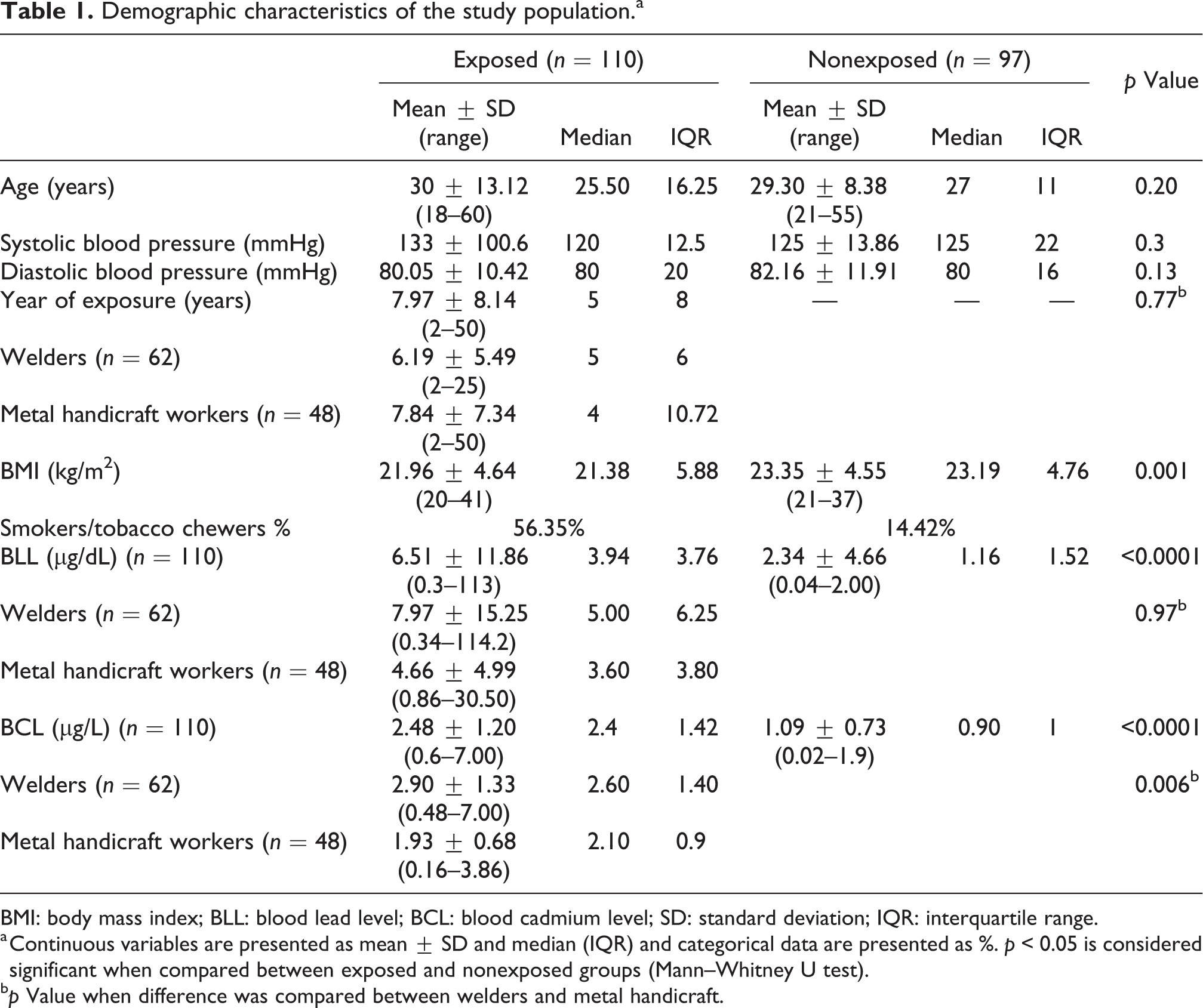
BMI: body mass index; BLL: blood lead level; BCL: blood cadmium level; SD: standard deviation; IQR: interquartile range.
a Continuous variables are presented as mean ± SD and median (IQR) and categorical data are presented as %. p < 0.05 is considered significant when compared between exposed and nonexposed groups (Mann–Whitney U test).
bp Value when difference was compared between welders and metal handicraft.
BLL and BCL in the study population
BLL and BCL were found to be significantly higher in the exposed group in comparison to the nonexposed group. The mean BLL in the exposed group was 6.51 ± 11.86 µg/dL and in the nonexposed group was 2.39 ± 4.66 µg/dL (p < 0.0001). The mean BCL in the exposed group was 2.48 ± 1.20 µg/L and in the nonexposed group was 1.09 ± 0.73 µg/L (p < 0.0001; Table 1). Furthermore, subgroup analysis based on their job profiles revealed a significant difference in the BCL between welders and metal handicraft artisans; however, no such difference was observed in BLL between these groups (Figure 1(a) and (b)). The median BCL level in welders was 2.60 µg/L, which was significantly greater than the 2.10 in metal handicraft artisans (p = 0.006). The confounding variables (age, BMI, and smoking) did not show a statistically significant relationship with metal levels and ILs in the study population.
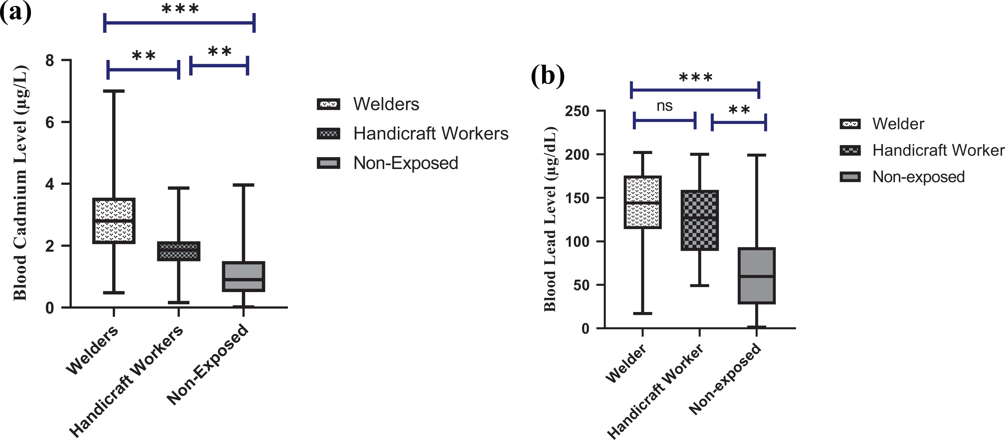
(a) Comparison of BCL in welders (n = 62), metal handicraft workers (n = 48), and nonexposed group (n = 97). BCL was significantly higher in welders in comparison to metal handicraft workers as well as nonexposed group. (b) Comparison of BLL in welders (n = 62), metal handicraft workers (n = 48), and nonexposed group (n = 97). Welders and metal handicraft workers did not show significant differences between each other but a significant difference was observed when compared with the nonexposed group. **p < 0.05; ***p < 0.0001; Kruskal–Wallis test was used to compare BLL and BCL between welders, metal-handicraft workers, and nonexposed group; BLL: blood lead levels; permissible limit: BLL: 5 µg/dL or greater be defined as elevated [CDC, 2001], BCL: blood cadmium levels; permissible limit: BCL: 0.3–1.2 µg/L [WHO, 1996].
Serum cytokine concentrations
The mean plasma IL-2, IL-4, and IL-10 levels were significantly different between the exposed and nonexposed groups, while IL-17 did not differ between the groups. In particular, IL-2 and IL-10 levels were significantly lower, and IL-4 was significantly higher in the exposed workers (Table 2). Among the exposed individuals, when compared between welders and metal handicraft artisans, the IL-4 level was found to be significantly higher in metal handicraft artisans, while IL-17 and IL-10 levels were higher in the welders (Supplementary Figure S1).
Serum cytokine levels in the exposed and nonexposed groups.a
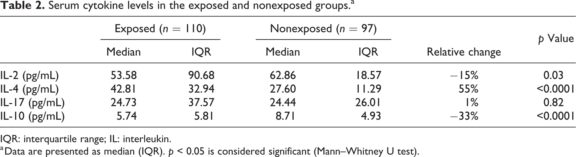
IQR: interquartile range; IL: interleukin.
a Data are presented as median (IQR). p < 0.05 is considered significant (Mann–Whitney U test).
Concerning correlations between the measured cytokines and metals level of the exposed workers (n = 110), Spearman’s analysis revealed no statistically significant correlation between the cytokine levels and the individual metals level (Table 3).
Correlation between BLL, BCL, and cytokine levels in the exposed group (n = 110).a
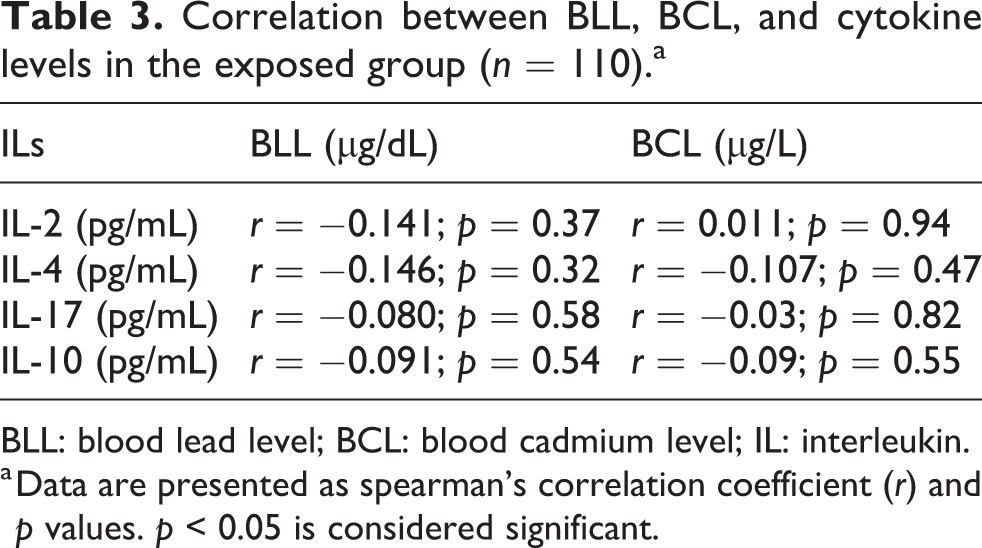
BLL: blood lead level; BCL: blood cadmium level; IL: interleukin.
a Data are presented as spearman’s correlation coefficient (r) and p values. p < 0.05 is considered significant.
However, in the combined group (exposed and nonexposed), BCL was found to be positively correlated with IL-17 (r = 0.15) and IL-4 (r = 0.15), while negatively associated with IL-10 (r = −0.15). BLL, on the other hand, had a negative correlation only with IL-2 (r = −0.15). Age and cytokine level did not have any significant correlation in the study participants (Table 4).
Correlations between cytokine levels, age, BCL, and BLL in the combined study population (n = 207).a
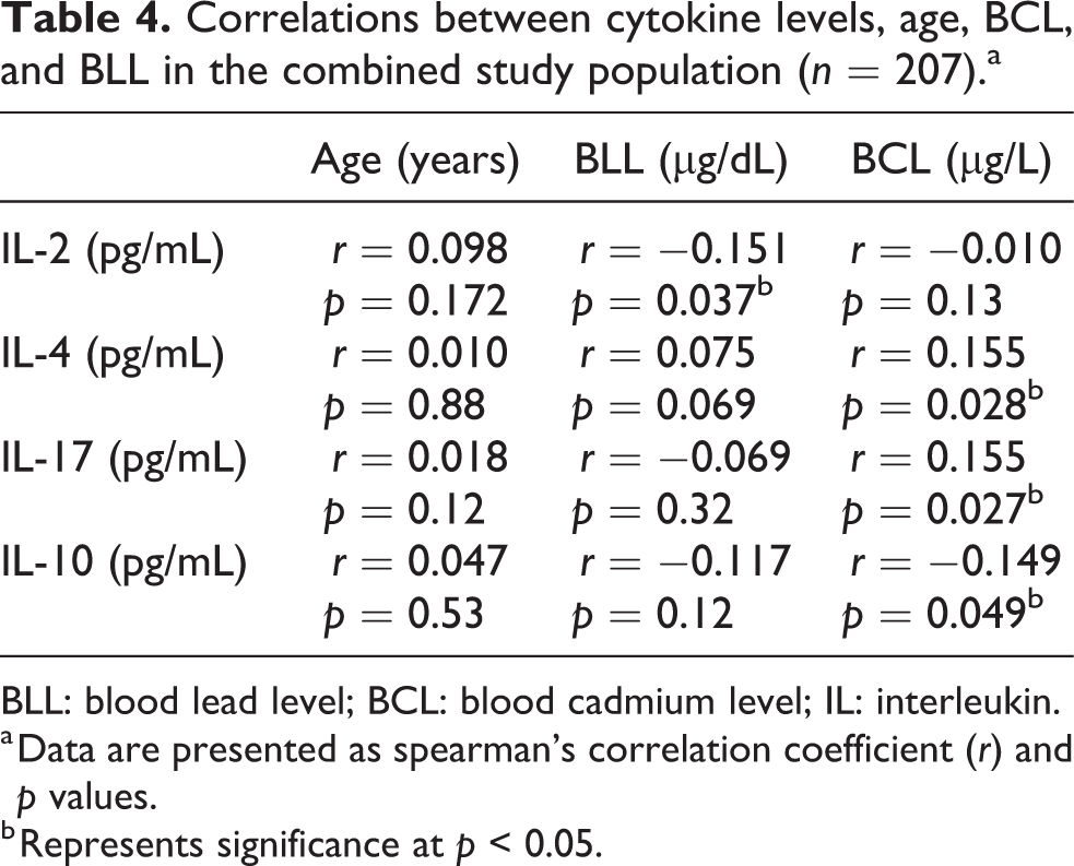
BLL: blood lead level; BCL: blood cadmium level; IL: interleukin.
a Data are presented as spearman’s correlation coefficient (r) and p values.
b Represents significance at p < 0.05.
The association of co-exposure to metals with ILs was examined using combined categorical variables classified as low and high groups. The cutoff value was set to the median level of Cd and Pb, which was 2.40 µg/L and 3.94 µg/dL, respectively. The study participants were divided into four subgroups as low Pb/Cd (n = 19), low Cd high Pb (n = 19), high Cd low Pb (n = 28), and high Pb/Cd (n = 44). The variations in ILs concentration by combined exposure level of Cd and Pb are presented in Table 5. Among the studied ILs, serum IL-2, IL-4, and IL-17 showed a significant difference between the groups. The IL-4 level was significantly lower in the high Pb/Cd group than the low Pb/Cd group. Serum IL-17 levels were significantly higher in the high Cd–low Pb group than the low Pb/Cd group, while IL-2 levels were significantly lower in the high Pb–low Cd group than the low Pb/Cd group (Table 5).
The concentration of cytokines by combined exposure levels of BCL and BLL in exposed workers (n = 110).a
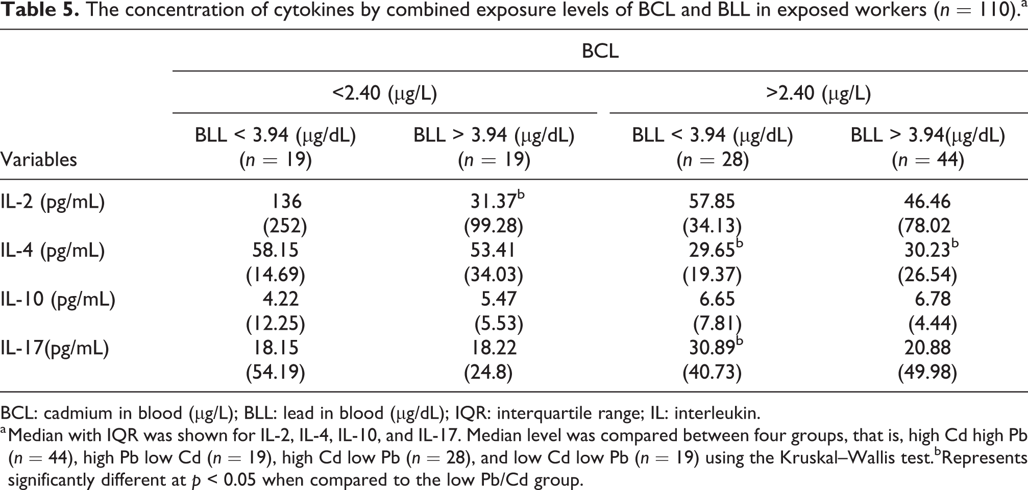
BCL: cadmium in blood (µg/L); BLL: lead in blood (µg/dL); IQR: interquartile range; IL: interleukin.
a Median with IQR was shown for IL-2, IL-4, IL-10, and IL-17. Median level was compared between four groups, that is, high Cd high Pb (n = 44), high Pb low Cd (n = 19), high Cd low Pb (n = 28), and low Cd low Pb (n = 19) using the Kruskal–Wallis test.bRepresents significantly different at p < 0.05 when compared to the low Pb/Cd group.
Discussion
Extensive use of Pb and Cd in industries poses direct and indirect health effects on human health. Epidemiological studies have evaluated the relationship between heavy metal exposure and immune dysfunction and reported conflicting results (Wu et al., 2018).
T cells are more susceptible to the toxic effects of Pb exposure, which may alter the development of T cells and the cytokine produced with a bias towards Th2 response (Fenga et al., 2017). While in vitro studies revealed an immunosuppressive effect of Pb, findings from in vivo studies suggest an immune stimulatory role of Pb (Colombo et al., 2004).
Cytokines secreted by the respective T helper (Th) cells are the immunomodulatory signals that mediate and regulate immunity, inflammation, hematopoiesis, and many other cellular processes, forming a cytokine network (Luis Muñoz-Carrillo et al., 2019). In the present study, serum levels of Th1 cytokine: IL-2, Th2 cytokine: IL-10 and IL-4, and a Th-17 cytokine: IL-17 were measured in workers occupationally co-exposed to Pb and Cd and compared with the nonexposed group. To the best of our knowledge, it is the first human study to evaluate the association of co-exposure of these metals with candidate ILs. Serum levels of IL-2, IL-4, and IL-10 were significantly different between the exposed and the nonexposed groups.
In contrast to the in vitro findings (Krocova et al., 2000), where Pb and Cd exposure resulted in a significant increase in IL-2 concentrations, the serum levels of IL-2 in our study’s exposed group were significantly lower when compared to the nonexposed group. However, similar findings of a decrease in IL-2 concentration in Cd-exposed primary T lymphocytes of BALB/c mice at different time intervals were reported by Pathak and Khandelwal (2008). Furthermore, a few other studies in occupationally exposed workers reported no alterations in IL-2 levels (Valentino et al., 2007; Yücesoy et al., 1997). In contrast, Colombo et al. (2004) demonstrated that a mixture of Pb and Hg inhibited IL-2 production while Cd stimulated IL-2 production in preactivated T cells. However, the mixture of all three metals did not have any effect on IL-2 production. On the contrary, on subgroup analysis, we found that the IL-2 levels were comparatively higher in the low Pb/Cd group, suggesting a possible inverse association between Pb and Cd and IL-2 levels in the exposed population. The weak negative correlation between IL-2 and BLL further corroborates the findings of prior human and experimental studies that have reported Pb-induced suppression of Th1 cytokines (IL-2 and IFN-γ) (Heo et al., 1998; 2007).
IL-4 is primarily secreted by activated Th2 cells. In vivo, Pb exposure resulted in significant IL-4 production in mice (Heo et al., 2006). Additionally, Iavicoli et al. (2006) suggested a dose-dependent change in the cytokine levels of different lymphocyte subsets on Pb exposure. They reported an increase in IL-4 levels with a profound decrease in IL-2 and interferon gamma at higher Pb concentrations. Reports on the effect of Pb on IL-4 levels in human subjects are ambiguous wherein significant decrease (García-Lestón et al., 2012), increase (Dobrakowski et al., 2016), or no change has been observed (Valentino et al., 2007). Moreover, a few experimental studies have demonstrated Cd-mediated changes in IL-4 levels, wherein inconclusive results have been reported (Hanson et al., 2012; Turley et al., 2019). IL-4 levels in Cd-exposed human subjects have not been explored extensively. Significantly higher IL-4 levels were noted in the exposed group in the current study. In further subgroup analysis, IL-4 was significantly higher in the low Pb/Cd group than in the high Pb/Cd group. The present study’s result agrees with the previous literature that states the effect of these metals individually or in combination resulting in a Th2 bias.
IL-17 is the mediator of the Th17 immune response and is primarily responsible for the activation of mononuclear phagocytes, the recruitment of neutrophils, and the induction of epithelial antimicrobial responses (Hemdan et al., 2013). The effect of metal or metal mixtures has not been explored concerning the Th17 subset. We came across only studies where Cd exposure in mice resulted in a significant increase in IL-17 messenger RNA (mRNA) expression, which did not translate into higher serum IL-17 levels (Djokic et al., 2015; Turley et al., 2019). In an extensive literature search, only Dobrakowski et al. (2016) reported IL-17 levels concerning occupational Pb exposure. They observed no effect on IL-17 levels on short-term Pb exposure, whereas increased IL-17 levels were noted in workers exposed for longer duration to Pb. We noted an increasing but nonsignificant trend in IL-17 levels in the exposed group in comparison to controls. Subgroup analysis revealed the highest IL-17 level in individuals with low Pb and high Cd levels, thereby suggesting that Cd may significantly affect the induction of IL-17 production.
IL-10 has pleiotropic effects in immunoregulation. It acts as an anti-inflammatory cytokine produced from Th2 cells and regulatory T cells and inhibits Th1 cells. Various experimental studies have reported conflicting results concerning IL-10 level in Pb-exposed animals (Kamińska et al., 2007; Krocova et al., 2000), whereas significantly higher IL-10 levels have been observed in occupationally Pb exposed workers (Valentino et al., 2007). Riemschneider et al. (2015) reported significantly lower IL-10 levels in murine macrophages exposed to Cd, whereas others have reported no significant difference in IL-10 mRNA levels in wound tissue between Cd treated and untreated mice (Mei et al., 2017). Odewumi et al. (2015) reported a dose-dependent decrease in IL-10 expression level following Cd exposure in human lung cancer cells. IL-10 level was found to be significantly lower in the exposed group in this study population. Furthermore, subjects in the low Pb low Cd group were found to have the lowest level of IL-10, while individuals in the high Pb high Cd group had higher IL-10 level. Higher IL-10 level in the high Pb/Cd group in this study could be attributed to the dose-dependent effect, wherein previous studies have reported an increase in IL-10 level at higher metal concentrations.
Furthermore, there are certain limitations to this study. To get a better idea about the co-exposure to the metal in occupational settings, exposure markers like metal load in the air and dust should be determined, which was beyond our scope. Besides, the presence of other particulate matter in the occupational environment may affect the studied variables, which could be a possible reason for no significant correlation between the measured metals and immune parameters in the study. Further, more studies with a panel of more extensive, comprehensive cytokines would give a better immune picture to reach a conclusive idea about the immune-modulatory effect of co-exposure to Pb and Cd in occupational settings.
In conclusion, we found that co-exposure to even low Pb and Cd levels may alter cytokine equilibrium. A significant increase in IL-4 and decreased IL-2 levels draws a parallel with previous findings suggestive of metal facilitating bias towards Th2-mediated immune responses. However, decreased IL-10 levels in the exposed group could be attributed to the observed range of Pb and Cd in the study population, as earlier studies have reported a dose-dependent increase in the IL-10 levels with higher doses of Pb and Cd. Furthermore, on subgroup analysis, higher IL-10 levels seen in the high Pb–high Cd group agrees with the dose-dependent effect in IL-10 levels. Collectively, Pb and Cd exposure in workers is associated with increased IL-4 and IL-17 and decreased IL-2 and IL-10, suggesting a pro-inflammatory immune response.
Supplemental material
Supplemental Material, sj-pdf-1-tih-10.1177_07482337211019172 - Effect of occupational co-exposure to lead and cadmium on selected immunomodulatory cytokines
Supplemental Material, sj-pdf-1-tih-10.1177_07482337211019172 for Effect of occupational co-exposure to lead and cadmium on selected immunomodulatory cytokines by Taru Goyal, Prasenjit Mitra, Preeti Singh, Shailja Sharma, Purvi Purohit and Praveen Sharma in Toxicology and Industrial Health
Footnotes
Authors’ note
Dr Prasenjit Mitra is now affiliated with Department of Biochemistry, Postgraduate Institute of Medical Education & Research, Chandigarh, India.
Author's contribution
Taru Goyal and Prasenjit Mitra have contributed equally as the first authors in this work.
Compliance with ethical standards
Research Involving Human Participants and/or Animals: This article involves the participation of human subjects with approval from the Institutional Ethical Committee (IEC), All India Institute of Medical Sciences, Jodhpur, Rajasthan India (AIIMS/IEC/2018/555).
Declaration of conflicting interests
The author(s) declared no potential conflicts of interest with respect to the research, authorship, and/or publication of this article.
Funding
The author(s) disclosed receipt of the following financial support for the research, authorship, and/or publication of this article: All-India Institute of Medical Sciences
Informed consent
Prior written informed consent was obtained from all subjects recruited in this study.
Supplemental material
Supplemental material for this article is available online.
References
Supplementary Material
Please find the following supplemental material available below.
For Open Access articles published under a Creative Commons License, all supplemental material carries the same license as the article it is associated with.
For non-Open Access articles published, all supplemental material carries a non-exclusive license, and permission requests for re-use of supplemental material or any part of supplemental material shall be sent directly to the copyright owner as specified in the copyright notice associated with the article.