
Abstract
Exposure to mobile phone radiation causes deleterious health effects on biological systems. The objects of this study were to investigate the effect of 900-MHz radiofrequency waves (RFW) emitted from base transceiver station antenna on intrapancreatic homocysteine (Hcy), tumor necrosis factor-α (TNF-α), and nerve growth factor (NGF) as predisposing factors involved in pancreatic beta cell damage. Thirty male rats (Sprague-Dawley, 200 ± 10 g) were randomly divided into the control (without any exposure) and exposed groups: short time (2 h/day), long time (4 h/day), and exposed to 900-MHz RFW for 30 consecutive days. On the last days of the experiment, animals were killed and pancreas tissue was dissected out for evaluation of serotonin, Hcy, TNF-α, and NGF. There was a significant decrease in the serotonin and NGF levels in the pancreatic tissue of exposed groups compared to the control group (p < 0.05). Also, the levels of serotonin and NGF in the long-time exposure were significantly lower than the short-time exposure (p < 0.05). However, levels of Hcy and TNF-α were significantly increased in the pancreas of exposed groups compared to the control groups (p < 0.05). Exposure to 900-MHz RFW decreased pancreatic NGF and serotonin levels and increased the proinflammatory markers (Hcy and TNF-α), which can be a predisposing factor for type 2 diabetes.
Introduction
A wide spectrum of electromagnetic fields (EMFs) is emitted by several sources such as communication equipment, radar, and base transceiver station antennas. Changes in lifestyles have dramatically increased lifetime exposure to EMF. Previous studies have demonstrated the deleterious health effects of exposure to 900-MHz radiofrequency waves (RFW) on the brain tissue and reproductive system (Azimzadeh and Jelodar, 2019; Azimzadeh and Jelodar, 2020).
A growing body of studies has suggested that overproduction of reactive oxygen species (ROS) and, consequently, oxidative stress as the main mediator of nonthermal effects of RFW on biological tissues (Ismaiil et al., 2019; Masoumi et al., 2018).
Serotonin (5-HT) is a monoamine, and approximately 5% of total body content is produced in serotonergic neurons of the central nervous system, whereas 95% is produced, stored, and released from enterochromaffin cells, enteric neurons, pancreatic cells, and adipose tissue in the gastrointestinal tract (Berger et al., 2009).
Serotonin acts as an autocrine/paracrine factor to alter beta cell mass and proliferation. It stores in granules together with insulin and has a functional role in the regulation of insulin secretion, and altered seratonin function is involved in beta cell dysregulation and eventually diabetes mellitus in humans (Kim et al., 2015). Serotonin along with insulin is released from beta cells and its secretion is regulated by increased glucose levels in human and mouse beta cells (Bennet et al., 2015; Cataldo et al., 2015). It has been suggested that intrapancreatic synthesized serotonin supports normal insulin secretion in an autocrine manner (Sumara et al., 2012). Studies have reported that exposure to the various spectra of EMF and extremely low-frequency magnetic fields (ELF-EMF) decreased insulin levels (Khaki et al., 2016; Mortazavi et al., 2016).
Nerve growth factor (NGF), as a classical trophic factor for nerve cells, is produced and secreted by adult rat pancreatic beta cells through autocrine/paracrine mechanisms, which plays a critical role in normal islet morphogenesis (Rosenbaum et al., 1998).
NGF is associated with systemic glucose metabolism and exerts trophic effects on beta cell survival, maturation, and insulin secretion (Chaldakov et al., 2009; Navarro-Tableros et al., 2004). It has been shown that a high level of glucose enhances NGF biosynthesis, and consequently, NGF promotes insulin secretion from cultured beta cells in humans and mice (Pingitore et al., 2016; Rosenbaum et al., 2001). Moreover, alteration of NGF in the islets may be involved in the etiology of diabetes (Kim et al., 2009).
Recently, vascular contractile cells were also suggested as a potential source of NGF in the pancreas with a functional role in glucose homeostasis and glucose-stimulated insulin secretion, which indicated a direct role for NGF signaling within pancreatic endocrine cells in controlling insulin secretion (Houtz et al., 2016). A significant increase of NGF level following exposure to 900-MHz RF has also been reported in testis tissue (Azimzadeh and Jelodar, 2019).
Homocysteine (Hcy) derived from the essential amino acid methionine and its increased serum and plasma levels is a predictive factor of high-risk mortality (Fratoni and Brandi, 2015). Hcy is a well-known and powerful proinflammatory factor that can stimulate the production of inflammatory cytokines such as TNF-α (Dalal et al., 2003). It has been suggested that the generation of ROS/oxidative stress is one of the main mechanisms through which Hcy exerts toxic effects (Li et al., 2008; Ma et al., 2018).
Moreover, previous studies reported that a high Hcy level is associated with glucose intolerance, insulin resistance, and impaired insulin signaling pathways through oxidative stress, which can lead to type 2 diabetes (Becker et al., 2003; Golbahar et al., 2007).
Tumor necrosis factor-α (TNF-α) is a master regulator of inflammation with a key role in the cytokine network, which regulates essential biologic functions (e.g. cell differentiation, proliferation, survival, apoptosis), and also mediates a broad spectrum of responses to stress and injury.
Although the major source of TNF-α production is immune cells (macrophages/monocytes), many other cell types including pancreatic acinar cells can produce, release, and respond to TNF-α (Hehlgans and Pfeffer, 2005). It has been reported that oxidative stress and proinflammatory cytokines such as TNF-α induce acinar cell injury and pancreatic inflammation (Powell et al., 2000).
Therefore, the present study aimed to evaluate the status of pancreatic tissue following exposure to 900-MHz RFW for short-time (2 h/day) and long-time (4 h/day) periods for 30 consecutive days by measuring intrapancreatic serotonin (an easier method to track insulin release), proinflammatory markers (Hcy and TNF-α), and NGF.
Materials and methods
Animal experiments
Twenty adult male rats (Sprague-Dawley, 200 ± 10 g) were randomly divided into three equal groups (n = 10): control group (without any exposure), short-time (2 h/day), and long-time exposed group (4 h/day) with free access to food and water. Animals were housed in polycarbonate cage with dimensions 42 × 26.5 × 15 cm3 without any metallic interfering materials in the animal room under controlled conditions: 12-h light/dark cycles and temperature between 20 ± 2°C. The irradiation of animals started at 9:00 a.m. for 30 consecutive days. All investigations were conducted in accordance with the “Guiding Principles for the Care and Use of Research Animals” approved by Shiraz University (S9431209, February 2018).
Signal generator and irradiation protocols
The 900-MHz RF exposure apparatus was made in the Department of Telecommunication and Electronics Engineering, Shiraz University. A simulator with a 12-cm antenna emitting 900-MHz RFW circularly with an average power density of 0.6789 mW/cm2 was used. The output of the device was monitored by a spectrum analyzer (FSH6, Rohde and Schwarz, Germany). The simulator was centrally placed 1 m away from the cages of the exposed group (short time and long time for 30 consecutive days) on the floor of the experimental room. The whole-body average specific absorption rate was 0.035 W/kg as it was measured 1 m away from the signal generator by SPM2 EMF meter (WPF18 Field Probe 300 kHz–18 GHz, Wave Control, Spain).
Harvesting of the pancreas and hormonal assay
On the last day of the exposure period, rats were anesthetized using a 2% diethyl ether-saturated cotton ball in a chamber for 3–5 min and euthanized by whole blood collection through heart puncture. The pancreas in all groups was rapidly harvested, homogenized, and centrifuged (a vicious circle that facilitate inflammation in the tissue; xg). Then, the supernatant was used to evaluate the concentration of investigating factors including Hcy (Diazyme, Shanghai, China), serotonin (IBL, Switzerland), TNF-α, and NGF by ELISA kits (Crystal Day Biotech Shanghai, China). The total protein concentration of the pancreas tissue was determined by the procedure, described by Bradford (Bradford, 1976).
Statistical analysis
All data are presented as mean ± standard error of the mean. The software of SPSS version 21.0 for Windows was used for statistical analysis of data. The analysis of variance (ANOVA) followed by post hoc Tukey’s multiple comparisons test was used to compare mean values between groups, and the significance level was set at p < 0.05.
Results
In this study, the amounts of all studied factors (serotonin, Hcy, TNF-α, and NGF) were measured within the pancreas tissue.
The mean of serotonin in the short-time (45.58 ± 0.89) and long-time (36.15 ± 1.06) exposed groups was significantly less than the control group (70.25 ± 1.48) (p < 0.05). Also, there was a significant difference between the two exposure groups (p < 0.05; Figure 1).
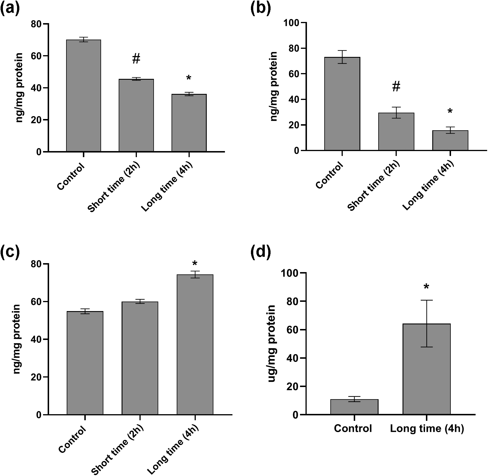
The impact of 900-MHz RFW on the (a) serotonin, (b) NGF levels in the pancreas tissue of rats (N = 10/each group), and (c, d) proinflammatory markers (Hcy and TNF-α). Short-time (2 h/day) and long-time (4 h/day) groups were exposed to RFW for 30 consecutive days. All data were represented as mean ± standard error of the mean. * represents a significant change compared to other groups (p < 0.05). # represents a significant change compared to other groups (p < 0.05). Hcy level in the short-time exposed group was missed due to a technical error. RFW: radiofrequency wave; TNF-α: tumor necrosis factor-α; Hcy: homocysteine; NGF: nerve growth factor.
Likewise, the mean of NGF levels showed a significant decline in the short-time (29.62 ± 4.29) and long-time (15.9 ± 2.55) exposed groups compared to the control group (73.09 ± 5.07) (p < 0.05). NGF level in the short-time exposed group was significantly lower than the long-time exposed group (p < 0.05; Figure 1).
However, the mean of TNF-α level showed a significant increase in the long-time exposed group (74.35 ± 1.85) compared to the short-time exposed (60.1 ± 1.11) and control group (54.87 ± 1.33) (p < 0.05) (Figure 1).
The mean Hcy level in the long-time exposed group (64.3 ± 16.51) was significantly higher than the control group (11.07 ± 1.84) (p < 0.05) (Figure 1). Due to laboratory technical error during the experiment, the data of Hcy level in the short-time exposed group were not collected.
Discussion
The key findings of this study were that exposure to 900-MHz RFW resulted in a significant decrease in serotonin and NGF levels accompanied by a significant increase in the proinflammatory markers (Hcy and TNF-α) in the pancreas tissue of rats.
Serotonin monitoring is often an easier method to track insulin release since it stores with beta cell granules and is released along with insulin (Robinson, 2009). Therefore, in this study, the level of serotonin was used as an index of insulin secretion from beta cells. To the best of our knowledge, this is the first report demonstrating the alteration of serotonin level in the pancreas tissue following exposure to RFW.
Contradictory results have been reported on the effect of EMF on insulin secretion. It has been reported that exposure to 900 MHz (6 h/day for 7 days) significantly decreased the mean insulin level (Mortazavi et al., 2016). Also, a significant decrease in insulin level following exposure to ELF-EMF (50 Hz, intensity of 3 mT, 4 h/day for 6 weeks), which was associated with the decreased area and perimeter of pancreatic islets, was reported (Khaki et al., 2015). On the other hand, some studies demonstrated the enhancement of insulin secretion following exposure to ELF-EMF (60, 50, and 5–20 Hz) (Destefanis et al., 2015; Gholampour et al., 2011; Sakurai et al., 2008) and the strong static magnetic fields used in magnetic resonance imaging (Sakurai et al., 2009). Variation in the results is linked with applied different experimental conditions and methodologies, in which the duration of exposure is the most important.
Alteration in the size of pancreatic islets and changes in the cell membrane electrical activity are the suggested mechanisms for the effects of EMF on the pancreas function and insulin release (Farashi et al., 2018). Oxidative stress is also a well-known mediator in the occurrence of pathological lesions and degenerative effects in the pancreatic endocrine (including beta cells) and exocrine cells (Farahna et al., 2014; Sakurai et al., 2008; Topsakal et al., 2017).
We have previously reported that exposure to 900-MHz RFW increased both TNF-α and IL-1β levels in testis tissue (Azimzadeh and Jelodar, 2019). Other studies have also reported an increase in both mRNA and protein levels of TNF-α and IL-1β following exposure to EMF (Makar et al., 2006; Wu et al., 2012). Hence, inflammation can be one of the mechanisms of action of EMF.
TNF-α mediates a wide spectrum of responses to stress and injury, and its intrapancreatic increase is one of the well-established mechanisms in the pathogenesis of acute pancreatitis (Malleo et al., 2007; Surbatovic and Radakovic, 2013; Werlang et al., 2017).
TNF-α, oxidative stress, and neutrophils generate a vicious circle that potentiates tissue damage (Malleo et al., 2007). Importantly, TNF-α is involved in the apoptosis and dysfunction of pancreatic beta cells (Rehman and Akash, 2016), which impaired insulin secretion and induced insulin resistance that is critically involved in the pathogenesis of type 2 diabetes mellitus through oxidative stress. Therefore, since TNF-α suppressed the insulin secretion and glucose-stimulated insulin secretion in both rat pancreatic islet and insulinoma cell lines (Sato et al., 2018), it can be suggested that in the present study, TNF-α is one of the potential inflammatory factors in decreasing serotonin levels (co-secreted with insulin granules) within pancreas tissue.
In the present study, the Hcy level in pancreas tissue was significantly increased in the long-time exposed group following exposure to 900-MHz RFW. Hcy, through the inhibition of nitric oxide synthesis and increase of oxidative stress, may cause endothelial damage and dysfunction (Pushpakumar et al., 2014). Several reports have demonstrated that high levels of Hcy disturb pancreatic microvascular circulation, which aggravated pancreatic damage and may be a primary reason for experimental and clinical acute pancreatitis (Dembiński et al., 2001; Lonardo et al., 1999). On the other hand, vascular occlusion due to hypoxia can lead to ROS production and oxidative stress especially in the beta cell (Solaini et al., 2010) accompanied by intensive apoptotic injury compared with alpha cells (Bloch et al., 2012). Interestingly, Hcy can inhibit insulin secretion in a dose-dependent manner possibly via alterations in beta-cell function (Patterson et al., 2006).
Moreover, it has been documented that long-term exposure to ELF-EMF (50 Hz, 1 mT, 4 h/day) induces oxidative/nitrosative stress in rat liver (Erdal et al., 2008); on the other hand, nitrosative stress impaired Hcy metabolism and triggered Hcy accumulation in acute pancreatitis (Rius-Pérez et al., 2020). Hence, we can suggest that the exposure to 900-MHz RF through impairment of Hcy metabolism and oxidative/nitrosative stress leads to an increase of intrapancreatic Hcy level. Therefore, increased TNF-α and Hcy levels within the pancreas tissue following exposure to 900-MHz RFW can cause oxidative/nitrosative stress and hypoxia through several pathways. It is worth mentioning that the reason for the further increase in Hcy than the TNF-α level compared to their corresponding control groups can be explained by two reasons. First, an increased TNF-α level causes microvascular injury, which along with oxidative stress and neutrophils, generates a vicious circle that provides favorable inflamed tissue. Second, oxidative/nitrosative stress impaired Hcy metabolism.
Considering high metabolic activity in the beta cells because of the great oxygen consumption for insulin release, particularly in response to high blood glucose levels (Gerber and Rutter, 2017) and lack of antioxidant enzymes, make them more susceptible to oxidative stress leading to a negative effect on their proliferation and development (Wang and Wang, 2017). Therefore, we can suggest that significant intrapancreatic increases in TNF-α and Hcy following exposure to 900-MHz RF may induce beta-cell dysfunction.
NGF is an important regulator of the synthesis and secretion of epidermal growth factor (EGF) and insulin by the pancreatic beta cells in an autocrine/paracrine manner (Gezginci-Oktayoglu et al., 2012). A brief exposure of beta cells to NGF increases insulin secretion. Also, acute blockage of NGF signaling or NGF withdrawal decreased the secretion of EGF and insulin from beta cells (Rosenbaum et al., 2001). In our study, NGF levels in the pancreas tissue significantly decreased following exposure to 900-MHz RFW. Besides, a suitable beta cell mass is needed to produce an optimal concentration of NGF and insulin, which exerts positive feedback for beta cell survival and function (Navarro-Tableros et al., 2004). Therefore, this suggests that a decrease in pancreatic NGF levels can lead to dysfunction of beta cells.
Activation of sympathetic fibers and the release of norepinephrine can be the cause of the decrease in NGF secretion from beta cells (Nisoli et al., 1996; Peeraully et al., 2004). Interestingly, RF induced significant increases in sympathetic activity (Messina et al., 2017), and therefore, we can suggest this pathway as another mechanism involved in the decrease in the pancreatic NGF levels.
NGF protects cells against oxidative stress, apoptosis, and exhibits antioxidant effects. We have recently published evidence showing an increase in NGF level in the testis tissue following exposure to 900-MHz RF (Azimzadeh and Jelodar, 2019), which is opposite to our present result in the pancreas. In the testis, there was no significant difference in Hcy levels (4.79 ± 0.17 in the control group vs. 6.01 ± 0.2 in exposed group). Therefore, the data suggest that the remarkable increase in the Hcy level in the present study intensified oxidative stress and decreased intrapancreatic NGF levels.
Nonetheless, future studies are required to evaluate the molecular alterations of the pancreatic proinflammatory biomarkers (TNF-α and Hcy) and NGF at the mRNA expression levels and also use the pancreatic beta cells or isolated islets to check glucose-dependent insulin secretion.
Conclusion
In this work, we provided new insights to understand the possible mechanisms for pancreatic damage as a predisposing factor for diabetes mellitus following exposure to 900-MHz RFW (4 h/day for 30 consecutive days). The decreased intrapancreatic serotonin level, which co-stores with insulin granules, and significantly increased levels of both proinflammatory factors, Hcy and TNF-α, within the pancreatic tissue accompanied by a significant decrease in the NGF level may contribute to post-RFW exposure of pancreatic beta cell dysfunction.
Footnotes
Acknowledgements
We acknowledge the Department of Telecommunication and Electronics Engineering, Shiraz University, for providing the signal generator. Also, we want to thank Dr Nazifi laboratory for his excellent technical support.
Declaration of conflicting interests
The author(s) declared no potential conflicts of interest with respect to the research, authorship, and/or publication of this article.
Funding
The author(s) received no financial support for the research, authorship, and/or publication of this article.