
Abstract
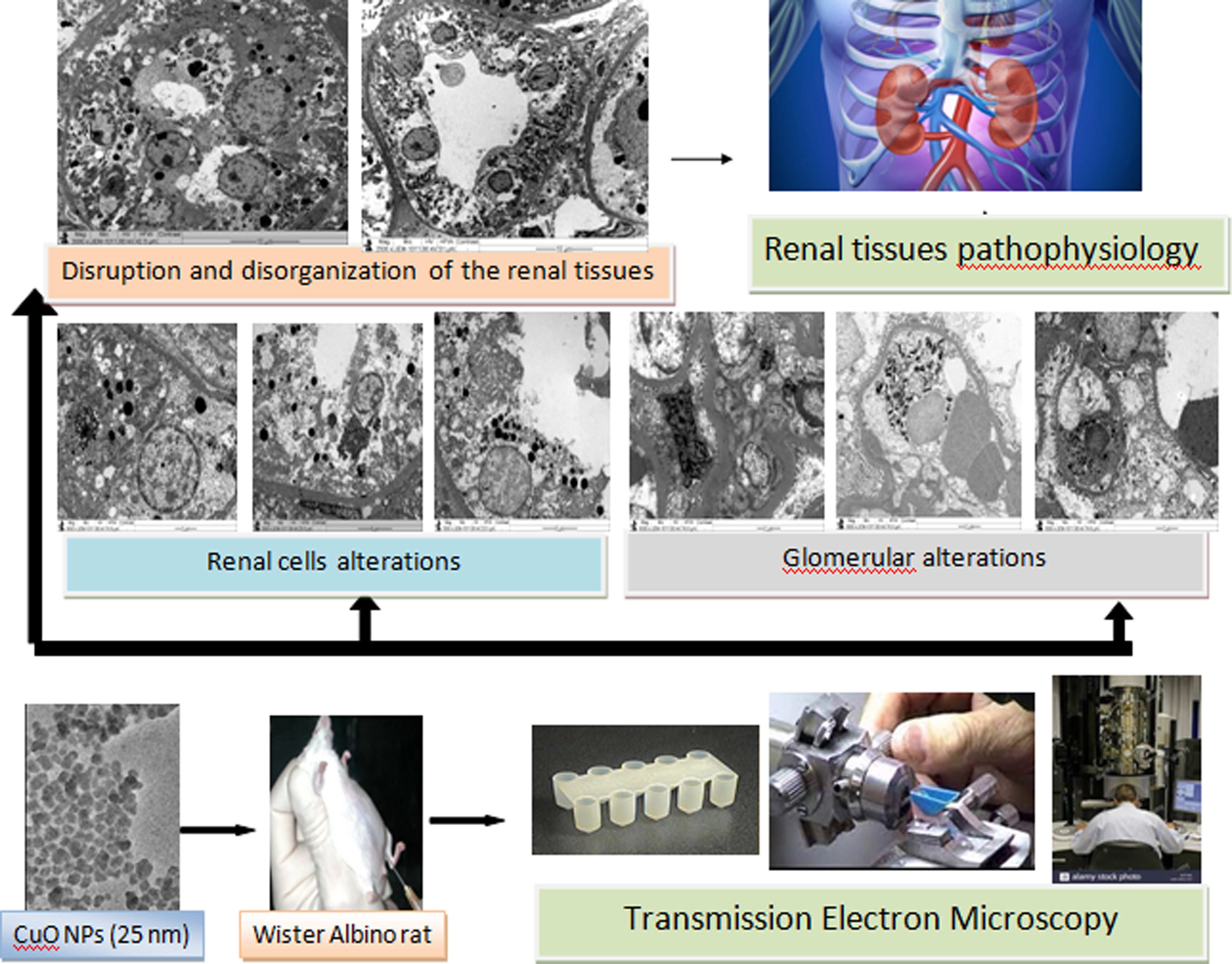
Copper oxide nanomaterials are used in many biomedical, agricultural, environmental, and industrial sectors with potential risk to human health and the environment. The present study was conducted to determine the renal ultrastructural damage caused by 25 nm CuO nanoparticles in renal tissues. Adult healthy male Wister Albino rats (Rattus norvegicus) were administered 35 intraperitoneal injections of CuO nanoparticles (2 mg/kg). Ultrastructural changes were evaluated using transmission electron microscopy techniques. The renal tissues of rats with subchronic exposure to CuO nanoparticles demonstrated glomerular alterations that included hypertrophic endothelial cells, dilated capillaries and occlusions, podocyte hypertrophy, pedicle disorganization, mesangial cell hyperplasia, and crystalloid precipitation. Moreover, the treated renal cells exhibited mitochondrial swelling and crystolysis, cytoplasmic vacoulization, lysosomal hypertrophy, apoptotic activity, endoplasmic reticulum dilatation, nuclear deformity, chromatin dissolution, and basement membrane thickening. In addition, disruption and disorganization of the renal cells microvilli together with cystolic inclusions were also detected. It was concluded from the present findings that CuO nanoparticles could interact with the components of the renal tissues in ways that could cause ultrastructural injury, suggesting renal tissue pathophysiology. Additional studies are suggested for a better understanding the nanotoxicity of CuO nanomaterials.
Introduction
Copper oxide nanoparticles (CuO NPs) are used in many biomedical, agricultural, environmental, commercial, and industrial applications. These nanommaterials (NMs) are widely used as anti-inflammatory, antioxidant, antidiabetic, anticancer, microbial warfare, and drug delivery agents and in osteoporosis-treatment drugs
Some studies have indicated that the kidneys are targets for the toxicity of CuO NPs (Lee et al., 2016; Privalova et al., 2014). Other reports indicated that CuO NMs demonstrated more toxicity than that of other metallic oxides such as TiO2, ZnO, Fe3O4, and Fe2O3 on cell viability due to reactive oxygen species (ROS) production, DNA damage, and rapid dissolution of nanocopper (Privalova et al., 2014). Cronholm et al. (2013) attributed the cytotoxicty of CuO NPs to their release of copper ions resulting from their rapid dissolution. Moreover, some studies demonstrated that nanocopper had mutagenic features and genotoxic actions (Akhtar et al., 2016; Alarifi et al., 2013). Other studies indicated that exposure to CuO NPs could induce marked degenerative alterations in the proximal convoluted tubules (PCTs) together with renal cell desquamation and urinary space widening (Privalova et al., 2014). In addition, CuO NPs were reported to induce histological and ultrastructural alterations including inflammation, apoptosis, genotoxicity, and mitochondrial damage together with oxidative stress, lipid metabolism, and energy metabolism disorders (Ahamed et al., 2010; Lei et al., 2015; Liao and Liu, 2012; Sarkar et al., 2019).
The widespread use of CuO NPs in many applications raises concerns about their adverse impacts on human health and ecosystems. Relatively little is known about the toxicological alterations on the cellular level that might be induced by exposure to these NMs. The current work was carried out to detect renal ultrastructural changes that might result from exposure to CuO NPs.
Materials and methods
Experimental animals
Two groups (control and CuO NPs treated) of male rats (Rattus norvegicus) of nine rats each were used in the present work. All animals under study were kept for acclimatization for 7 days under the proper conditions (temperature, 24 ± 1°C; light/dark cycle, 12 h each) with ad libitum food and drinking water.
CuO nanoparticles
The copper oxide NPs were obtained from Aldrich (Sigma Company, Burlington, Massachusetts, USA). According to the manufacturer certificate, these NPs had a surface area of 29 m2/g, a size of 25 nm, and a boiling point 2567°C. The size of these NMs was confirmed by TEM techniques.
Experimental protocol
Copper oxide NPs were disaggregated by ultrasonication for 20 min before being diluted with sterile normal saline at 37°C before injection. The final NPs solution was prepared in a way that each member of the treated rats under study could receive daily the requested dose (2 mg/kg bw) in 0.4 mL of solution. The dose was chosen after previous MTT assay reports and nanotoxicity investigations targeted in vivo and in vitro toxicity of CuO NPs toxicity (Ahamed et al., 2010; Naz et al., 2020). Each member of the CuO NPs treated group was administered 35 intraperitoneal injections over 7 weeks (daily injection in five successive days/week). The injection of animals on each day was conducted at 10 a.m. Each of the control group was exposed to 0.4 mL of the vehicle at 37°C with the same procedure.
The rats were cared for and treated in accordance with the guide of Canadian Council which is followed by Jerash University. Moreover, the study protocol was approved by the ethical committee of Jerash University (reference number JU/2/87/2020). At Termination, the rats were anesthetized by using ketamine/xylazine at 80/8 mg/kg (cocktail in normal saline) via intraperitoneal route, dissected for kidneys excision and euthanized by vital organs (heart and lungs) removal.
Ultrastructural processing and examination
Tissue biopsies were taken from the kidneys of all rats under study, transferred immediately to a pool of cold (4°C) 0.1 M phosphate buffer and chopped into pieces approximately 1 mm in length. The tissue blocks were then primary fixed by perfusion in 0.1 M phosphate buffer containing 2.5% glutaraldehyde (pH, 7.4; temperature, 4°C) for 6 h. The tissue cubes were then double rinsed in the buffer used in mincing (0.1 M phosphate buffer), post fixed in cacodylate buffer containing 2% osmium tetroxide at 22°C for 3 h and rinsed in three changes 0.1M cacodylate buffer, one of which was over night.
The samples were then dehydrated rapidly at 4°C in an acetone ascending series (50%, 70%, and 95% for 10 min each change; two changes (100%) for 10 min each) before being embedded in Epon-Araldite resin in polymerized polyethelene wells at 60°C for 72 h for curing (Hayat, 2000). Semithin sections (700 nm thick) and ultrathin sections (60 nm thick) were cut from the polymerized blocks by using a Leica EM UC6 ultramicrotome and a glass or diamond knife.
Semithin sections were stained with toluidine blue (0.1%) while the ultrathin ones were mounted on hexagonal copper grids (200 mesh) and stained with (0.5%) uranyl acetate-lead citrate double stain. Stained semithin sections were examined by light microscope while the stained ultrathin ones were evaluated for renal ultrastructural changes using a Joel TEM (80 KV, JEM-1011, Japan).
Results and discussion
Control kidneys
The control kidneys showed normal ultrastructural features of the glomeruli, renal tubules, and renal interstitial components. In addition, the control rats demonstrated normal ultrastructures of the glomerular capillaries, the proximal and distal convoluted portions of the renal tubules together with the collecting tubules. Moreover, the glomerular tuft, trilaminar basement membrane, amorphous mesangium, mesangial cells, visceral cells (podocytes), fenestrated endothelial cells, and the slight pores exhibited normal and well-contacting ultrastructural components (Figures 1(a) and (b)). The renal cells demonstrated normal mitochondria, endoplasmic reticulum (ER) with typical apical microvilli of the PCTs and normal basement membrane (Figure 1(c)). Moreover, the renal cells showed spherical nuclei, euchromasia, and basal cell membrane enfolding (Figure 1(d)). (a–d): Transmission electron micrographs of control kidneys showing. (a) Portion of normal capillary tuft (block white arrow) and glomerular capillary, gc with normal red blood cells, RBC. (b) Normal podocytes (star) with normal pediciles (left block arrow) and mesangial cell (mc) embedded in an amorphous matrix. (c) Normal appearance of renal cell with normal nucleus (N), euchromasia and trilaminar basement membrane (white arrow). (d) Normal endoplasmic reticulum (quad arrow), mitochondria (m) embedded in plasma membrane enfolding and normal basement membrane (BM).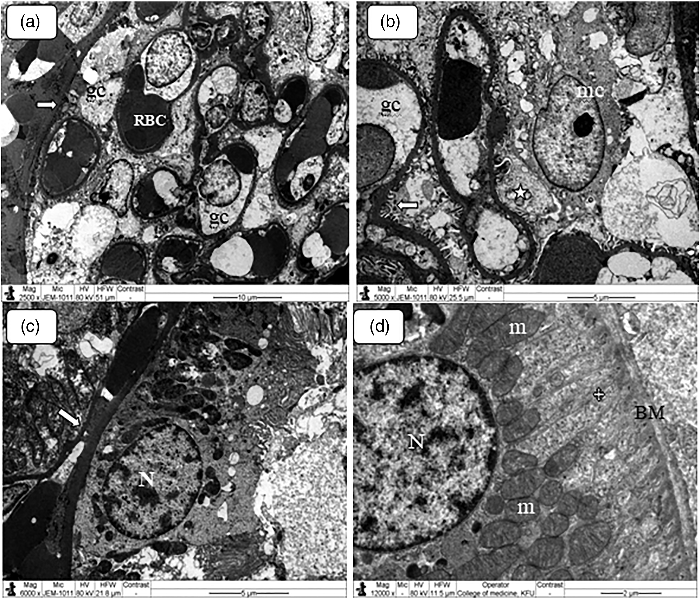
Kidneys of rats exposed to 25 nm CuO NPs
The kidneys of CuO NPs-treated animals showed glomerular and tubular ultrastructural alterations. The glomerular capillaries of rats exposed to 25 nm CuO NPs exhibited the following renal ultrastructural changes:
Endothelial cell enlargement: Hypertrophic endothelial cells were demonstrated by some glomerular capillaries of CuO NPs-treated rats (Figure 2(a)). Lysosomal structure hypertrophy was also demonstrated by some enlarged endothelial cells. These cells play an essential role in controlling vascular relaxation, contraction, and the flow of electrolytes and fluid into and out of the urinary space with unique function in filtration of the glomeruli. Endothelial cell enlargement may indicate interfering with glomerular blood flow and filtration. The glomerular endothelium contained pores of about 60 nm, and most of its surface was covered by a thin membrane (Scott and Quaggin 2015). (a–f): Transmission electron micrograph of ultrathin sections in the kidney of rats subjected to CuO NPs demonstrating glomerular alterations. (a) Enlarged endothelial cell (ec) crowded with many lysosomes. Note the degenerated cellular debris in the remaining part of the capillary (5-point star) and in the mesangium (*). (b) Glomerular mesangium filled with degenerated cellular debris (explosion). Note the mesangial cell (mc) and the parietal cell (block arrow). (c) Podocytes hypertrophy (arrows). (d) Podocytes disorganization (block arrows) and debris accumulation in the urinary space (us). (e) Crystals precipitation (quad arrows). (f) Mesangial cells hyperplasia (block arrows).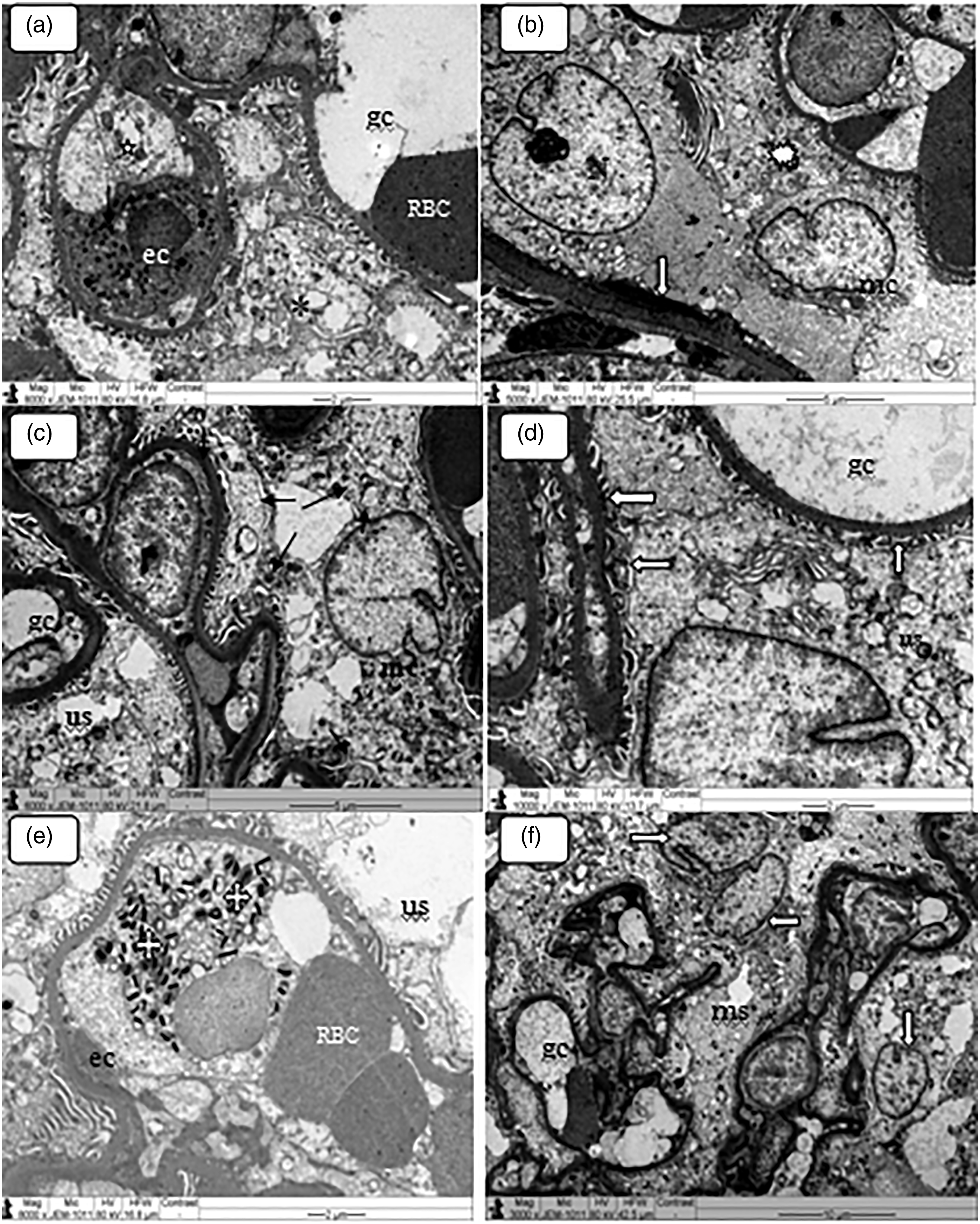
Glomerular capillary occlusion: The glomerular capillaries of rats administered CuO NPs contained cellular debris consisting of membrane-bound cytoplasm and degenerated cells (Figure 2(b)). This might be the result of the expansion of mesangial cells that reduced the filtration area or dysfunction of the filtration barriers especially the glomerular basement membrane (GBM) and the podocytes.
Podocyte hypertrophy: The kidney of rats exposed to CuO NPs exhibited occasional podocyte hypertrophy together with discretion and disorganization of pedicles (secondary foot processes) (Figure 2(c)). Moreover, some degenerated podocytes showed widening and reduction in number of pedicels, shrinkage, and granulation of the nuclei (Figure 2(d)). This deposition may indicate membranous glomerulonephritis as suggested by some previous reports (Pavelka et al., 2010). Podocytes are specialized satellite epithelial cells and considered as part of the glomerular barrier with contractile activity of their pedicles by opposing glomerular capillary distension (Quaggin and Kreidberg 2008).
Crystalloid precipitation on the glomerular capillaries: Some glomerular capillaries demonstrated precipitation of crystals (Figure 2(e)). To our knowledge, this is the first report of NMs precipitation in the glomerular tufts. This may cause irreversible glomerular lesions as indicated by some antiviral agents used in AIDS patients as crystals in glomerular capillary lumens and with foscarnet for cytomegalovirus disease where crystal precipitation was observed in the glomeruli and in proximal tubules (Philipponnet et al., 2015). Moreover, glomerulonephritis was reported to be induced by crystalline nephropathy (Sharma et al., 2019).
Mesangial cell hyperplasia: Some glomeruli of rats exposed to copper oxide NMs demonstrated mesangial cells hyperplasia (Figure 2(f)). This finding may indicate a need to produce extracellular matrix. Mesangial cells are modified pericytes with phagocytic activity and capable of secreting inflammatory mediators. These cells have contractile features that play a crucial role in the regulation of the glomerular filtration rate by changing resistance in the glomerular tufts (Almansour et al., 2019). Mesangial hyperplasia was reported in renal injury including metabolic and immunological disorders (Metwaly and Al-Quraishy 2011). In addition, this alteration may indicate renal injury and glomerulosclerosis together with phagocytic activity to remove protein aggregates that lodge within the glomerular filtrate.
Glomerular capillary widening: The glomeruli of rats receiving CuO NPs showed enlargement of glomerular capillary tufts (Figures 3(a) and (b)). This alteration is a feature of many disorders including diabetes mellitus (Awad et al., 2007). (a–e). Transmission electron micrograph of ultrathin sections in the kidney of control and CuO NPs treated rats demonstrating. (a) Control glomerular capillary. (b) Treated rat glomerular capillary widening (stars). (c) Control GBM (down arrow). (d) Treated rat GBM thickening (block arrow). (e) Urinary spaces widening (quad arrows). Note the glomerular crystal precipitation (explosion), the parietal cell (block arrow), and the podocytes (arrows). GBM: glomerular basement membrane.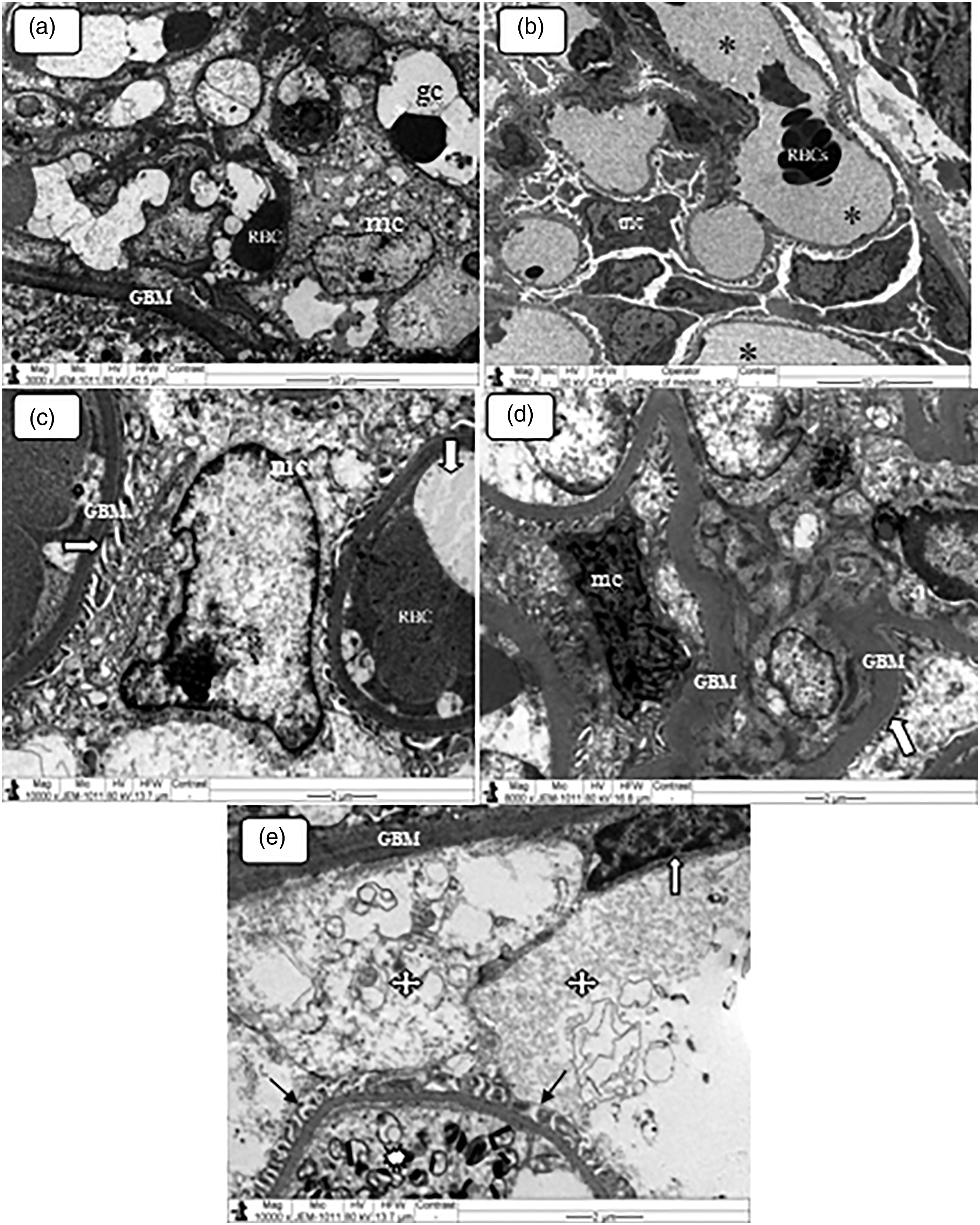
Glomerular basement membrane thickening: Occasional glomeruli of CuO NPs-treated rat demonstrated thickened GBM (Figures 3(c) and (d)). This membrane consists of fused membranes of the endothelial cells and podocytes with glycoproteins components (Faul et al., 2008; Scott and Quaggin 2015). It controls water and solutes permeability but prevents the passage of macromolecules and blood cells (Miner 2012). The thickening of GBM is seen in nephrites and diabetic glomerulosclerosis indicating unhealthy podocytes and proceeding towards albumenuria (Marshall 2016). This finding may indicate podocyte dysfunction where these cells play essential role in the function of the GBM.
Urinary space widening: Compared with the control rats, the glomeruli of rats administered CuO NPs demonstrated urinary space widening with granular electron-dense materials accumulation (Figure 3(e)). This finding might be a direct consequence of hyperfilteration of the injured glomeruli. However, this ultrastructural changealso might be caused by the thickening causing urine accumulation in the urinary space. Some reports indicated that urinary space widening could induce proteinurea (Marshall, 2016).
On the other hand, rats exposed to CuO NPs showed the following alterations in the ultrastructural features of the renal cells:
Cytoplasmic vacuolization: The renal cells of some PCTs showed cytoplasmic vacuoles of variable sizes (Figure 4(a)). Some of these vacuoles demonstrated flocculent materials. This alteration can be transient or irreversible and often accompanies cell death (Shubin et al., 2016). Cytoplamic alterations can affect cell organelles especially the ER, lysosomes, mitochondria, and Golgi apparatus. This change resulting from CuO NPs may indicate alterations in the permeability of the renal cell plasma membrane causing fluid accumulation and cytoplasmic necrosis. (a–f): Transmission electron micrograph of ultrathin sections in the kidney of rats subjected to CuO NPs treatment demonstrating renal cells alterations. (a) Cytoplasmic vacuolation (v) (b) Lysosomal hypertrophy (arrows). (c) Degenerative changes (star). Note the prominent nucleoli (block arrow). (d) Mitochondrial swelling and cristolysis (m). Note microvilli diroganization (white asterisk). (e) Cytoplasmic inclusion (block arrow). Note the thickening of the renal cell basement membrane (BM), cytoplasmic vacuolation (v) and lysosomes hyperplasia (arrows). (f) Intranuclear inclusions accumulation (block arrow). Note the precipitation of black deposits in some lysosomes (white stars).
Lysosomal accumulation: Variable sized lysosomal structures crowded with minute electron-dense materials were seen. Some of these structural membranes demonstrated phagocytosis (Figure 4(b)). This alteration may indicate degradation of the remnants and debris of organelles of the cells injured due to CuO NPs exposure. Some previous reports confirmed lysosomal damage induced by chronic and acute subjection to other NMs (Almasour et al., 2019). Lysosomal disruption may lead to the liberation of their hydrolytic enzymes into the cytoplasm causing marked lysis and dissolution of the cell organelles (Metwaly and Al-Quraishy 2011).
Degenerative changes: Some cells of the proximal tubules showed loss and lysis of organelles with electron dense deposits accumulation (Figure 4(c)). Degeneration is a result of ion and fluid homeostasis imbalance that lead to an increase of intracellular water (Schrand et al., 2010). This alteration might indicate subacute renal cells injury induced by CuO NMs that might result from the interaction of these NMs with cellular proteins. Moreover, NMs including copper oxide ones demonstrated corona as a result of their interaction with the targeted cells proteins (Duran et al., 2015).
Mitochondrial damage: Some renal cells of the PCTs demonstrated swelling and cristolysis (Figure 4(d)). This finding may indicate that chronic exposure to 25 nm CuO NPs could induce mitochondrial injury. This damage might result from the alterations in renal cells osmolarity changing the integrity of the mitochondrial membranes (Galvan et al., 2017). Moreover, mitochondrial crystolysis and swelling may impair oxidative phosphorylation. The mitochondrial injury induced by CuO NMs may stimulate the production of ROS and affect cellular respiration as a result of mitochondrial membrane depolarization. However, some reports indicated mitochondrial membrane and cristae injury by metal and metal oxide NPs (Katsnelson et al., 2015). Mitochondria dysfunction plays a crucial role in the etiology of some diseases. Swelling of the mitochondria may indicate solutes and water entrance into the mitochondrial matrix interfering with oxidative phosphorylation and/or the electron transport system in the mitochondrial cristae (Pavelka 2010). The mitochondrial function impairment leads to ATP depletion and cellular dysfunction (Ulicna et al., 2012).
Cytoplasmic inclusions: Some cells of the PCTs demonstrated crystalloids cytoplasmic inclusions, which are most likely a biomolecular condensation (Figure 4(e)). These cytoplasmic inclusions might be the result of the renal cells pathophysiology caused by CuO NPs toxicity (Mosaheb et al., 2005).
Intranuclear inclusions: Nuclear inclusions were seen in some renal cells of the PCTs epithelial lining (Figure 4(f)). The intranuclear inclusion is a sort of adaptive response and often accompanies nuclear invagination (Jarrar and Mahmoud, 2000).
Endoplasmic reticulum dilatation: Occasional renal cells of the PCTs of rats exposed to copper NMs treatment exhibited ER dilatation (Figure 5(a)). This finding may suggest that copper oxide NPs could cause ER injury that might be caused by disturbances of the redox reactions carried out in the ER membranes. The ER is a multifunctional organelle in lipids biosynthesis, protein folding, and calcium homeostasis. Endoplasmic reticulum dilatation is considered as a sign of premature death of the cell (Almansour et al., 2019). (a–f): Transmission electron micrograph of ultrathin sections in the kidney of rats subjected to CuO NPs demonstrating. (a) Endoplasmic reticulum dilatation (block arrows). (b) Chromatin lysis (white asterisk). Note the cytoplasmic degenerative changes. (c) Apoptotic activity (block arrow). Note the lysosomal hyperplasia (arrows) and in the necrotic changes in the other cell (quad arrow). (d) Control renal cell basement membrane (block arrow). (e) Basement membrane thickening (block arrow) in comparison with the control renal cell. Note lysosomal hyperplasia. (f) PCTs microvilli clumping and swelling (explosion). Note the hydropic vacuolation (v) and cytoplasmic degeneration (quad arrow).
Renal cell nuclear alterations: Chromatin lysis was demonstrated by occasional renal cells of rats exposed to CuO NMs (Figure 5(b)). In addition, some renal cells exhibited nuclear envelope irregularity and swelling. Moreover, some renal cells demonstrated prominent nucleoli as an indication of renal cell activation (Elmore, 2007).
Apoptotic activity: Some PCTs lining cells exhibited apoptotic activity (Figure 5(c)). This change is usually associated with programmed cell death related to ATP production reduction, oxidative stress, and mitochondrial damage (Elmore, 2007). Some reports demonstrated that exposure to CuO NPs (20 nm and 100 nm) could induce oxidative stress and apoptosis (Chen et al., 2011; Regoli and Giuliani , 2014).
Renal cell basement membrane thickening: Compared with the control subjects, the renal tubules exhibited basement membrane thickening (Figures 5(d) and (e)). This finding was less prominent in the distal convoluted tubules (DCTs) than the proximal ones and the collecting tubules. This abnormality might result from the accumulation of lucent materials accumulation in the subendothelial spaces of the membrane (Neumann et al., 2004).
PCTs microvilli alterations: The microvilli of some renal cells of rats subjected to CuO NPs showed swelling, disruption, disorganization, attenuation, and clumping of fused microvilli (Figure 5(f)). In addition, cellular debris was present within the tubular lumen. Renal cells microvilli are sensitive to toxins and drugs and react to toxins by ballooning in the renal tubules lumen (Nigam et al., 2015).
Renal tubule lumen narrowing: Some renal tubules demonstrated lumen narrowing resulting from apical cytoplasmic bulges of the renal cells towards the lumen (Figure 6(a)). Some reports indicated that nanocopper materials could cause renal cell fragmentation and deposition in the lumen of PCTs (Lee et al., 2016; Wang et al., 2016). (a–b): Transmission electron micrograph of ultrathin sections in the kidney of rats subjected to CuO NPs demonstrating. (a) Lumen narrowing (quad arrow) filled with cellular debris. Note the prominent of lysosomal hyperplasia in the renal cells (arrows) together with nuclear envelop irregularity (block arrow↓) and prominent nuleoli. (b) DCT demonstrating cytoplasmic degenerative vacuolation (v), nuclear and prominent nuleoli. (b) DCT demonstrating cytoplasmic degenerative vacuolation (v), nuclear.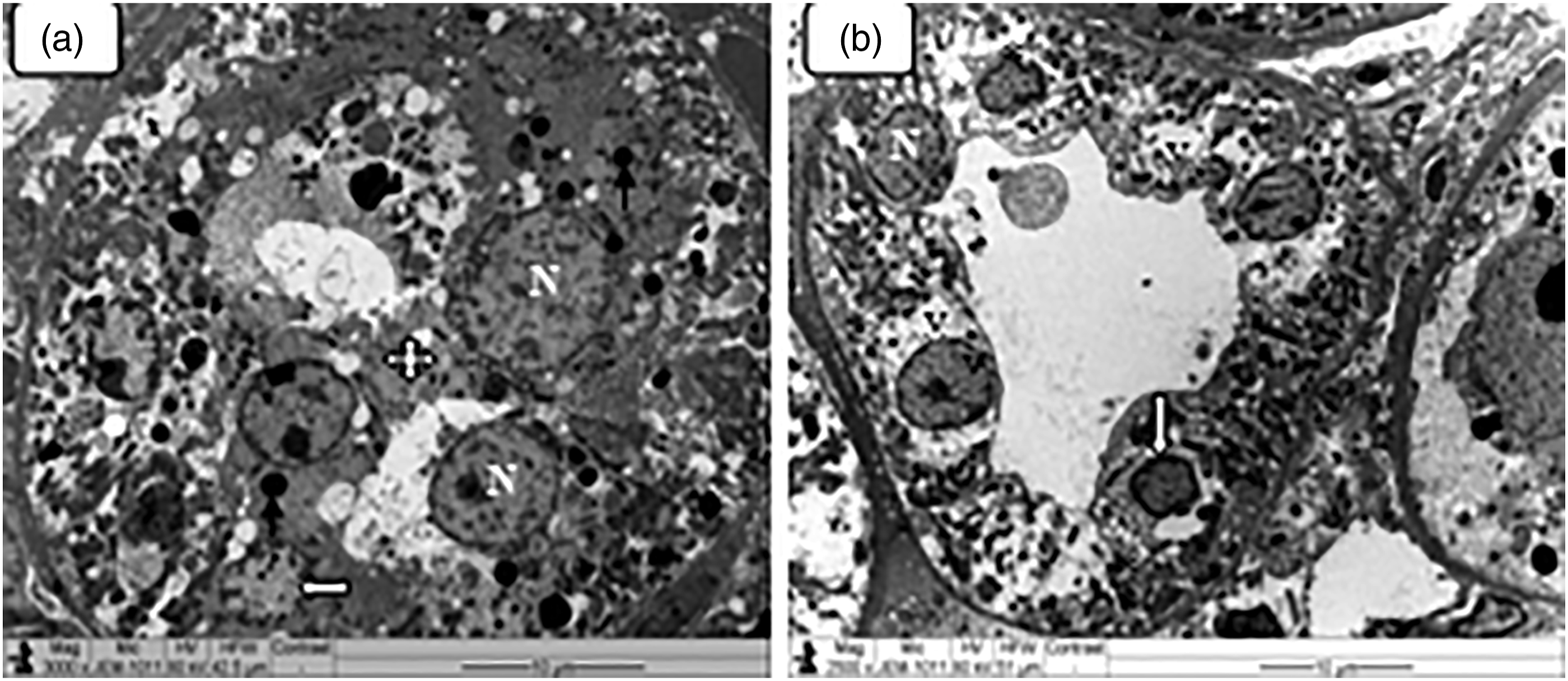
The Distal renal tubule epithelial lining exhibited occasional morphological changes, specifically cytoplasm vacuolation as well as nuclear renal cells alterations with little or no lumen narrowing (Figure 6(b)). However, CuO NPs-treated animals demonstrated no considerable ultrastructural alterations in the Henle’s loop, and only minor focal ultrastructural degenerative changes were seen in the collecting tubules.
The results of the present work showed that chronic administration of 25 nm CuO NPs could induce ultrastructural damage to renal tissue components. Previous investigations reported that CuO NMs could stimulate renal oxidative stress, disordering the function and structure of the kidney by affecting signalling proteins and renal-cell nuclear receptors with reduction in the activity of CYP450s (Xu et al., 2018). The renal damage caused by nanocopper might be the result of regeneration from ROS, induced oxidative metabolism, and lipid peroxidation from chronic exposure to CuO NPs (Chang et al., 2012; Kaviani et al., 2019). In addition, alterations in the hematological components and plasma biochemistry are reported elsewhere (Prialova et al., 2014). Marked retention of CuO NPs in the renal tissue with significant alterations in the kidney mass and biochemical indices, mainly serum total proteins, albumins, globulins, bilirubin, and alkaline phosphatase together with urinary coproporphines, albumin, ∂-ALK, and creatinine, were also reported (Lee et al., 2016; Prialova et al., 2014). Moreover, it was reported that CuO NPs could lower glutathione levels but elevate alkaline phosphatase (El-Kassas and Okbah 2017). Also, some studies reported that oxidative dissolution of small-sized NMs could induce the production of hydrogen peroxide causing oxygen depletion as a result of ROS cellular generation (Liu et al., 2010).
Our findings demonstrated that chronic exposure to CuO NMs induced ultrastructural renal damage with effects on the ultrastructural features and potential function and metabolism of the renal tissues. The renal damage induced by CuO NPs could be associated with oxidative stress induced in the nephron ultrastructure by these NMs. The present findings suggest that CuO NPs may have variable toxicokinetics on the renal tissue components. More efforts are needed to elaborate the toxicity and pathogenesis of nanocopper materials on human health and the environment.
Footnotes
Author’s note
The authors alone are responsible for the content and writing of the article.
Acknowledgments
The authors are grateful to the Deanship of Scientific Research at King KhalidUniversity, Abha, KSA, under grant No (R.G.P.2/87/42). In addition, we are grateful to Jerash University for the ethical approval of the project and putting all facilities under our disposal.
Declaration of conflicting interests
The author(s) declared no potential conflicts of interest with respect to the research, authorship, and/or publication of this article.
Funding
The author(s) received no financial support for the research, authorship, and/or publication of this article.