






Abstract
As forensic radiology sees an exponential gain in popularity, postmortem computed tomography (PMCT) is increasingly being used in the appropriate setting, either as preautopsy guidance or as part of complementary virtual autopsy protocol. Many articles have expounded the value it adds to forensic pathology in the general setting and the appropriate technical parameters to be used for optimum benefit. We aim to put forth a concise review on the role of PMCT specifically in trauma and the pitfalls to be aware of. Reviews have shown that presumed cause of death in trauma have been proven by autopsy to be wrong in about 30% cases. Radiology applied to postmortem investigation in unnatural deaths and more specifically in trauma shares many semiotic features with emergency radiology. Therefore, in the near future, emergency radiologists might be required to integrate this type of imaging in their regular practice. Although the predominant drawbacks are time-dependent, PMCT also has some difficulty in differentiating antemortem and postmortem events. However, in many such scenarios, PMCT and autopsy play a complementary role in arriving at conclusions, and we believe understanding the benefits and role in trauma is imperative considering the expanding usage of PMCT.
Introduction
Trauma accounts for 9% mortality despite computed tomography (CT) playing a crucial role in prompt and accurate assessment of trauma patients. 1 Postmortem computed tomography (PMCT) in the context of trauma has been shown to help in identifying the cause of death commonly occult to the health-care professionals when only relying on the clinical examination, therefore improving completeness of trauma registries. 2 Postmortem computed tomography also complements forensic investigations in the context of unexplained or iatrogenic causes of death and helps format guidelines for future management.
Traditionally, the role of identifying the factors resulting in death has always been the role of coroners and forensic pathologists. In the last decade, PMCT has evolved as an adjuvant modality to assess injuries and in some scenarios as a reasonable alternative for postmortem evaluation. Computed tomography also has the advantages of widespread availability, short acquisition time, and overall better radiological understandability compared to modalities such as ultrasound or magnetic resonance imaging. Postmortem computed tomography semiology in trauma overlaps with emergency and trauma radiology CT imaging. Select scenarios may find benefit in additional PMCT angiography to identify vascular injury or assess integrity of vascular devices. The advantage of PMCT is to offer a nonaltered evaluation of the corpse at the time of imaging. On the other hand, postmortem dissection has drawbacks of inadvertent external contamination, spread or leakage of body fluids, and displacement or rupture of medical devices such as endotracheal tube cuff.
In this article, we review various studies which have evaluated PMCT and autopsy and try to identify the optimum method for PMCT imaging as well as the potential advantages and disadvantages in various trauma scenarios. We hope this increases the confidence of radiologists and the involved trauma teams in adopting PMCT in trauma-related mortality.
Background
The steady decline of regular autopsy led to an increased interest toward PMCT. In many jurisdictions across the world, although a coroner has the right to call for an autopsy, it may, at times, have to be performed against the wishes of the next of kin who are unwilling due to personal or religious reasons. Postmortem computed tomography is of additional value in such trauma-related deaths to document and evaluate the injuries. Many trials have compared PMCT and autopsy and have shown their complementary roles. 3,4 In a review by Scholing et al, 5 the highest agreement between the 2 methods was shown for gunshot wounds, with PMCT correlating best with autopsy in identification of thoracic region injuries. A recent review by Wijetunga et al however did not demonstrate a similar conclusion where 70% lung injuries were missed on PMCT. 6 Although a moderate degree of overall correlation was noted, PMCT was shown to fall short in many areas (such as certain solid organ injuries, source of hemorrhage, etc). Studies have shown highest correlation between PMCT and autopsy to be in lower limb injuries with lowest correlate for thoracoabdominal trauma, also highlighted by Wijetunga et al. 6,7 Postmortem computed tomography on one hand while being better than autopsy in detection of skeletal injuries and air embolism performed inferior to autopsy in identifying soft tissue and vascular trauma, 5,7,8 especially when using noncontrast acquisitions. In the study by Wijetunga et al, PMCT failed to recognize all brainstem and cardiac injuries as well as 65% subdural bleeds, stressing the drawback of solely relying on PMCT. 6,7 Failure to identify vascular injury is thought to be primarily due to cardiovascular collapse and lack of contrast use. As in a standard trauma imaging protocol, studies have shown improved detection rate on performing CT angiography and this may, in future, be a routine addition to PMCT. 3,9 Leth’s and Ibsen’s higher rates of detection of solid organ injuries are presumed to have been due to the use of experienced forensic radiographer and dedicated regional scanning. 10 Such conditions are unlikely to be met with in the majority of PMCT scans, thereby limiting the detection capability of PMCT relative to autopsy. Postmortem computed tomography is acknowledged to be less sensitive in identifying small extra-axial bleeds with the agreement varying between different studies. Although a high percentage of head trauma including contusions, extra-axial bleeds, and brainstem injuries were missed in Wijetunga’s analysis, there was a high correlation in the study by Legrand et al 6,7 where cases were analyzed by a neuroradiologist with forensic training using a dedicated separate head and neck acquisition as opposed to being part of a whole-body protocol.
Recommended Technical Parameters
Postmortem computed tomography protocoling utilizes the fact that radiation dose and motion artifacts have no bearing on postmortem imaging. Similar protocol to antemortem imaging is usually used with whole-body CT imaging covering the head, spine, thorax, abdomen, pelvis, and limbs. Although most centers opt not to administer contrast, 11 contrast-enhanced PMCT has been described using different vascular approaches and injection techniques. 12 Optimally, the body is placed in a supine position in an artifact-free body bag to prevent soiling and contamination, and the head is held in midline position by restraining band. Ideally, the arms should be placed close to the body for head and neck imaging and above the head for thoracoabdominal scanning. However, these conditions might not be met depending on the corpse status at the time of imaging (eg, rigor mortis, advanced putrefaction, etc). Acquisition of scout images is also useful in case the body has to be repositioned prior to scanning. An initial scan may be obtained prior to repositioning to avoid possible alterations in PMCT findings brought on by the displacement. Ideal positioning requires isocentric location of the spine to avoid noise and image degradation. Gascho et al have described a series of ideal prescan acquisition parameters and postscan reconstruction parameters for optimum image quality. 13 The ability to disregard tube current and use higher milliampere settings coupled with dose modulation for uniformity provides for ideal image quality in PMCT. In addition, thinly collimated slices and small pitch further decrease artifacts and optimizes spatial resolution. Like in clinical radiology, general PMCT acquisition is best at 120 kVp setting, although different parameters can be used to overcome artifacts or optimize soft tissue contrast. Dual-energy CT (DECT) provides additional advantage of metallic artifact reduction and identification of chemical composition of internal constituents and foreign bodies (eg, metallic prosthesis or projectile) and therefore can be considered when using DECT capable scanners.
Reconstruction using extended CT scale allows for improved characterization of metallic foreign bodies. Choice of filter depends on the area where improved resolution is sought after, with smooth filters for soft tissue, hard ones for bone, and medium for cartilaginous components.
Postmortem CT Angiography
One of the rapidly evolving adjuncts to PMCT is the use of postmortem CT angiography. Of various techniques and materials tested, one of the most accepted protocols is by Grabherr et al, 3 which advocates cannulation of the femoral artery and vein through an inguinal incision. A specially constructed perfusion device (Virtangio Machine, Fumedica AG, Switzerland) is then connected, through which an oil-based contrast agent of very specific viscosity mixed with paraffin is injected (Angiofil Macro, Fumedica AG). The special nature of the contrast agent prevents diffusion of contrast into the capillary space and body cavities and remains physically separate from blood.
Multiphase sequential imaging with arterial, venous, and dynamic study is then performed, and a finding identified on 2 phases is deemed to be of significance and nonartifactual. With evidence pointing to a 64% to 81% jump in detection of findings on addition of angiography to standard postmortem CT protocol, the inclusion of multiphase angiography is validated and is being increasingly performed. 3
In special circumstances involving bloated or obese corpses, algorithms allow an extended field of view to be developed and utilized to improve spatial resolution. The use of small voxels in thin sections allows high-resolution 3-dimensional (3D) postprocessing with volume rendering (VR) and cinematic rendering (CR) gaining importance with their visual effectiveness especially in medicolegal cases, helping the nonmedical personnel to understand the sustained injury.
Primary Identifiable Causes of Death
Highest mortality from trauma happens in the early minutes to hours after the traumatic event. Many studies have pointed out that an overwhelming 50% to 60% death are categorized as immediate and include those within minutes to 1 hour after arrival in hospital. 14,15 The top 2 causative factors in immediate and early deaths have been highlighted in multiple studies, 14,15 being irreversible neurologic injury (50%) and exsanguination (35%), respectively. A multitrauma center trial of 17 trauma patients defined cause of death as one or more injuries involving an organ system not compatible with life. 11 In presence of hemothorax or hemoperitoneum, identifying the exact site of extravasation was not deemed necessary with presence of internal hemorrhage attributed as etiology.
Advantages of PMCT
Road Traffic Accidents and Blunt Trauma
Postmortem computed tomography has an indisputable role in identifying fracture sites and patterns, localizing foreign bodies, and showing gas distribution. A retrospective cohort study of 52 pretrauma deaths revealed PMCT to be particularly superior to autopsy in detecting bony injuries of the upper extremity, with PMCT identifying an additional 30 injuries missed on autopsy. 6 Identifying skeletal injuries, especially fractures in certain anatomic regions such as the cervical spine and pelvis are better recognized on PMCT (Figure 1). An analysis of 453 patients revealed that occipital condyle fractures were probably the most overlooked injuries at autopsy following road traffic accidents. These injuries are on the contrary superiorly detected by PMCT and correlate well with the mechanism of collision 16 involving rotation associated with lateral bending, which helps in trauma scene reconstructions for medicolegal purposes. Many skeletal injuries are not identified at autopsy unless there are overt physical signs, the mechanism of injury requiring a more detailed analysis to avoid damage to the corpse, or because an anatomic area may not be considered to be of relevance. Many pelvic fractures may be missed on autopsy as there are often no clear pointers to injury. 7,17
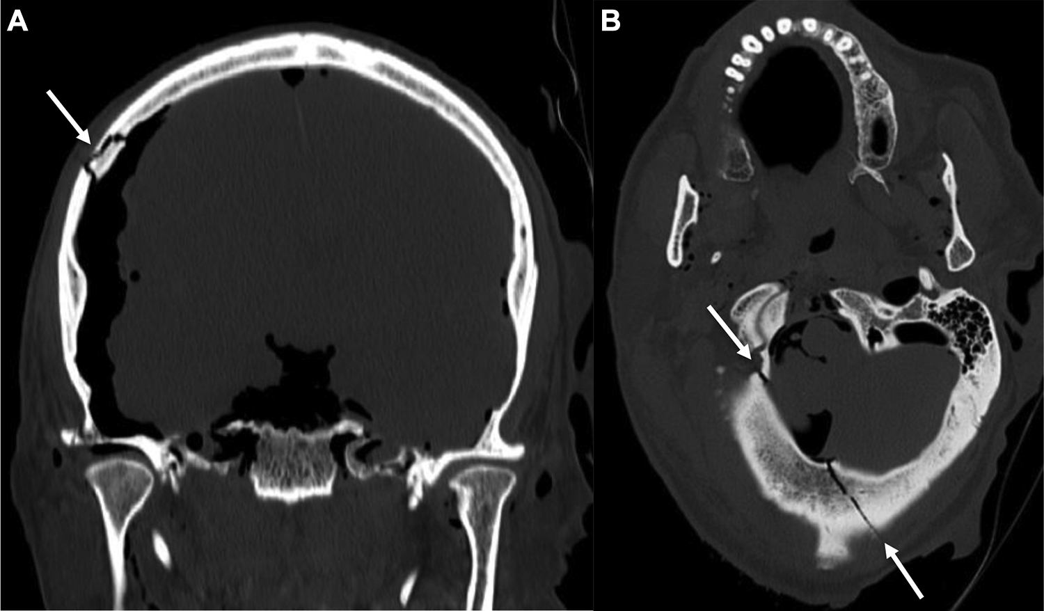
A and B, CT head with bone reformats demonstrating multiple skull fractures (arrows) extending to the foramen magnum with additional findings of pneumocephalus and soft tissue air within masticator spaces. CT indicates computed tomography.
Some studies have shown PMCT and autopsy to have similar capabilities in depicting brain injuries, while some have shown PMCT to be superior in detecting epidural and subdural hematomas. In some scenarios, PMCT holds superior value in detection of cerebral contusions and in pointing toward diffuse axonal injury which can be missed on autopsy. 7 Postmortem computed tomography was also shown to be better at showing intraventricular hemorrhage and pneumocephalus, which were identified in 47 cases on PMCT and missed on autopsy in a study by Legrand et al 7 (Figure 2).
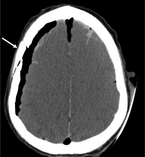
Axial PMCT of the head shows a right parietal bone fracture (arrow) as well as the associated subarachnoid hemorrhage and pneumocephalus. PMCT indicates postmortem computed tomography.
Gas accumulation within the body, resulting in pneumothorax, pneumomediastinum, air embolism, or pneumorachis, is superiorly evaluated by PMCT 6 (Figures 3 and 4), where the very nature of an autopsy procedure nullifies the value of the findings by iatrogenic introduction of air within the corpse. 11 Some techniques may be performed during autopsy to identify the presence of air, such as opening the pericardial sac while keeping the organ submerged, or using an aspirometer to detect gas embolisms, although time-consuming and requiring advanced skills. 10
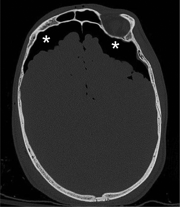
Pneumocephalus with Fuji sign (stars) is easily seen on PMCT while the same will need special techniques to be documented on autopsy. PMCT indicates postmortem computed tomography.
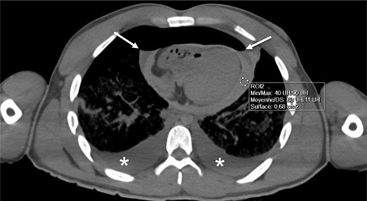
Hemopneumopericardium (arrows) and bilateral hemothorax (stars). The autopsy demonstrated a full-thickness laceration through the right ventricle and bilateral costophrenic recess. Note the bilateral concave menisci (arrows) at the air-blood interface excluding a tension pneumopericardium, which would be impossible to identify on conventional autopsy. The autopsy revealed 310 mL and 2800 mL in the pericardium and pleural cavities, respectively.
Internal Ballistics
A bullet trajectory can be virtually reconstructed with PMCT by following the bullet and shrapnel fragments and blood and gas locules while considering potential ricochet paths. Especially in the skull, inward or outward beveling of the bone identifies the entry and exit wounds, respectively, as shown by Thali et al 18 (Figure 5). Elsewhere in the body, entrance and exit wound usually do not have beveled margins, and bone, bullet fragments, and organ injuries may be used to presume the trajectory. When several bullets strike the body with crossover trajectories, ballistic analysis is significantly limited, if not impossible to perform.
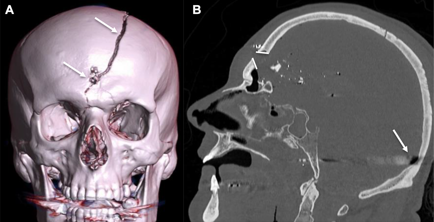
Virtual ballistics. A, 3D volume rendering demonstrates the entry wound and associated frontal bone fracture (arrows). B, On the sagittal bone reformat, the beveled edges of the frontal bone fracture (divergent lines) confirm the entry point with inward displacement of multiple bone fragments. The bullet completed its course in the occipital region creating streak artifacts (arrow). 3D indicates 3-dimensional.
Nonmetal jacket bullets tend to fragment at the first impact and shrapnels are also frequently deposited under the skin of the entrance wound. After the shot, there is constant loss of acceleration of the bullet with significant dissipation of energy at the time of the impact. Moreover, after the impact, the majority of nonarmored bullets fragment all along the trajectory. If energy and acceleration are not completely dissipated, the bullet can spurt out from the body. In the absence of an exit wound, a coarse hyperattenuating piece of bullet (main core) should be searched in the body, often away from the entrance orifice. A pellet may even enter the bloodstream and migrate to distant parts before causing death, known as projectile embolus.
When supposing the virtual path, if the exit wound or the main core of the bullet is located in a noncongruent position with respect to the entrance wound, potential ricochets should be taken into account (Figure 6). Three-dimensional reconstruction with different multiplanar views or video of the VR allows the investigators to visualize the supposed path of the projectile.
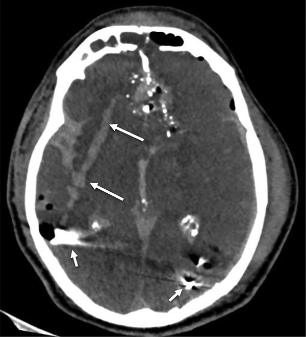
Virtual ballistics. The internal trajectory is clearly identified within the brain parenchyma with secondary hemorrhagic changes (long arrows), with fragmented bullet fragments lodged adjacent to the left occipital and right parietal bones (short arrows).
Penetrating Trauma
Injuries produced by a sharp object present a well-defined traumatic discontinuity of tissues. Dissecting these tissues disturbs and may alter the injury track, whereas PMCT can display the depth and direction of the track without disturbing anatomical congruity.
Gaping wounds on the cutaneous surface are well depicted, most notably with 3D reconstructions (Figure 7). Nongaping wounds are more difficult to demonstrate but can be detected by searching for linear interruption of skin and subcutaneous fat (Figure 8). It is also possible to describe blade track using indirect signs as blood infiltrating the subcutaneous tissue and introduction of gas bubbles (Figure 8). It is important to consider that a path may not always corresponds to the real depth of the stab or to blade thickness, and therefore, inferring the size and type of knife is not possible. 19 Depending on the skin and underlying tissues elasticity, a given knife could produce different depths and lengths of wound, not congruent with its thickness and blade length.
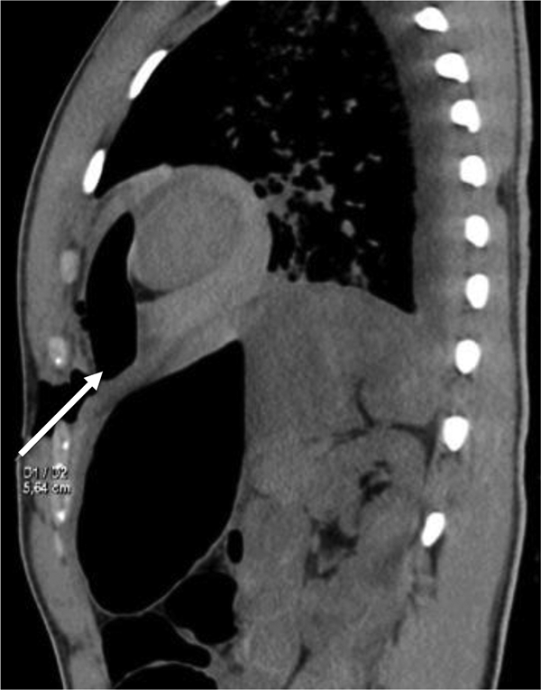
Twenty-one-year-old male patient with stabbing injury to the chest. Sagittal PMCT displays the depth and trajectory of stab wound extending to the pericardium (arrow). A hemopneumopericardium is also present. PMCT indicates postmortem computed tomography.
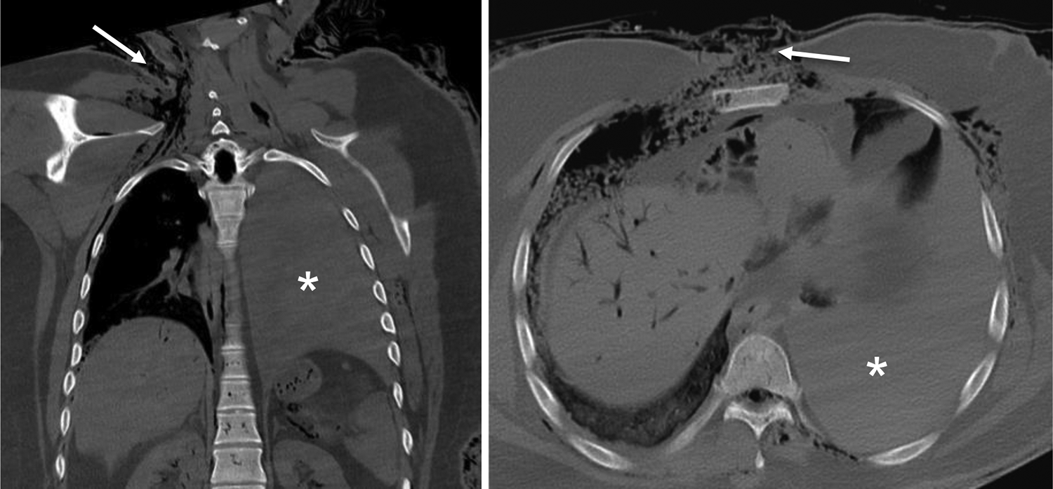
Small defects in the skin of a decomposed body can be difficult to identify. PMCT is a great adjunct to guide forensic pathologists to the entry point of the stab wound (arrows). Note the massive left hemothorax (stars). PMCT indicates postmortem computed tomography.
All injured organs along the stab path can be showed in two-dimensional mode and multiplanar reformat plans, which can perfectly match the orientation of the stab channel (Figure 7). Postmortem computed tomography is also helpful to identify broken-off fragments of the weapon used.
Child Abuse
Postmortem computed tomography is also an ideal tool to rule out significant injury in unexplained sudden infant deaths considering its noninvasive full-body evaluation to assess for nonaccidental injuries or other natural causes of death. Additionally, PMCT can provide an approximate dating of injuries helpful in cases of suspected repetitive child abuse (Figure 9). Proisy et al in their study of 47 cases of sudden infant death were able to identify the injury in child abuse cases with a high concordance with autopsy 20 (Figure 10).
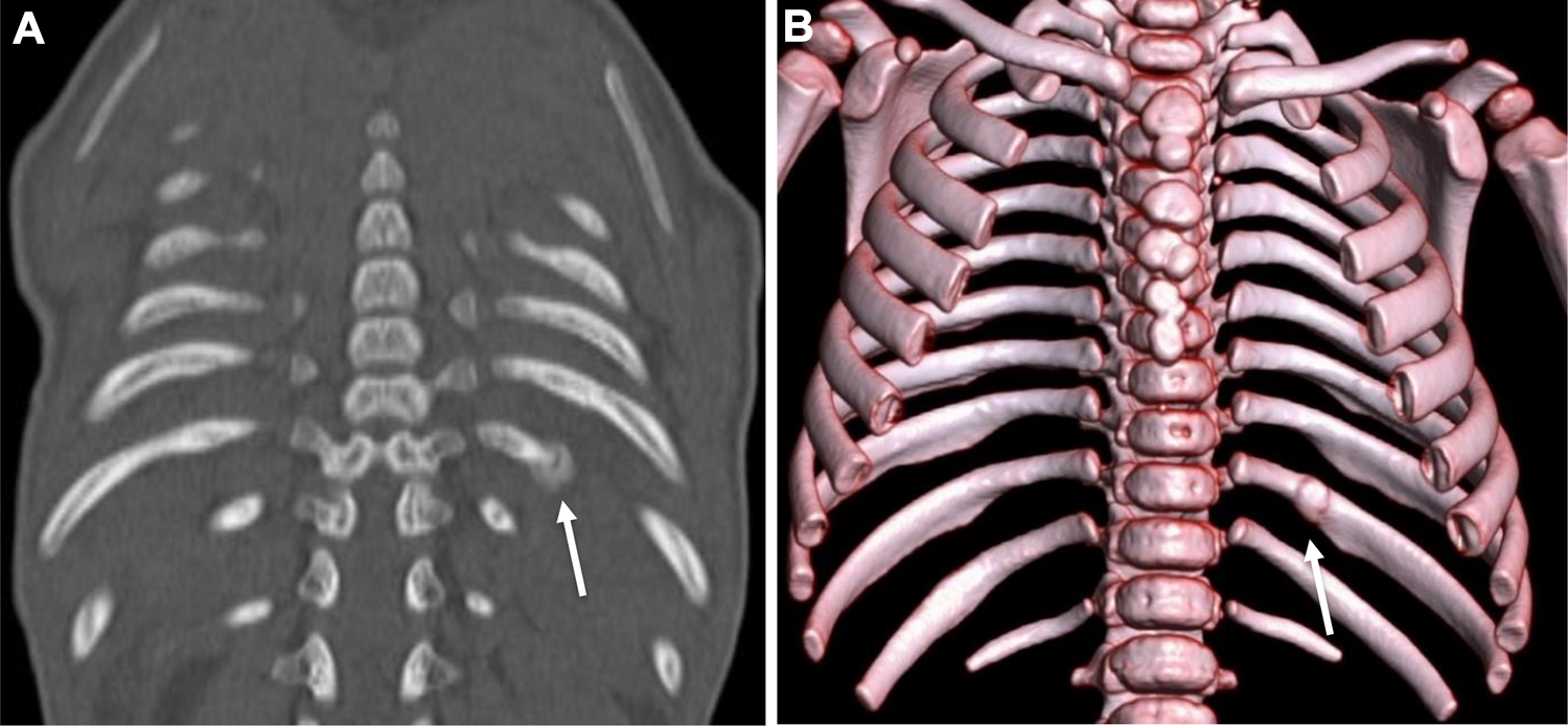
A, Coronal and (B) 3D volume rendering bone reformats demonstrating a subacute to chronic fracture of the posterior left 10th rib, missed during conventional autopsy. This was suggestive of episodes of nonaccidental injury prior to infant’s death, therefore implying a long history of child abuse. 3D indicates 3-dimensional.
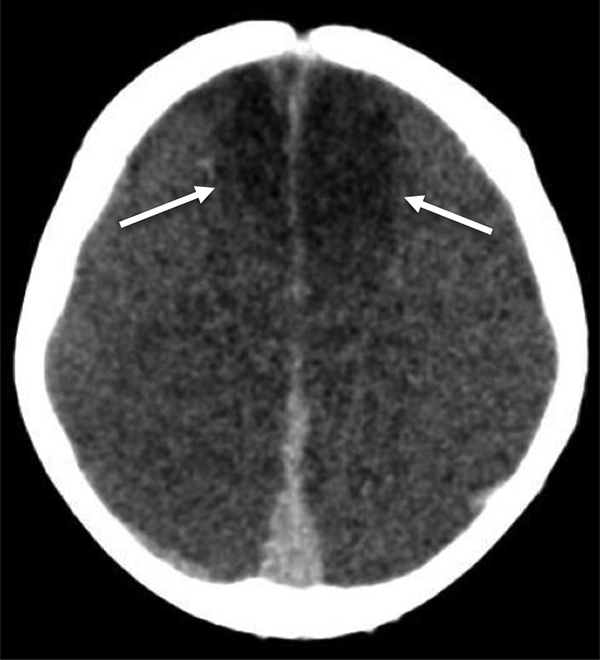
PMCT evaluation of an 8-month-old boy. Large bilateral frontal contusions (arrows) without hemorrhage or skull fracture likely resulting from severe shaking. PMCT indicates postmortem computed tomography.
Identifying Misplaced Resuscitative Devices
Postmortem computed tomography is excellent at evaluating the position of medical devices placed during resuscitation, offering a hindsight to first responders, emergency physicians, and trauma surgeons. In a study by Steenburg et al 2 evaluating 38 PMCT cases, 47.7% of the patients had at least 1 support medical device that was either malpositioned or suboptimally positioned, including endotracheal tubes, chest tubes, thoracostomy needles, and intraosseous lines (Figure 11A and B). Suboptimal positioning of such devices may contribute to delayed or failed resuscitation and PMCT therefore offers invaluable feedback to first responders and clinical teams involved in management of acute trauma cases.
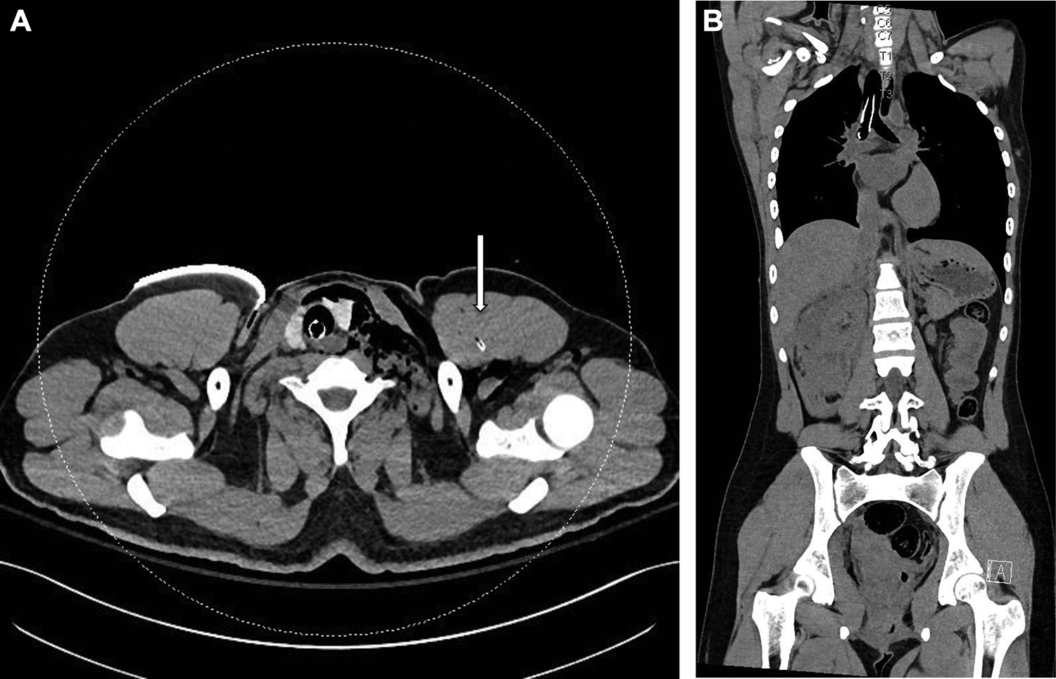
A and B, Misplaced resuscitative device. A, Postmortem CT of road traffic accident victim with massive bilateral hemopneumothoraces and failed resuscitation shows misplaced chest tube with tip within the pectoralis muscle (arrow). B, Another case of motor vehicle collision with misplaced endotracheal tube in the right main bronchus. CT indicates computed tomography.
Radiological Identification of Unknown Trauma Victims
In severe trauma or fire, extreme disfiguration may result in difficulty identifying the body. Figure 12 depicts the evisceration of a badly disfigured unidentified fire victim who was later identified by comparing with antemortem imaging. Radiological identification can be carried out by comparing antemortem imaging (if available) to PMCT to identify distinctive anatomical landmarks (Figure 13) or prosthesis (Figure 14) a patient may be known to have.
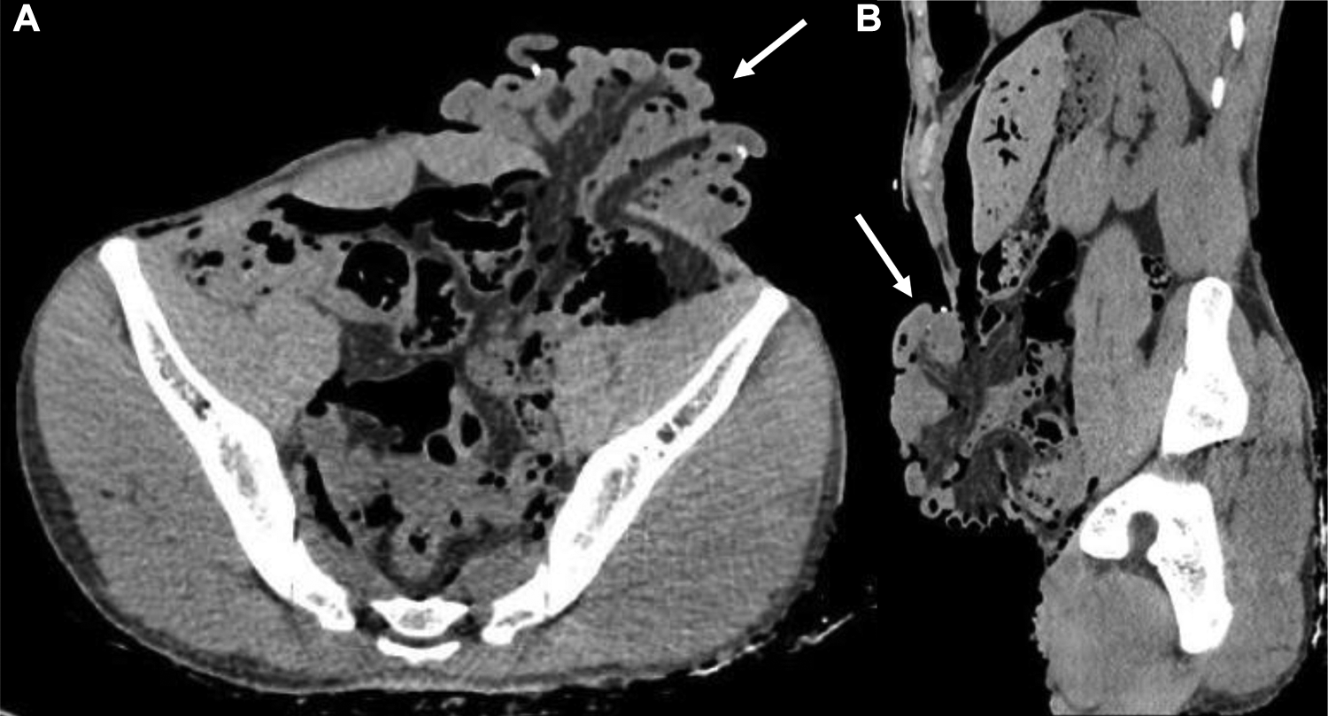
PMCT of a fire victim. A, Axial and (B) sagittal PMCT images demonstrating evisceration of multiple bowel loops secondary to an increased intra-abdominal pressure when a corpse is submitted to elevated temperatures. PMCT indicates postmortem computed tomography.
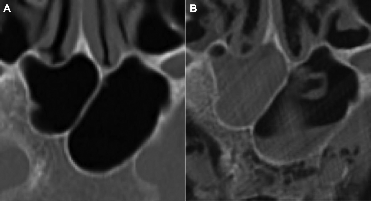
Victim identification. Comparison of (A) antemortem and (B) postmortem CT studies demonstrating identical sphenoid sinus morphology and allowing adequate identification of the deceased person, such as in this case of fire victim trapped in a car. CT indicates computed tomography.
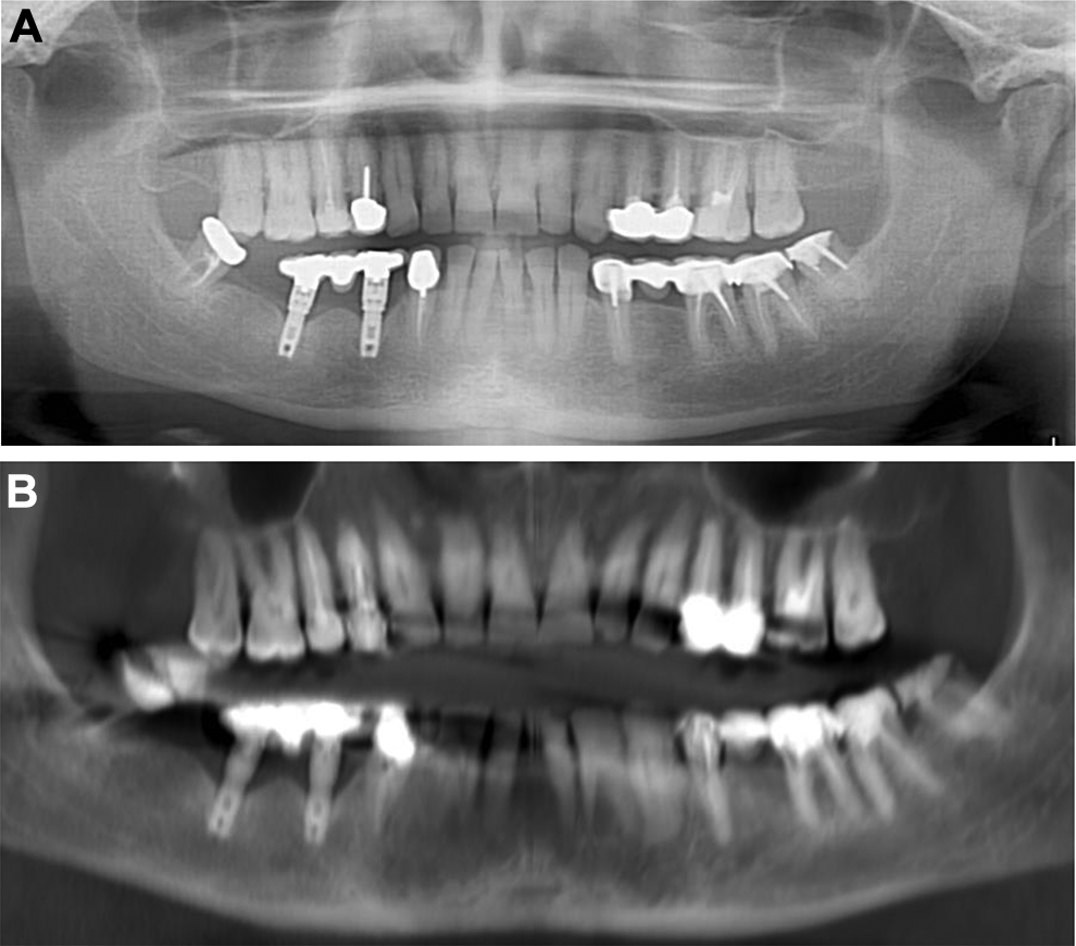
Victim identification. A, Antemortem and (B) postmortem imaging demonstrating a perfect match between the dental implants leading to accurate identification of a victim unidentifiable by external examination only.
These include assessing the length of long bones, using unique anatomical landmarks or prosthesis to serve as a reference for identification. Radiological identification is a user-friendly, fast, cost-effective, and accurate alternative to conventional DNA testing to identify an unknown person.
Photorealistic Presentation of Findings
Volume rendering is the standard post-processing technique used to display 3-dimensional anatomy. Cinematic rendering is a recently introduced 3D reconstruction technique depicting anatomy true to scale with realistic color depictions of injuries, resulting in better anatomical detail and identification of pathology. 21 Both techniques are best used to display skeletal trauma, vessel pathology, and superficial injuries (Figure 15). Compared to VR, CR is a more photorealistic representation, gives smoother contour to vessels, with increased contrast resulting in easier understanding of complex fractures (Figure 16). Pattern of injuries is better depicted, making it easier to match with the causative weapon. This has been shown in a recent study by Ebert et al 22 where 10 PMCT cases were reconstructed in both VR and CR and 48 questionnaires evaluating both were analyzed. A statistically significant number found the injuries realistic and easier to understand on CR. 22 The disadvantage of CR is the higher computational power and longer time needed to render the final image. The biggest impact of these models is in the legal setting where they serve as evidence presentation in court and may hold significant impact on juries involved in issuing verdicts based on forensic evidence.
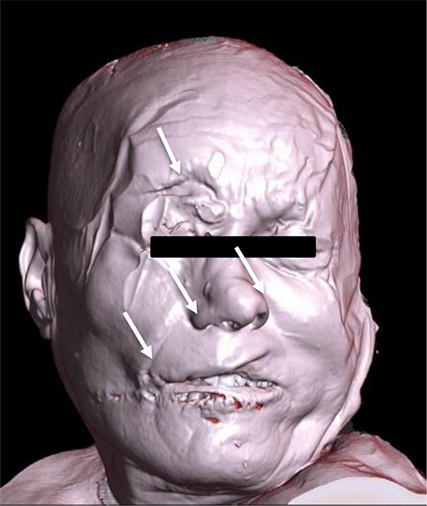
PMCT imaging of a 75-year-old male patient beaten to death with a baseball bat. 3D surface rendering image demonstrating photorealistic depiction of the superficial injuries and had excellent correlation with the autopsy specimen. PMCT indicates postmortem computed tomography; 3D, 3-dimensional.
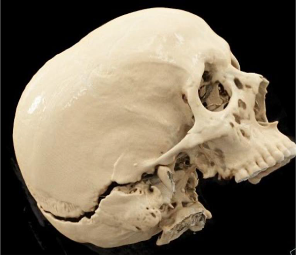
PMCT of a male patient following a motorcycle accident. 3D cinematic rendering image of the skull displays an unusual fracture separation of the skull base (arrows), highlighting the severity of the trauma. PMCT indicates postmortem computed tomography; 3D, 3-dimensional.
Advantages of Conventional Autopsy Over PMCT
Postmortem computed tomography has been shown to be less sensitive in assessment of vascular injuries, representing the second most prevalent cause of mortality in trauma. Although PMCT has been shown to identify hemorrhage as cause of death, it is limited in identification of the source of hemorrhage, despite use of contrast-enhanced acquisitions. 10 Conventional autopsy has been shown to be more sensitive demonstrating the associated arterial injury in majority of cases, 11 in up to 90% of cases in the analysis by Wijetunga et al. 6,17
Postmortem computed tomography displays most extra-axial cerebral collections comparably to autopsy but is less sensitive in cases of thin extra-axial bleed. The analysis by Wijetunga et al revealed that autopsy correctly depicted 9 subdural bleeds which were missed on PMCT. Legrand et al in their review of head trauma identified 3 epidural bleeds which were not seen on PMCT. 5 The study also hypothesized that the failure of PMCT to recognize 7 brainstem injuries and 9 brain contusion was likely due to poor SNR and beam hardening artifact. 6
In cases of suffocation and strangulation, autopsy offers distinct advantages. 11 Postmortem computed tomography is good at demonstrating osseous and cartilaginous injuries related to hanging and strangulation. However, the benefits of direct examination outweigh the overall sensitivity of PMCT. Of the 14 hanging cases analyzed by Willaume et al, osteocartilaginous lesions were found on PMCT in only half the analyzed cases whereas external examination correctly identified positive findings. Physical examination and direct visualization of ligature marks and neck bruising are usually not depicted on PMCT scan 23 (Figure 17). The advantage of direct examination is also depicted in the suboptimal depiction of the specific findings related to an entry wound (Figure 18).
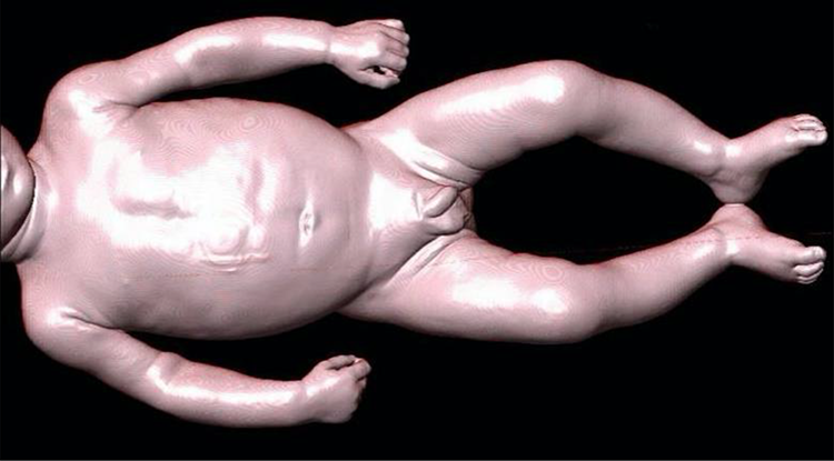
Limitation of PMCT in assessment of superficial injuries. An 8-month-old male patient evaluated by PMCT. 3D surface rendering of whole-body is relatively unremarkable, while physical examination depicted numerous superficial areas of bruising. PMCT indicates postmortem computed tomography; 3D, 3-dimensional.
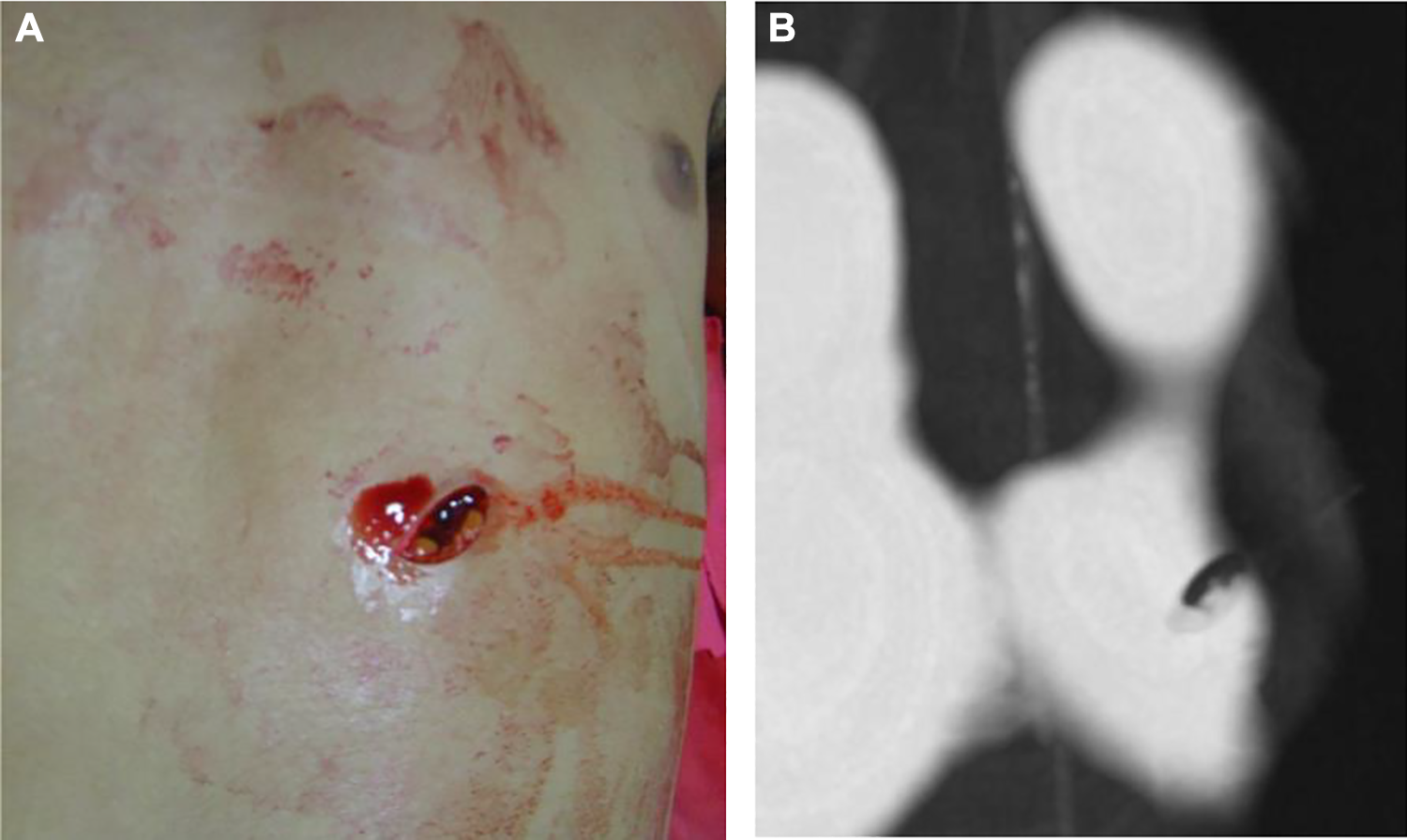
A and B, Although surface rendering produces a reliable reproduction of the stab wound, it falls short of the autopsy in analyzing the margins, shape, and depth of the entry wound.
Lung analysis has also been shown to be more accurate on autopsy compared to PMCT. In a recent series, autopsy identified 20 lung injuries which were missed on PMCT and a review failed to reveal any findings in 12 of these, while 6 of the lung contusions were deemed to represent atelectasis or aspiration rather than air space opacification. 6
Solid organ injury is often commonly better recognized under direct examination compared to noncontrast PMCT. Contusions not just to solid organs but to brain are also more effectively picked up on autopsy. 10 In Wijetunga et al study, despite careful second-look examination after failure to recognize 11 cardiac and 11 liver injuries, radiologists were unable to identify any of the cardiac injuries and only 2 of the liver injuries on additional review of images. 6
Pitfalls
The radiologist should be aware of the changes the deceased body undergoes in order to avoid false-positive and false-negative interpretations. Although it is known that gas accumulates as part of body decomposition, it is more important to know how to differentiate gas secondary to injury or medical intervention from postmortem gas accumulation. This is especially vital in penetrating trauma cases and in suspected gas embolism. It has been found that intravascular gas from decomposition changes is usually symmetrical in distribution. 24 Putrefactive gas usually originates in the intestinal vessels before migrating. Egger et al after studying distribution of gas in 119 postmortem cases concluded that gas initially appears in solid organs and close to 50% in their study had gas in the cardiac chambers and 40% within the liver. They observed that gas appeared in the right heart chambers and within the cavity itself prior to appearing within the myocardium (Figure 19). They also concluded that gas formed sequentially in the abdominal veins and arteries after appearing in the heart and liver. Gas formation begins within a fairly variable time frame (5-48 hours after death), pointing out the unreliability of predicting gas formation based on time after death. 25 An additional factor that favored postmortem air was resuscitation status. Intravascular gas was consistently shown to be present in those who had gone resuscitation versus those who had not (72% vs 47%, P = .013) in a study by Wagensveld et al. 26
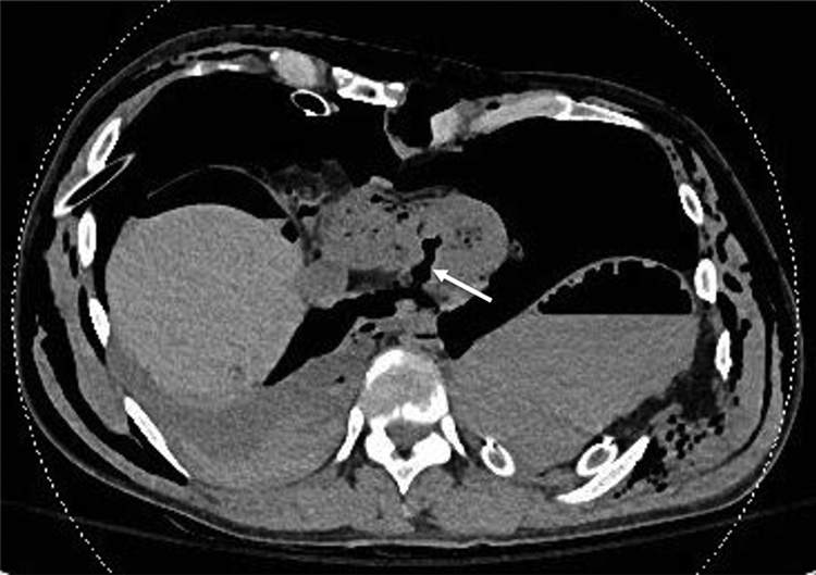
PMCT of a 44-year-old male patient following motorcycle accident. Noncontrast image of the chest demonstrating extensive injuries and a collapsed heart, typically seen in the context of rapid exsanguination. Additionally, extensive air within the cardiac chambers and a full-thickness left ventricle laceration were seen (arrow). PMCT indicates postmortem computed tomography.
False-positive venous sinus thrombosis and subdural bleeds may be diagnosed on PMCT when pitfalls are not recognized when evaluating PMCT of the brain. In a study of 50 patients who had both antemortem and postmortem CT scans, Takahashi et al in a study of 208 cases demonstrated high attenuation of the superior sagittal sinus in 80% of cases they studied, with similar findings of the transverse sinus and tentorium in 48%. 27 Smith et al also described hyperattenuation of the cortical veins adjacent to the falx. 24 These postmortem findings are usually symmetrical as opposed to asymmetric in case of primary pathology. Cerebral autolysis can simulate diffuse cerebral edema, traumatic brain injury, and acute infarction due to effacement of the corticomedullary junction, cerebral hypoattenuation, and effacement of extra-axial spaces. Both Panda et al and Levy et al studied cerebral autolysis changes, seen on PMCT within 6 hours to 24 to 48 hours following death. 28,29 Delayed imaging and freezing of the body for preservation may lead to artifacts simulating edema leading to misreading of brain images. 30
Aortic injuries imply a high degree of impact and may point to an identifiable cause of death. Intramural thrombus may be simulated by hematocrit effect where there is dependent layering of hyperdense cellular contents and supernatant low attenuation plasma. The hyperattenuating aortic wall may parallel the appearance of an intramural hematoma mimicking pathology. This was explained by Christie et al who postulated this being due to contraction of the aortic wall, loss of pressure, and low density of luminal contents from hematocrit effect and dilution after massive infusion. 31 Of 100 cases reviewed, hyperattenuation of thoracic aorta was seen in 90% cases by Wagensveld et al. 26 The vanishing artery sign is seen in cases of severe hemorrhage where rapid exsanguination causes collapse of the aorta or pulmonary arteries (Figure 20). Livor mortis from dependent accumulation of fluid and simulation of pulmonary contusions, pleural collections, and liver contusions may misguide the reading radiologist. This often depends on the amount of time that has passed prior to scanning the patient. Interestingly, Levy et al found that although increased attenuation within solid organs preferentially occurs within the lungs, liver, spleen, and kidneys, autolysis changes in fact happen earliest within adrenals and pancreas but these however retain their normal imaging appearance. 29
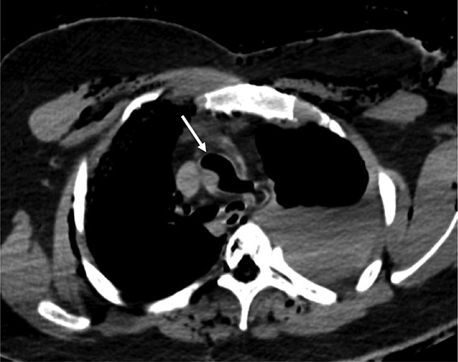
“Vanishing aorta sign.” PMCT image showing a partially collapsed aorta containing gas, typically seen in sudden death with rapid blood loss in the absence of clot formation to maintain the aortic shape. Posttraumatic vascular gas is typical in stab or gunshot wounds. However, distinction with intravascular gas decay cannot be made as the alleged postmortem interval in this case was deemed to be 10 days. PMCT indicates postmortem computed tomography.
While assessing superficial wounds, skin folds, or artifacts should not be mistaken for penetrating tracks (Figure 21). When there is extensive subcutaneous emphysema and contamination, ancillary findings cannot be relied upon to make this distinction.
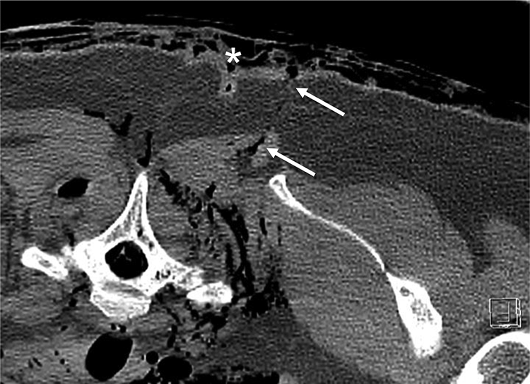
Skin fold (star) may be mistaken for stab wound (arrows) and distinguishing the two might be difficult in extensive superficial trauma. Identification of overlying skin defect and gas tracking within the deeper subcutaneous tissues is key.
Conclusion
Postmortem computed tomography is a useful adjunct to autopsy helping in guiding conventional autopsy and finding injuries potentially missed clinically. In cases where there is no identifiable cause of death, PMCT is a viable alternative that closely matches an autopsy examination. Ability of PMCT to pinpoint the trauma findings provides answers to the trauma team and helps to identify shortcomings in their approach. Although autopsy retains an advantage in identifying injuries incompatible with life, PMCT has established superiority in facial and osseous injuries and in identifying vascular and abnormal gas accumulation. Virtual ballistics and its 3-dimensional capability are also beneficial in photorealistic depiction of complex traumatic injuries, recreating physical scene and presenting the same in a legal setting. Postmortem computed tomography will definitely play a greater role and see routine usage as improved familiarity of radiologists in image interpretation results in improved detection rates.
Footnotes
Authors’ Note
The manuscript is unpublished and not currently under consideration by another journal.
Acknowledgment
The authors thank the University Hospital of Nîmes (France) for the excellent autopsy and PMCT images which are produced here with permission.
Declaration of Conflicting Interests
The author(s) declared no potential conflicts of interest with respect to the research, authorship, and/or publication of this article.
Funding
The author(s) received no financial support for the research, authorship, and/or publication of this article.