
Abstract
Cerebral vasculopathy is an important but underrecognized complication of neurofibromatosis type 1. Over a 10-year period, we retrospectively assessed the prevalence, clinical manifestations, management, and outcome of cerebral vasculopathy in children with neurofibromatosis type 1. Magnetic resonance imaging (MRI) of the brain was performed on 78% of the patients (312/398) of which 46% (143/312) had magnetic resonance angiography of the intracranial arteries; 4.8% (15/312) had cerebral vasculopathy. Approximately half were asymptomatic at presentation; none had neurologic deficits. Cerebral vasculopathy included moyamoya changes (7) and stenosis/occlusion of major intracranial arteries (8). On follow-up (mean 4 years), 2 patients developed radiologic progression; 1 was treated with aspirin alone, whereas another underwent revascularization surgery. Although cerebral vasculopathy in neurofibromatosis type 1 may be asymptomatic at presentation, there may be radiologic and clinical progression leading to morbidity and mortality. Magnetic resonance angiography should be considered with brain MRI for early detection and timely intervention of cerebral vasculopathy.
Neurofibromatosis type 1 is the most common autosomal dominant condition affecting the nervous system with an estimated incidence of 1 in 2500 to 3500 individuals independent of ethnicity, race, and sex. 1,2 Although Von Recklinghausen first described neurofibromatosis type 1 in 1882, 3 formal diagnostic criteria were established only in 1987 by the National Institutes of Health (NIH) Consensus Development Conference. 4
Vasculopathy is a significant but underrecognized complication of neurofibromatosis type 1, affecting arterial and venous blood vessels of all sizes. 5,6 Neurofibromin, the protein product of the neurofibromatosis type 1 gene, is expressed in endothelial and smooth muscle cells of blood vessels and is likely to be involved in the pathogenesis of neurofibromatosis type 1 vasculopathy. 7 Clinical manifestations of neurofibromatosis type 1 vasculopathy may stem from stenosis or occlusion of the vessels resulting in cerebral or visceral infarcts, aneurysms resulting in hemorrhage, or arteriovenous fistulae. 8 Most individuals with neurofibromatosis type 1 vasculopathy are asymptomatic, 9 although an increased mortality has been reported in patients with neurofibromatosis type 1 and vasculopathy. 10 Characteristic neurofibromatosis type 1 vascular lesions have been described in the entire arterial tree, but involvement of the renal arteries with consequent hypertension is most common. 9,11 The cerebral vascular lesions are patchy in distribution, but multiple vessels can be involved. There are few studies of cerebral vasculopathy in neurofibromatosis type 1 in children. 12 –14 In this study, we review our experience of cerebral vasculopathy in children with neurofibromatosis type 1 in order to improve knowledge and raise awareness among caregivers of this association.
Methods
Study Subjects
An institutional review board–approved retrospective chart review was performed on all children ≤18 years of age identified with neurofibromatosis type 1 over a 10-year period (2000-2010) at the Children’s Hospital, Cleveland Clinic. The information was gathered from our clinic database. All patients included met National Institutes of Health diagnostic criteria for neurofibromatosis type 1. Data collected included sex, age at the time of diagnosis of neurofibromatosis type 1, age at the time of diagnosis of vasculopathy, clinical manifestations of vasculopathy, neuroimaging findings, treatment for vasculopathy, and outcome. Exclusion criteria included stroke from other causes, cerebral arterial dissection, incidental radiologic findings like developmental venous malformations, and a previous history of cranial irradiation that could be etiologically related to the vasculopathy.
Neuroimaging
The majority of the subjects (78%) in our study underwent magnetic resonance imaging (MRI) of the brain with or without magnetic resonance angiography. The studies were reviewed independently by a neuroradiologist blinded to the clinical presentation. MRI of the brain was performed as routine screening for intracranial complications of neurofibromatosis type 1 based on the treating physicians’ discretion or based on the neurologic symptoms (headache, seizures, suspicion of intracranial tumors) during the study period. Patients underwent neurovascular imaging studies, including magnetic resonance angiography, computed tomographic angiography, or conventional angiography of the intracranial vessels either with MRI of the brain or later if there was a suspicion of intracranial vasculopathy on MRI of the brain. Vasculopathy was defined as any abnormality of the intracranial vascular system that could not be defined as a normal variant. The studies were reviewed to detect stenosis and/or occlusion of the intracranial arterial system, abnormal collateral arterial supply, intracranial aneurysm, or evidence of arteriovenous malformation or arteriovenous fistulae. Those patients with comorbid optic nerve gliomas were identified as they have been reported to have an increased incidence of cerebral vasculopathy. 14
Statistical Analysis
Descriptive statistics were used in this study.
Results
Study Subjects
During the study period, 398 children with a confirmed diagnosis of neurofibromatosis type 1 were identified from our clinic database. Of these patients, 312 (78%) underwent MRI of the brain as routine screening for intracranial complications based on the treating physicians discretion or based on the neurologic symptoms (headache, seizures, suspicion of intracranial tumors). One hundred forty-three (46%) of these 312 children had magnetic resonance angiography. Fifteen (15/312, 4.8%) had cerebral vasculopathy identified by neuroimaging. All these patients had magnetic resonance angiography and 4 had additional neuroimaging studies (computed tomographic angiography/conventional angiogram) to better define the vascular anatomy. None had intracranial hemorrhage, aneurysm, or arteriovenous fistulae.
Clinical Characteristics
The clinical features in the 15 children with vasculopathy are summarized in Table 1. The mean age at diagnosis of neurofibromatosis type 1 was 4.3 ± 3.5 years (1-12 years) with 8 boys. The mean age at the time of diagnosis of their vasculopathy was 11.7 ± 7.3 years (2-18 years). The indications for neuroimaging were headache (5), seizures (2), brain tumor (1), and screening for intracranial complications (7). None of our patients had focal neurologic deficits or complications attributable to their vasculopathy at presentation or at the time of the radiology follow-up (mean 3.8 ± 2.9 years; 1-10 years). One child was treated with aspirin as primary stroke prevention therapy, 1 underwent revascularization surgery (encephaloduroarteriomyosynangiosis procedure), and 1 patient died 2 years after diagnosis from a thalamic glioma. The remaining 12 children are being clinically followed with no specific treatment. None of our patients received any form of cranial irradiation.
Clinical Characteristics of Children With Neurofibromatosis Type 1–Associated Cerebral Vasculopathy
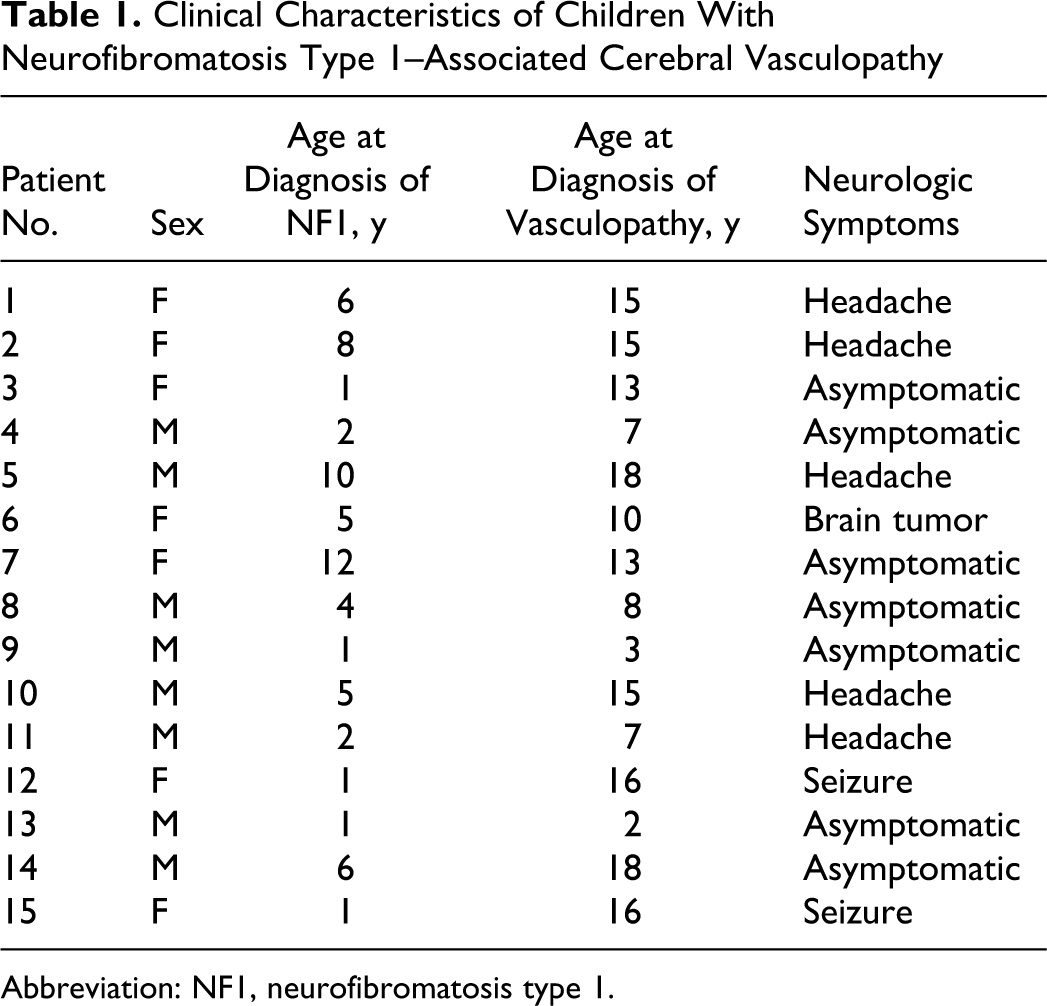
Abbreviation: NF1, neurofibromatosis type 1.
Neuroimaging Findings
Three hundred twelve (78%) of 398 children with neurofibromatosis type 1 had MRI of the brain. In addition, 143 (46%) of these patients had underwent magnetic resonance angiography of the intracranial vasculature. Of the 15 children with vasculopathy, 10 had magnetic resonance angiography at the time of initial MRI and 5 had magnetic resonance angiography subsequently performed to confirm vasculopathy. Neuroimaging findings are summarized in Table 2. Stenotic lesions were noted in all children. There were no aneurysms, arteriovenous malformations, or arteriovenous fistulae. One child had an old cerebral infarct, whereas the rest had no ischemic or hemorrhagic lesions. Seven children had moyamoya changes (unilateral 5 and bilateral 2) [Figures 1 and 2]); 3 had >1 major intracranial artery involvement (internal carotid artery, middle cerebral artery, or posterior cerebral artery); 3 had isolated supraclinoid internal carotid artery involvement (occlusion 1 and stenosis 2); and 2 had isolated vertebral artery involvement (occlusion 1 and stenosis 1).
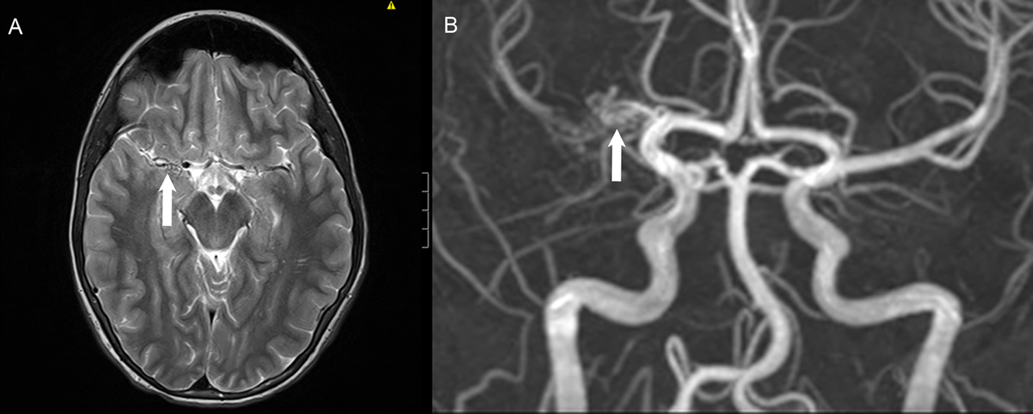
MRI of brain on T2-weighted image showing loss of flow void signal along the course of the right middle cerebral artery proximal segment being replaced by mesh of vessels (A); corresponding magnetic resonance angiography showing occlusion of middle cerebral artery with moyamoya changes (B).
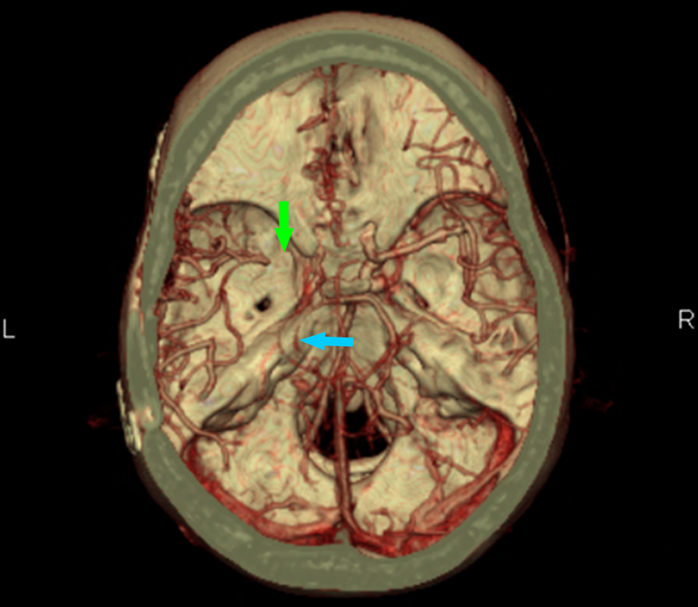
Computed tomographic angiogram showing multiple vessel occlusions: left middle cerebral artery occlusion (green arrow), left posterior cerebral artery severe stenosis (blue arrow), and left distal internal carotid stenosis (without arrow).
Neuroimaging, Follow-up, and Treatment of Children With Cerebral Vasculopathy
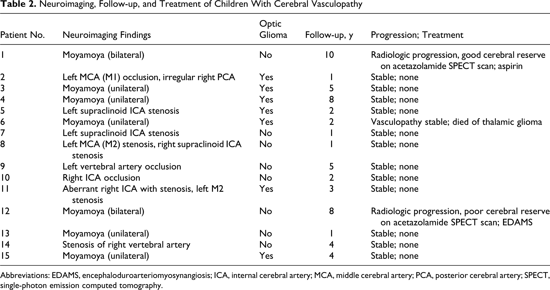
Abbreviations: EDAMS, encephaloduroarteriomyosynangiosis; ICA, internal cerebral artery; MCA, middle cerebral artery; PCA, posterior cerebral artery; SPECT, single-photon emission computed tomography.
Optic glioma was noted in 7 children with imaging findings of vasculopathy (47%).
The mean duration of radiology follow-up was 3.8 ± 2.9 years (1-10 years). There was no radiologic progression of vasculopathy in 13 children. Two had radiologic progression on serial magnetic resonance angiographies without development of clinical symptoms or signs. Both had moyamoya and underwent cerebral perfusion reserve studies with acetazolamide single-photon emission computed tomographic (SPECT) scans. One showed good reserve and was treated conservatively with aspirin for primary stroke prevention, whereas the other had poor cerebral reserve and so underwent revascularization surgery.
Discussion
This study focuses on cerebral vasculopathy in children with neurofibromatosis type 1. Vasculopathy has long been associated with neurofibromatosis type 1, but the precise incidence is unknown. Symptomatic involvement is uncommon, 9 and most affected patients remain asymptomatic throughout life. 10 However, when detected early in childhood, this might have a relevance in later life as these patients are already at risk of increased morbidity and mortality given their inherent vascular abnormalities.
Available modalities for evaluation of both intracranial and extracranial vasculature include conventional angiography, magnetic resonance angiography and computed tomographic angiography, each with advantages and disadvantages. Conventional angiography is the gold standard but has associated risks, including a 0.7% risk of stroke. 15 Computed tomographic angiography involves the use of contrast and high-dose ionizing radiation as opposed to magnetic resonance angiography, which does not involve ionizing radiation; however, these are both noninvasive examinations. Conventional angiography has the highest spatial resolution, allowing for depiction of small vessel abnormalities with decreasing spatial resolution in computed tomographic angiographic imaging and a further decrease with magnetic resonance angiography. 15
Cerebrovascular manifestations of neurofibromatosis type 1 are rare with earliest recognized vascular abnormalities involving the renal arteries and to a lesser extent the celiac artery, iliac arteries, and the abdominal aorta, studied initially with conventional catheter-based angiography. 16 Vascular abnormalities of the intracranial circulation encompass stenoses, occlusions, ectasia, fusiform aneurysm formation, or arteriovenous fistulae involving the anterior and/or posterior circulation. Moyamoya disease may occur with supraclinoid internal carotid artery narrowing. The vasculopathy may also involve the extracranial carotid and vertebral arteries. Angiographically, the affected vessels have proximal narrowing, complete narrowing, and poststenotic dilatation. 17 MRI, adopted for widespread clinical use in the early 1980s, is advantageous not only for its lack of ionizing radiation but also because alterations in normal-appearing flow voids of the circle of Willis can be detected and characteristic collateral flow voids seen in moyamoya disease can be detected. 18
Three recent studies have addressed the issue of cerebral vasculopathy in children with neurofibromatosis type 1. Rosser et al 12 reported that of 353 patients with neurofibromatosis type 1, 316 (90%) underwent MRI as part of routine screening and 8 (2.5%) patients were identified with an abnormality of the cerebral vasculature. In the study by Cairns et al, 13 144 (21%) of 698 patients underwent MRI for a clinical indication and 7 (5%) were identified with cerebrovascular dysplasia. The most recent study by Rea et al 14 noted that of 419 patients with neurofibromatosis type 1, 266 (63%) underwent neuroimaging for a clinical indication and 17 (6%) were identified with arteriopathy. In the present study, of 398 children with neurofibromatosis type 1, MRI was performed in 312 (78%) either as a part of routine screening or because of the presence of neurologic symptoms. Fifteen (4.8%) children had cerebral vasculopathy, which is consistent with the previously reported prevalence of 2.5% to 6%. Rea et al’s prevalence of cerebral vasculopathy may be an underestimate as only 63% of their patients with neurofibromatosis type 1 had an MRI and a much lesser proportion had magnetic resonance angiography (35/266, 13%). 14 In comparison, 78% of our patients had MRI and almost half had magnetic resonance angiography (143/312, 46%) lending credence to the true prevalence of cerebral vasculopathy. However, ours and previous studies being retrospective in nature, there is always a selection bias affecting the results and true prevalence.
The mean age at the time of presentation of cerebral vasculopathy in previous studies was between 5.2 and 7.3 years. 12 –14 In comparison, the diagnosis of cerebral vasculopathy in our series, however, was made at a mean age of 11 years, which was older than the other published series. Of interest also in the study by Rea et al was the presence of optic glioma in 13 of the 17 (76%) patients with arteriopathy, the majority having extensive tumors. 14 As optic glioma 8 is usually diagnosed during the first 6 years of life and the majority of their patients with arteriopathy were younger than age 6 years, they postulated that extensive optic glioma may trigger the formation of specific growth factors, leading to abnormal vascular endothelial proliferation. 14 In patients with neurofibromatosis type 1, optic pathway gliomas apparent on MRI are present in 15% of patients before 6 years of age. In the present study, about 50% of patients had optic glioma, making the association of vasculopathy and optic glioma significant and rife for further study.
The pathogenesis of neurofibromatosis type 1–related vasculopathy are poorly understood but are likely related to neurofibromin. The NF1 gene is large, spanning approximately 335 kb of the chromosome 17q11.2.43 and its protein product neurofibromin shares functional homology and sequence with a group of Ras-GTPase-activating protein. 19,20 Loss of neurofibromin activity results in unopposed Ras activity and subsequent activation of several important downstream signaling intermediates, leading ultimately to increased cell growth. 21
Neurofibromin has been demonstrated in the endothelial layer of rodent and bovine cerebral and renal arteries as well as the aorta. The putative hypothesis is that the loss of neurofibromin expression in endothelial cells leads to excessive proliferation of the vascular endothelial and/or smooth muscle cells. 7 In addition, neurofibromin serves to maintain the integrity of the endothelial cell layer and its mutation alters this integrity, leading to unopposed proliferation of the vascular smooth muscle cells. Neurofibromatosis type 1–associated vasculopathy does not affect all arteries in an affected patient, although all the arteries have the same constitutional mutation of the NF1 gene. The putative mechanisms are either a “second hit” mutation of the normal NF1 allele or somatic mutation at another locus leading to vascular changes. 5 In addition, interaction between host and environmental factors may play a role in the development of selective vasculopathy.
The pathology of neurofibromatosis type 1 vasculopathy was first described by Reubi in 1945. 22 Subsequently Sobata et al in 1988 classified neurofibromatosis type 1–associated cerebral vasculopathy into 3 groups: stenotic, aneurysmal, or both. 23 They noted that children usually had stenotic lesions (89%), with the remainder being aneurysmal formation only. Almost all children had stenosis/occlusion of the intracranial arteries in the 3 recent pediatric studies, 12 –14 a finding supported by our study. Moyamoya disease (Japanese word meaning “hazy, cloud of smoke drifting through the air,” referring to the angiographic appearance of the distal collateral network) is characterized by chronic progressive stenosis of the distal internal carotid artery and, less often, stenosis of the proximal anterior cerebral artery and middle cerebral artery, the basilar artery, and the posterior cerebral arteries. 24 Moyamoya disease is known to occur in both neurofibromatosis type 1 and in non–neurofibromatosis type 1 patients. Moyamoya disease can also develop secondarily to radiation-induced changes from the treatment of head and neck cancers 24 ; however, none of our patients had radiation therapy. In patients with neurofibromatosis type 1, moyamoya changes usually affect the anterior circulation and are usually unilateral, a finding also noted in our study. In our cohort, 47% (7/15) had moyamoya changes, which is less compared to the 2 recently published series (70%-76%). 13,14 As reported in previous studies, we also found that the internal carotid artery is the most commonly affected artery either alone or in combination with other major intracranial arteries (middle cerebral artery second most common followed by ACA, posterior cerebral artery and vertebral artery). None of our patients had intracranial aneurysms consistent with the experience of earlier investigators. 23
Children with neurofibromatosis type 1–associated cerebral vasculopathy can present with weakness, involuntary movements, headaches, or seizures as a result of cerebral ischemia from thrombosis or embolism of the intracranial vessels. 8 However, the majority of patients with neurofibromatosis type 1 cerebral vasculopathy are clinically asymptomatic. 12,13 In our series, the majority were clinically asymptomatic (n = 7), 5 had headache as a presenting symptom, 2 had seizures, and 1 had a known supratentorial glioma. Interestingly, none of our patients had focal neurodeficits on examination. Rosser et al 12 reported focal deficits in 12% and Cairns et al 13 in 29%, whereas Rea et al 14 reported a high figure of 47%. This apparent clinicoradiologic dissociation strengthens the advocacy for performing neuroimaging in neurofibromatosis type 1 for early detection of cerebral vasculopathy. There may be a lag of several months to years between the radiologic manifestations and the development of ischemic symptoms or signs secondary to neurofibromatosis type 1–associated cerebral vasculopathy. Furthermore, there may even be radiologic progression without the development of clinical symptoms. This underscores the importance of close clinical as well as radiologic surveillance. In our cohort, close follow-up showed 2 children with radiologic progression but no clinical symptoms. Similarly Rosser et al found 1 patient with clinical progression, Cairns et al found 3 with radiologic progression, and Rea et al found 5 with radiologic progression and 1 with clinical progression. 12 –14
There is no consensus about the management of neurofibromatosis type 1–associated vasculopathy as this complication has not been frequently reported. The options are based on the nature and location of the lesion and range from medical management with an antiplatelet agent to surgical intervention with revascularization procedures for moyamoya disease. However, it makes sense that treatment with an antiplatelet agent like aspirin may reduce the risk of clot formation in narrowed arteries where high flow is the main mechanism of platelet-rich clot formation. In the study by Rea et al, 7 patients were on aspirin, of which 5 were already symptomatic. In our study, 1 patient with evidence of radiologic progression was commenced on aspirin. As more data emerge in this field, a well-planned multicenter study in the future can address this issue and also help develop guidelines for the medical management of neurofibromatosis type 1 vasculopathy. Rea et al reported 6 patients in their series having had revascularization procedure, whereas Cairns et al reported 1 patient and Rosser et al reported 3 patients, respectively, having undergone revascularization procedures. 12 –14 Two of our patients had radiologic progression of their moyamoya vasculopathy, 1 of which had poor cerebral perfusion reserve on an acetazolamide SPECT scan and so underwent a revascularization procedure.
Given the rare nature of cerebral vasculopathy in neurofibromatosis type 1, multicentered studies will be needed to develop guidelines for the medical and or surgical management of neurofibromatosis type 1 vasculopathy. With increasing awareness of neurofibromatosis type 1 cerebral vasculopathy in children, the issue of management and optimal monitoring and follow-up of these lesions becomes important. Frequently, the vasculopathy is detected in asymptomatic individuals, or individuals being scanned for unassociated neurologic complaints, further complicating decision making. There is conflicting opinion regarding universal screening with MRI and/or magnetic resonance angiography in neurofibromatosis type 1 patients. Current published guidelines for the diagnosis and management of patients with neurofibromatosis type 1 do not advocate routine neuroimaging in asymptomatic patients. 25 26 On the contrary, the American Heart Association, in a scientific statement published in September 2008 on the management of stroke in infants and children, recommended that routine vascular screening with magnetic resonance angiography may be considered in children with relatively common and high-risk disorders such as neurofibromatosis type 1, Down syndrome, and sickle cell disease known to cause moyamoya syndrome. 24
Recommending routine screening with brain MRI/magnetic resonance angiography for children with neurofibromatosis type 1, however, has many inherent problems especially in terms of timing, need for potential sedation/anesthesia, resources, and logistics. However, it must be borne in mind that without neuroimaging, many cases of cerebral vasculopathy may be missed and allowed to progress over time radiologically and clinically into ischemic or hemorrhagic strokes that may have been potentially averted if detected early.
The imaging findings of neurofibromatosis type 1–related vasculopathy may be subtle and can easily be overlooked. Raising awareness of this specific complication will lead to early detection, closer and better follow-up, and timely intervention. Moreover, in many patients with neurofibromatosis type 1, these complications may be missed when only routine MRI studies are performed without adding magnetic resonance angiography. Although adding magnetic resonance angiography to MRI adds to the cost, scan time, and anesthesia time (in small children), it has the benefit of better delineation of the intracranial vasculopathy and can be a guide for future interventions. In the case of young children, performing both the studies is logistically helpful in that it obviates need for repeat anesthesia. As there is no consensus currently regarding management of neurofibromatosis type 1 cerebral vasculopathy consideration should be given to adding magnetic resonance angiography to MRI brain (performed for any indication) in children for early detection of cerebral vasculopathy.
Limitations of the present study include its retrospective nature as well as the small number of patients. Another limitation was lack of uniformity among patients with neurofibromatosis type 1 who underwent magnetic resonance angiographic examination in addition to MRI brain. Future prospective longitudinal studies that evaluate the clinical characteristics and neuroimaging protocols in patients with neurofibromatosis type 1 cerebral vasculopathy will provide important data that will help determine optimal imaging guidelines in children with neurofibromatosis type 1.
Conclusion
Cerebral vasculopathy is an underrecognized complication of neurofibromatosis type 1. The majority of the patients are asymptomatic at the time of detection of this complication. However, the natural history, long-term progression, and prognosis have not been well studied. The current treatment options for preventing ischemic stroke in stenotic lesions range from antiplatelet agents to revascularization procedures in patients with moyamoya disease. However, to date there is no consensus regarding the management of this complication in neurofibromatosis type 1. As more data emerge in this field, a well-planned longitudinal multicenter study can address these issues and help formulate guidelines for the optimal management and monitoring of these patients. In children with neurofibromatosis type 1 who are undergoing neuroimaging studies consideration should be given to adding magnetic resonance angiography to these studies for better delineation of the intracranial vascular abnormality. This will allow earlier detection of cerebral vasculopathy, closer and better follow-up, and when necessary timely intervention to prevent complications.
Footnotes
Acknowledgment
The work was done at the Pediatric Neurology Center, Cleveland Clinic, Cleveland, Ohio. The work was presented as a poster at the Child Neurology Society Annual Meeting in 2011 at Savannah, Georgia.
Author Contributions
PSG collected and organized the data and wrote the first manuscript (including the first draft). TME reviewed the neuroimaging findings independently. MM conceptualized the study. MM, ADR, TME, and NRF verified the results and revised the manuscript at all stages.
Declaration of Conflicting Interests
The authors declared no potential conflicts of interest with respect to the research, authorship, and/or publication of this article.
Funding
A. David Rothner, MD, received research grants from GlaxoSmithKline, Merck, and AstraZeneca and received consultant fees from AstraZeneca and M.A.P. The other authors had nothing to disclose.
Ethical Approval
This study was approved by the Institutional Review Board at the Cleveland Clinic.