






Abstract
Concussion is a known risk in youth soccer, but little is known about subconcussive head impacts. The authors provided a prospective cohort study measuring frequency and magnitude of subconcussive head impacts using accelerometry in a middle school–age soccer tournament, and association between head impacts and changes in (1) symptoms, (2) cognitive testing, and (3) advanced neuroimaging. A total of 17 youth completed the study (41% female, mean 12.6 years). There were 73 head impacts >15g measured (45% headers) and only 2 had a maximum peak linear acceleration >50g. No youth reported symptoms consistent with concussion. After correction for multiple comparisons and a sensitivity analysis excluding clear outliers, no significant associations were found between head impact exposure and neuropsychological testing or advanced neuroimaging. The authors conclude that head impacts were relatively uncommon and low in acceleration in youth playing a weekend soccer tournament. This study adds to the limited data regarding head impacts in youth soccer.
Soccer is the most popular sport worldwide, with 265 million players (22 million youth) 1 and a 2-fold increase in US participants from 1990 to 2014. 2 Soccer provides clear benefits for cardiovascular fitness 3 and bone health. 4 However, as with any sport, there is an injury risk, and concern has been raised about head injuries. 5 Soccer is unique in that players use their head to propel the ball, and such head impacts include a potential for injury. Players also incur head impacts from colliding with each other, the ground, or objects such as the goal post, and concussions do occur (0.73/1000 Athletic exposures [AEs] for girls’ soccer and 0.41/1000 AEs for boys’ soccer, compared to 0.94/1000 AEs for football, all during high school game play). 6 Limitations on heading in youth have been proposed with the goal of decreasing concussion risk 5 ; however, sufficient evidence to support a scientific recommendation regarding heading in youth soccer does not exist. A recent study 7 reported the most common mechanism of concussion in soccer was player-to-player contact, rather than contact with the ball. Unfortunately, there is little data regarding heading in youth soccer, or how this contributes to head impact exposure. One prior study examined head impacts in middle school–age soccer, 8 but only studied 45 minutes of scrimmage play. To our knowledge, the frequency and magnitude of “subconcussive” head impacts (ie, head impacts that do not result in concussion) in the middle school age group have not been studied, nor the proportion due to heading. Such information could prove useful in determining the risk associated with heading in youth soccer.
Several studies have suggested that physiologic changes due to subconcussive head impacts may be measureable, reporting changes in imaging after exposure to head impacts using diffusion tensor imaging and resting state functional magnetic resonance imaging (MRI). 9 -15 Other imaging approaches such as pseudo-continuous arterial spin labeling have not been used to examine subjects after sports-related head impacts but have been shown to be sensitive to mild traumatic brain injury. 16 Additional studies have explored the effect of head impact exposure on function (neuropsychological testing), but results have been mixed, with some authors reporting changes 17 -19 and others finding no differences. 20 -25 Only a few investigators have specifically evaluated the impact of an acute bout of soccer playing, and again results were mixed. 26 -29 None of these studies were conducted with middle school–age soccer players.
We designed a prospective cohort study to examine a short and intense period of soccer playing (a weekend soccer tournament), preceded and followed by neuropsychological testing and imaging. The short time period of the study ensured that no other factors would affect the outcomes in the subjects. Our primary goal was to objectively measure the frequency and magnitude of head impacts during a weekend youth soccer tournament. Our exploratory goal was to examine the association between head impact exposure and functional and anatomic measures, including neuropsychological testing and neuroimaging.
Methods
Participants
We recruited youth from 5 male and female club soccer teams in the Seattle area participating in tournaments in the summer of 2014. Athletes were excluded if they wore braces as these interfere with MRI quality. This study was approved by the University of Washington Institutional Review Board and all athletes and parents completed written informed assent and consent respectively prior to participation.
Overview
We utilized a prospective cohort design, measuring head impacts that occurred during a weekend soccer tournament using an accelerometer device called the xPatch, and exploring the association between head impact exposure and pre and post measurements of neurocognitive function, neuroimaging, and self-report of symptoms (Figure 1). Athletes were brought into the imaging center less than a week before and after the tournament to complete pre- and post-exposure assessments: concussive symptom questionnaires, 30 neuropsychological testing, and neuroimaging. Athletes and parents completed baseline questionnaires regarding demographic characteristics and concussion history. Athletes wore the xPatch during all games in the soccer tournament (3-6 games). Research assistants attended games to apply and remove xPatches, and observe collisions that occurred. The timing of collisions and whether or not they were headers was recorded for cross-comparison with xPatch data events.

Timeline of study events for Weekend youth soccer tournament (WEST), Seattle, WA, 2014.
Imaging Procedures
MRI scans were completed on a 3-Tesla Phillips Achieva MR system employing a vendor-supplied 32-channel head coil. Imaging lasted 30 minutes and 59 seconds and included a localizer sequence, a 1 mm isotropic MPRAGE (time to echo = 3.2 milliseconds, time to response = 7 milliseconds, slices = 145, image matrix = 192 × 192, duration = 3:49), 1 mm isotropic 3-dimensional T2-weighted image (time to echo = 239.2 milliseconds, time to response = 2500 milliseconds, slices = 145, image matrix = 192 × 192, duration 3:12), 64 direction diffusion sequence with b = 1000 seconds/mm2 and 6 non-diffusion-weighted images for diffusion tensor imaging analysis (time to echo = 68.3 milliseconds, time to response = 7500 milliseconds, 2 mm thickness, image matrix = 112 × 112, duration 9:16), pseudo-continuous arterial spin labeling obtained with label duration = 1525 and postlabel delay time of 1625 milliseconds (time to echo = 13.5 milliseconds, time to response = 5000 milliseconds, 5 mm thickness, image matrix = 96 × 96, duration = 4:39), and 2 acquisitions of resting state functional MRI (time to echo = 25 milliseconds, time to response = 2000 milliseconds, 3 mm thickness, image matrix = 64 × 64, duration = 4:24 each). Details regarding the methodology used for processing each imaging modality are included in the supplemental digital content.
Neuropsychological Testing
A neuropsychological battery was developed to assess cognitive areas most affected by brain injury. Working memory and reaction time were assessed using the Immediate Post-Concussion Assessment and Cognitive Testing (ImPACT), 30 a computerized test that yields 5 composite scores: impulse control, reaction time, memory composite score verbal, memory composite score visual, and visual motor composite score. Executive function and episodic memory were evaluated using 2 tests from the National Institutes of Health Toolbox 31 : Dimensional Change Card Sort Test and Picture Sequence Memory Test. Dimensional Change Card Sort Test assesses executive function by asking subjects to match a series of bivalent test pictures to the target pictures. Picture Sequence Memory Test measures episodic memory by asking youth to recall increasingly lengthy series of pictures. The King-Devick test was included as this has been used as a screening test for concussive injury with high sensitivity, 32 and it is thought to measure visual motor function and reaction time. All tests were administered by trained research assistants.
Concussion Symptoms
Concussion symptoms were measured using the 22-item Post-Concussion Symptom Scale completed by youth self-report youth during the assessments before and after the tournament.
Exposure
Head impact exposure was measured using the xPatch (X2biosystems.com), an adhesive-mounted device that measures acceleration of head impacts. The xPatch is 1 cm × 2 cm and is mounted with single-use adhesive behind the ear (Figure 2). It contains a triaxial accelerometer with which it measures linear acceleration along 3 axes. It also contains a 4.2v battery and a small memory chip. The xPatch measures continuously at a frequency of 1 kHz, and records when it senses an impact greater than a preset level of 10g, at which point the device saves 10 milliseconds prior to that impact as well as 90 milliseconds after, providing X, Y, and Z coordinates of linear acceleration at 1-milliseconds intervals. Peak linear acceleration for each impact can then be calculated. As the xPatch can also record non-hits (such as might occur by tapping the device), data were cross-checked using sideline observer information. Only hits that were both measured by the device and confirmed by an observer were included. The technology in the xPatch is identical to that in the xGuard (a mouthguard), which has previously been validated by an independent biomechanics laboratory at Stanford University. 33 We also performed a separate validation study comparing xPatch measurements from a Hybrid III headform to a criterion standard (a laboratory-grade triaxial linear accelerometer, Endevco Model 7268C, see supplemental digital content for images and further description). These data were then used to adjust the xPatch estimates to the criterion standard using the following equation: adjusted peak linear acceleration = (measured peak linear acceleration + 1.3066) / 7.625. Two head impact exposure measures were calculated for each player in a manner similar to that of McAllister and colleagues 34 : (1) frequency (number of impacts >15g) and (2) magnitude (maximum peak linear acceleration of a single impact).

Illustration of attachment of the xPatch behind the ear for the Weekend Youth Soccer Tournament (WEST), Seattle WA.
Analysis
After developing a database of confirmed head impacts, we calculated the frequency (number >15g) and magnitude (maximum peak linear acceleration) of head impact exposure measured during the weekend tournament. We graphed both variables to examine normality; calculated the mean, median, and range; and examined the proportion due to heading and nonheading. We then conducted an exploratory analysis of associations between head impact exposure and post-pre concussion symptoms, neuropsychological testing, and neuroimaging. Regions tested for each imaging modality are included in the supplemental digital content. Imaging data were analyzed using Spearman rank given non-normality within the data. Neuropsychological testing and concussion symptoms were analyzed using linear regression with robust assumptions. We corrected for multiple comparisons using both false discovery rate and Bonferroni methods, and found nearly identical results; thus, we report results with Bonferroni correction. Results that remained significant after Bonferroni correction were further tested for robustness using a sensitivity analysis excluding outliers (defined as data points more than 2 standard deviations from the mean). All analyses were conducted using Stata, version 12 (StataCorp, Inc) with alpha = 0.05. Most youth completed follow-up imaging and cognitive testing within 2 days of their final game, but 1 youth completed testing on day 7. Sensitivity analyses removing this youth did not significantly alter the results, and thus the final results contain these data.
Results
We enrolled 17 youth (7 female, 10 male): 77 were approached, 39 were not interested, 15 were excluded because of braces, and 7 declined as a result of scheduling. Average age was 12.6 years (1.0 standard deviation), and additional characteristics are shown in Table 1. Youth played 3 to 6 games and no youth reported symptoms consistent with a concussion. There were 73 head impacts >15g recorded by the xPatch and confirmed through observation (Table 2), almost half (45%) of which were due to heading. Heading appeared to be less common in the female players, with only 3 of 7 (43%) female players having observed headers during the weekend tournament whereas all of the male players (10 of 10) had at least 1 header. We did not analyze gender in greater detail as this was not a primary aim of the study and the numbers were small. The range for frequency of head impact exposure (number of head impacts >15g) was 0 to 24, mean 4.2, and median 3. The magnitude of head impact exposure (maximum peak linear acceleration) was right skewed, with 90% of impacts <30g, median 18.3g, and only 2 head impacts >50g. The highest maximum peak linear acceleration of a single impact was 115g. This impact was observed by a research assistant and was caused by a youth’s jaw hitting the ground during a fall.
Subjects Who Participated in the Weekend Youth Soccer Tournament Study (WEST) in Seattle, WA, 2014.
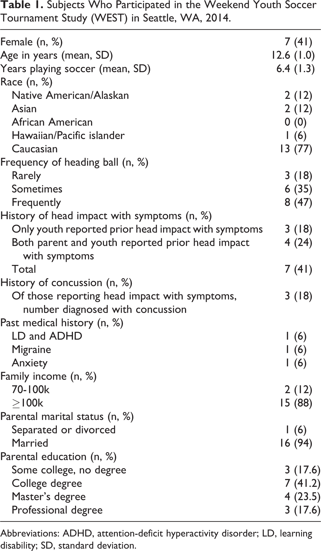
Abbreviations: ADHD, attention-deficit hyperactivity disorder; LD, learning disability; SD, standard deviation.
Head Impact Exposure of Subjects Who Participated in the Weekend Youth Soccer Tournament Study (WEST) in Seattle, WA, 2014.
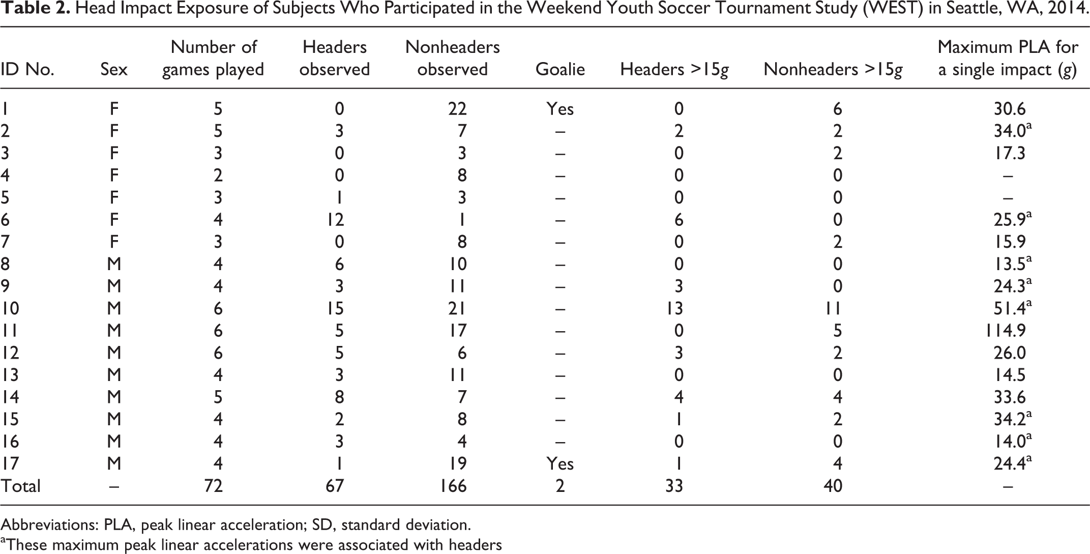
Abbreviations: PLA, peak linear acceleration; SD, standard deviation.
aThese maximum peak linear accelerations were associated with headers
No youth reported significant change in concussion symptoms, including the 2 youth who were outliers in terms of frequency of head impact exposure (the youth who sustained 24 head impacts >15g) and magnitude (the youth who sustained a head impact of 115g). Box plots of the 2 head impact exposure variables, number of head impacts >15g (frequency), and maximum peak linear acceleration (magnitude, see supplemental digital content to view these graphs) show these 2 subjects as outliers. There were no significant changes in neuropsychological testing after the tournament compared to baseline (Table 3). Associations between head impact exposure and neuropsychological testing are reported in Table 4. The directionality of associations was as expected for Picture Sequence Memory Test but was in the opposite direction for memory composite score visual (ie, the subject who experienced the large head impact performed better). Both of these associations were not robust to a sensitivity analysis removing head impact exposure outliers.
Neuropsychological Testing and Concussion Symptoms in the Weekend Youth Soccer Tournament Study (WEST) in Seattle WA, 2014.
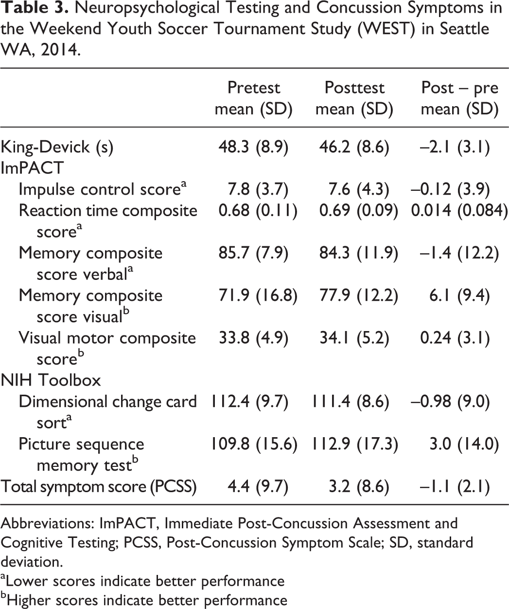
Abbreviations: ImPACT, Immediate Post-Concussion Assessment and Cognitive Testing; PCSS, Post-Concussion Symptom Scale; SD, standard deviation.
aLower scores indicate better performance
bHigher scores indicate better performance
Associations Between Neuropsychological Testing, Concussion Symptoms, and Head Impact Exposure (HIE) in the Weekend Youth Soccer Tournament Study (WEST) in Seattle, WA, 2014.
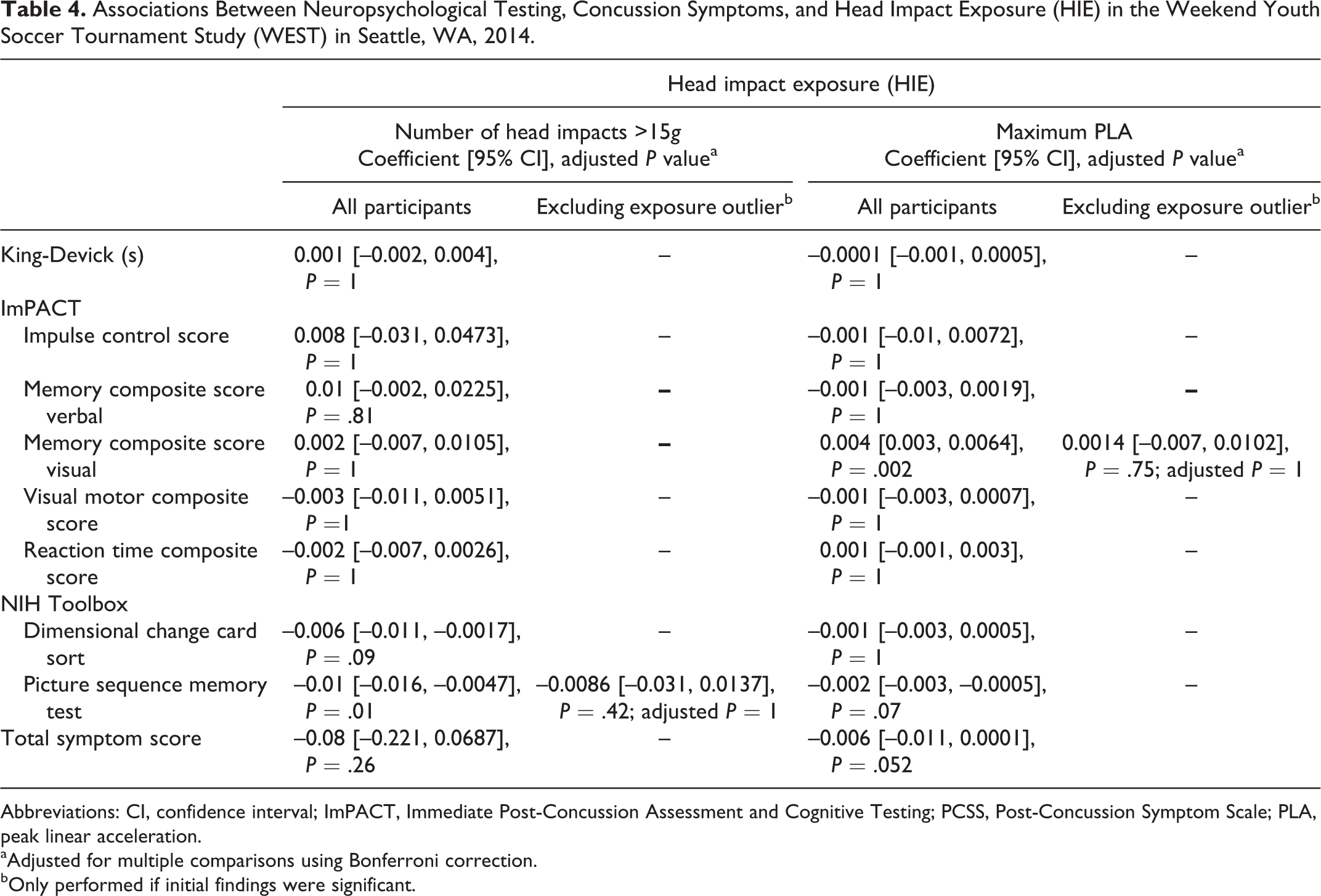
Abbreviations: CI, confidence interval; ImPACT, Immediate Post-Concussion Assessment and Cognitive Testing; PCSS, Post-Concussion Symptom Scale; PLA, peak linear acceleration.
aAdjusted for multiple comparisons using Bonferroni correction.
bOnly performed if initial findings were significant.
All MRI scans were read by a board-certified neuroradiologist (JA) and there were no findings indicative of acute brain injury. Associations between head impact exposure and imaging are reported in Table 5. Although one diffusion tensor imaging region was significant after Bonferroni correction, this was not robust to a sensitivity analysis removing head impact exposure outliers. No regions or tracks from the other imaging modalities (resting state functional MRI and pseudo-continuous arterial spin labeling) were significant.
Associations Between Head Impact Exposure (HIE) and Advanced Imaging in the Weekend Youth Soccer Tournament Study (WEST) in Seattle, WA, 2014.
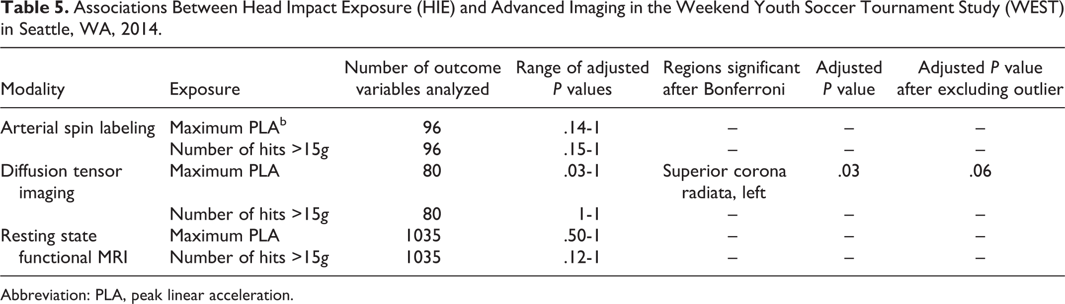
Abbreviation: PLA, peak linear acceleration.
Discussion
In this study of 17 middle school–age soccer youth playing a weekend soccer tournament, subjects experienced a wide range in frequency of head impacts, with some experiencing no head impacts >15g, and 1 individual sustaining 24 such impacts. Magnitude of head impacts (maximum peak linear acceleration of an individual impact) was right skewed with a median of 18.3g and only 2 hits >50g. No player reported symptoms consistent with concussion. In an exploratory analysis examining the association between head impact exposure and functional and anatomical outcomes, a few of the outcome variables were significantly associated with head impact exposure, but these were not robust to a sensitivity analysis removing outliers. To our knowledge, this is the first study to objectively measure frequency and magnitude of head impacts during a youth soccer tournament. This is also the first study to examine the relationship between head impact exposure in youth soccer and functional measures, including neuropsychological testing and advanced neuroimaging.
One of the strengths of this study was the ability to quantify all of the head impact exposure that occurred during the study time period. In addition, the frequency of head impacts varied substantially, with several subjects experiencing no head impacts or collisions and others quite a few. The primary limitation of our study was the small sample size. Given both the small sample size and the relatively low amount of head impact exposure, it is not surprising that we found no association between head impact exposure and functional or anatomical outcomes. However, we did have a few youth who sustained a greater frequency or magnitude of impacts and they did not report any symptoms, show any deficits on cognitive testing, or have any significant changes on imaging. We also acknowledge that we cannot generalize our results beyond a youth soccer tournament. Head injuries occur in youth soccer, and youth sustain concussions during this sport. However, given the limited amount of data regarding youth soccer and the recent concerns generated regarding heading, it is reassuring that we were not able to find measureable differences in functional or anatomic outcomes, even in players who headed the ball frequently. Finally, we must highlight that our sample was predominantly (77%) Caucasian and middle-high socioeconomic status, and we cannot generalize our findings to lower socioeconomic status or non-Caucasian youth. Future studies will be needed to understand whether there are any differential effects of subconcussive head impacts by race or socioeconomic status.
We chose 2 head impact exposure variables for this study: number of head impacts >15g and maximum peak linear acceleration of a single impact. These 2 variables were chosen as they parallel previous studies 34 and represent frequency of hits and magnitude of hits. Although some have opted for a cumulative measure, we find such a measure blends the effects of exposure and thereby is less meaningful (ie, 10 hits of 10g is not equivalent to 1 hit of 100g). We chose a relatively low acceleration threshold (15g) in order to capture all head impacts experienced by youth, and similar cutoffs have been used previously. 35,36 Although no clear threshold for injury has been suggested, it is clear that greater forces are more likely to result in greater injury.
Hanlon and Bir 8 performed the only other study of objectively measured head impact exposure in youth soccer and reported a similar level of head impact exposure to that in our study: less than 4 hits per hour per individual, with a maximum peak linear acceleration of 62.9g and most hits below 40g. However, they only studied youth during a 30- to 60-minute scrimmage, only included female subjects, and did not examine functional outcomes. Kaminski and colleagues 21 examined neuropsychological testing before and after a soccer season and found no deficits in function compared to control subjects. Rieder and colleagues 28 examined the impact of 1 week of heading on neuropsychological testing and found no differences between subjects and controls. Munce and colleagues 23 evaluated head impact exposure in a season of youth football for 22 youth and reported much greater head impact exposure (6183 impacts in 27 practices and 9 games, with 2% greater than 80g), but they also did not find significant associations between head impact exposure and functional outcomes.
It is difficult to compare our imaging findings to others in the literature, as methods are heterogeneous. Two studies reported imaging changes due to subconcussive hits in soccer, 11,12 but both examined youth with a lifetime of head impact exposure and compared their imaging results to a control group, rather than post-pre. 12 Davenport and colleagues 10 found diffusion tensor imaging changes in 24 nonconcussed high school football players after 1 season of football, but they examined the total number of abnormal white matter regions, rather than specific regions. Abbas and colleagues 14 reported resting state functional MRI changes in 10 high school football players during 1 season of football, but they imaged youth 9 times during the season to explore changes over time and did not collect objective measurements of head impacts.
Recent concern has been expressed about the effect of “subconcussive” head impacts in soccer, 5 and questions have been raised about the safety of heading in youth soccer. The goal of our study was to examine head impacts in youth soccer starting with the exposure incurred during a weekend soccer tournament. With public concern about the potential danger of subconcussive head impacts leading to long-term brain injury, an increasing number of studies are conducting imaging studies after head impact exposure in subjects who report no clinical symptoms. Our findings suggest some caution is needed in these explorations. Many of these studies utilize similarly small sample sizes, and these types of samples are extremely susceptible to outlier driven associations. Further studies are needed to better understand (1) the true rate of head impacts in youth soccer and (2) whether a clear relationship exists between subconcussive head impacts and functional outcomes both acutely and after chronic exposure.
Footnotes
Acknowledgments
We thank Sandy Poliachik, PhD, and Chris Budech, BA, who assisted with the resting state functional MRI analysis, all the athletes and families who participated in our study, and the coaches and soccer associations who supported our research.
Author Contributions
FPR, SPDC, SF, CLMD, JA, and AR-R conceptualized and designed the study, oversaw data collection and analysis, and reviewed the final manuscript. NE assisted with the analysis of the diffusion tensor imaging data. AVP assisted with the analysis of the resting state functional magnetic resonance imaging data. ES and MH recruited and consented subjects, attended games to apply accelerometer devices and record observed collisions, conducted neuropsychological testing, and participated in writing and editing of the final manuscript. MSV was strongly involved in conceptual design and methods for the study. RPC assisted with design of the study and data collection, completed validation study for device used for exposure measurement, participated in processing and analysis of exposure data, and reviewed the final manuscript. SD processed data and carried out analyses, as well as participating in interpretation of results and having a substantive role in writing and editing the final manuscript. CCS assisted with design of the study, oversaw data collection, data processing, and analysis, and participated in writing and editing the final manuscript. SPDC, SF, CLMD, JA, and AR-R assisted with data collection, processed data, oversaw analysis and interpreted results, and wrote, reviewed, and revised the final manuscript. FPR, SPDC, SF, CLMD, JA, AR-R, NE, AVP, MSV, and RPC revised the final manuscript.
Declaration of Conflicting Interests
The authors declared no potential conflicts of interest with respect to the research, authorship, and/or publication of this article.
Funding
The authors disclosed receipt of the following financial support for the research, authorship, and/or publication of this article: This study was funded by the Seattle Pediatric Concussion Research Collaborative, Seattle Children’s Research Institute.
Ethical Approval
This study had ethical approval. The IRB number is UW IRB #47543.

References
Supplementary Material
Please find the following supplemental material available below.
For Open Access articles published under a Creative Commons License, all supplemental material carries the same license as the article it is associated with.
For non-Open Access articles published, all supplemental material carries a non-exclusive license, and permission requests for re-use of supplemental material or any part of supplemental material shall be sent directly to the copyright owner as specified in the copyright notice associated with the article.
