






Abstract
Central neurocytomas are well-differentiated tumors of neuronal origin. These are relatively uncommon in the pediatric population. Anaplastic features reflected by brisk mitotic activity, microvascular proliferation, necrosis, and MIB-1 labeling index >2% or 3% have been proposed to indicate aggressive behavior. Because of its rarity, there is paucity of data regarding the histologic spectrum and outcome of central neurocytomas in children. With this short series, we describe our observations of the clinicopathologic characteristics and outcome of this tumor in children over a 5-year period.
Keywords
Central neurocytoma is a relatively uncommon, well-differentiated tumor of neurocytes classically arising within the ventricles, near Foramen of Monro. 1 The tumor commonly arises in young adults with 2/3rd of patients between the age group of 20 to 40 years. 1 As it is uncommon in the pediatric age group, the exact data regarding its incidence in children is not known. Histologically, these are defined by a uniform population of monotonous round to oval cells that demonstrate ultrastructural and immunophenotypic features of neuronal differentiation. The background stroma consists of neuropil that may be organized into cell-free zones resembling rosettes. Designated as WHO grade II, the criteria for “atypical” category are not established yet. Brisk mitotic activity, microvascular proliferation, necrosis, and MIB-1 labeling index >2% or 3% have been noted as features of anaplasia in a few studies. 2 -4 The clinical course of these tumors is usually benign, largely dictated by the extent of resection as an important parameter for prognosis.
The literature regarding adjuvant therapy in pediatric central neurocytoma, especially after near total excision, is sparse due to the rarity of disease. The long-term secondary effects of radiation in children could outweigh the chances of recurrence after near total excision. We present our experience on pediatric central neurocytoma with a detailed evaluation of clinical, radiologic and pathologic findings, diagnosed over the past 5 years. The follow-up and survival data was correlated with the above parameters.
Materials and Methods
All the cases of central neurocytoma diagnosed in the past 5 years were retrospectively reviewed, and examples of central neurocytoma arising in the pediatric age group (≤18 years) were included in the study. Six children with central neurocytoma were operated at PGIMER between 2012 and 2016. Five were available and one was lost to follow-up. A detailed clinical, radiologic, and pathologic analysis was performed. Hematoxylin-eosin (H&E) stained sections were evaluated and tumors were further categorized with immunohistochemical panel consisting of antibodies to glial fibrillary acidic protein, synaptophysin, neurofilament protein, and Ki-67. The imaging data were retrieved and evaluated in detail. The survival data were retrieved from the neurosurgical records at the last follow-up. The decision to subject the children to adjuvant radiotherapy was partly dependent on the extent of tumor excision as well as its histopathology.
Results
Clinical and Radiologic Features
Demographic, clinical features and radiologic findings are detailed in Table 1. There were 4 male and 2 female patients, with median age at first diagnosis being 12.5 years (age ranged from 4 to 17 years). Five children presented with features of raised intracranial pressure, which was the most common clinical presentation in our study. The other symptoms were seizure in 2, visual deterioration in 2, and walking difficulty in the single child. Three children had tumors located primarily in the lateral ventricle, and the rest had parenchymal lesions extending into the adjacent lateral ventricle. On magnetic resonance imaging (MRI), the tumors were T1 iso- to hypointense and T2 iso- to hyperintense, with mild to moderate heterogeneous contrast enhancement. One had clear evidence of intratumoral bleed (Figures 1 –3). There was no evidence of recognized genetic predisposing features or other tumor syndromes in any of the patients.
Clinical, Surgical, Imaging, and Histopathologic Details of 6 Patients of Neurocytoma.

Abbreviations: EVP, endovascular proliferation; GTE, gross total excision; HPE, histopathologic examination; ICP, intracranial pressure; NTE, near total excision; RT, radiotherapy.
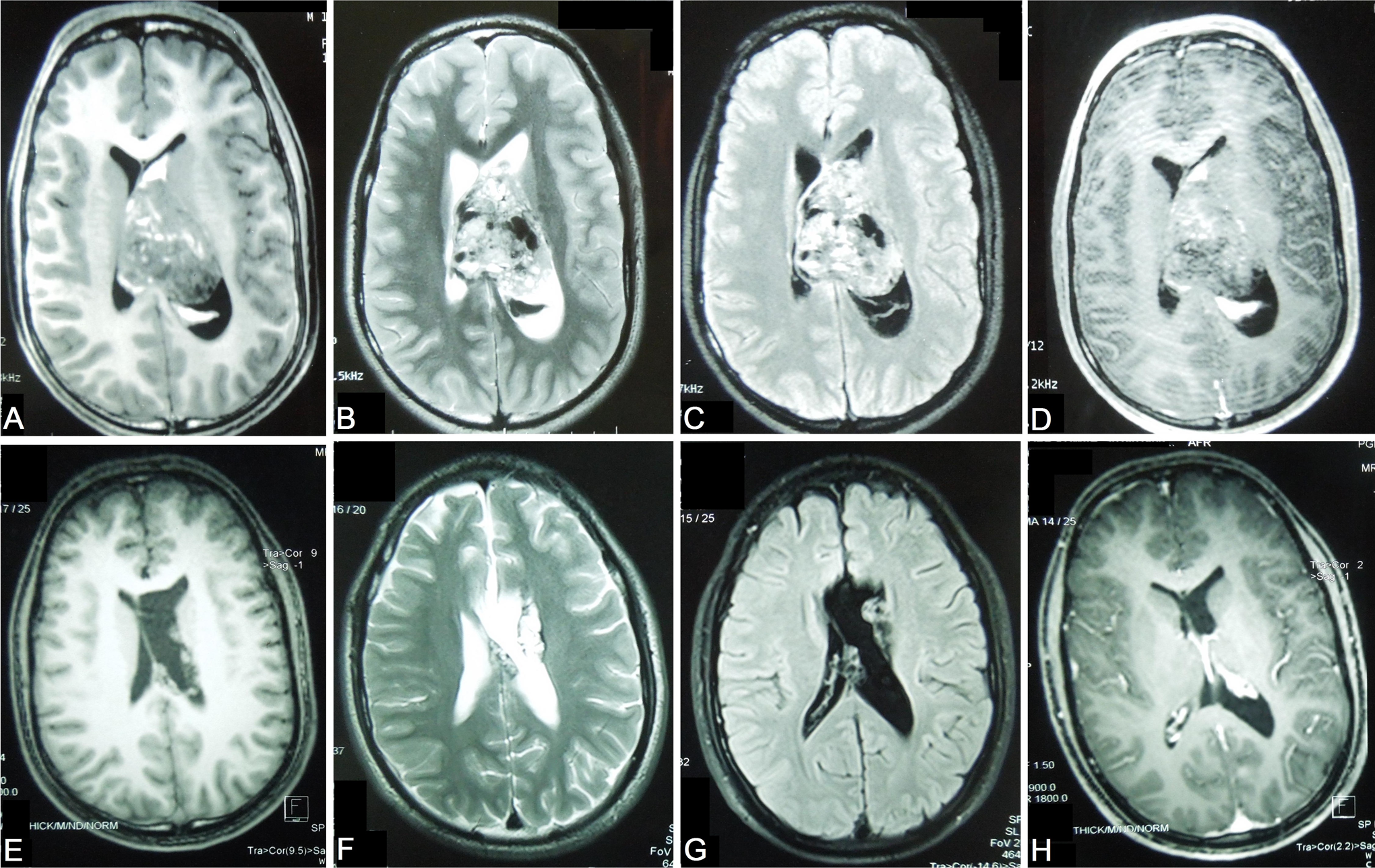
(A-D) T1, T2, FLAIR, and contrast images showing heterogeneously enhancing mass with areas of bleed in lateral ventricle. (E-H) Images after tumor excision showing postoperative changes. FLAIR, fluid-attenuated inversion recovery.
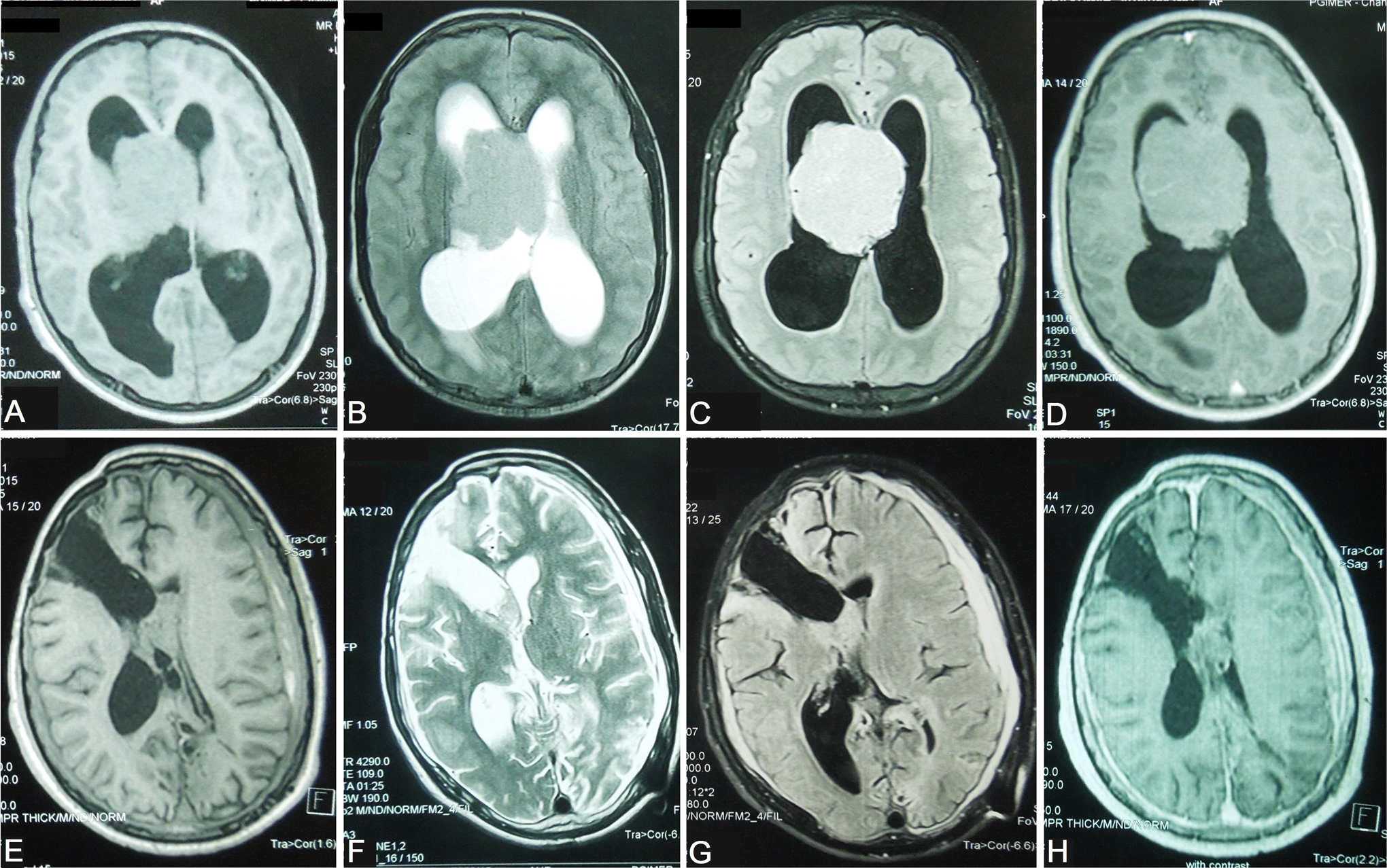
(A-D) T1, T2, FLAIR, and contrast sequence showing mildly enhancing lesion in lateral ventricle. (E-H) Postoperative images depicting right transcortical transventricular trajectory with no significant residual tumor, and postoperative artefacts. FLAIR, fluid-attenuated inversion recovery.
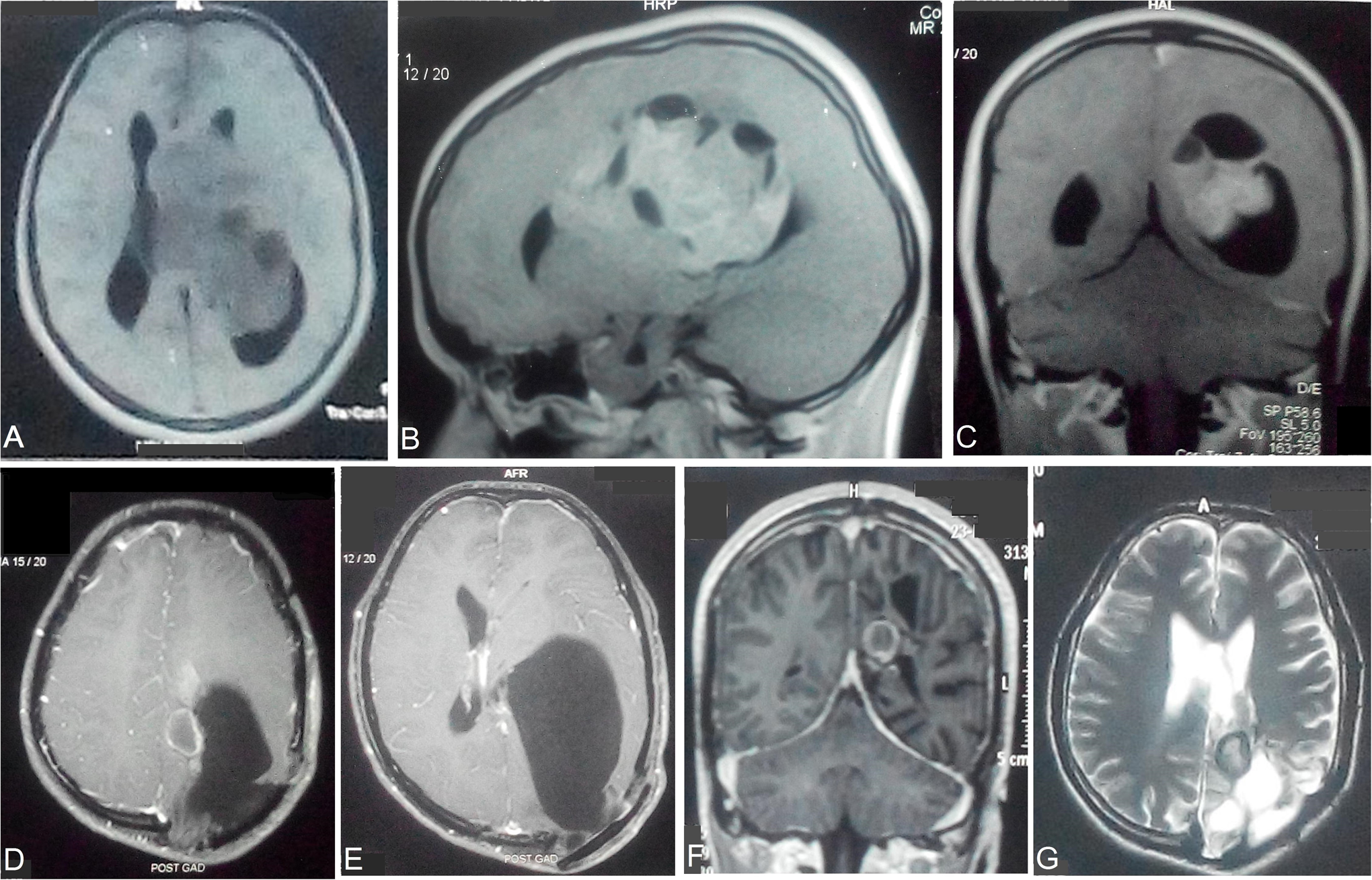
(A-C) T1 and contrast images demonstrating enhancing mass occupying the left lateral ventricle. (D, E) Postoperative T1 contrast sequence at 2 years showing small enhancing residual tumor with enlarged cystic component in lateral ventricle. (F, G) Decompressed cyst following cystoperitoneal shunt insertion.
Surgical Details, Outcome, and Follow-Up
The tumor was approached using a transcortical, transventricular corridor in all children. In a single child (case 4), the initial surgery was abandoned as a result of profuse bleeding; 3 months later, he underwent tumor excision by an interhemispheric transcallosal approach. Intraoperatively, the tumor was soft to firm in consistency and showed moderate to high vascularity. Three had near total excision; gross total excision could be achieved in others.
The decision to subject the children to adjuvant radiotherapy was partly dependent on the extent of tumor excision as well as its histopathology. Two children (cases 1 and 5) were planned for radiotherapy on the basis of increased mitotic figures on tumor histopathology. One child (case 5) completed 54 Gy of adjuvant radiotherapy. The other child (case 1) with residual disease expired at 3 months before radiotherapy could be initiated. The cause of death was probably acute hydrocephalus. There were 2 other children (cases 3 and 6) who underwent near total excision of tumor. Of the 2, one (case 3) underwent radiotherapy and the other (case 6) did not consent for it. This child (case 6) who had the longest follow-up of 4 years presented with wound bulge and enlarged cystic component at 2 years follow-up and required cystoperitoneal shunt for decompression (Figure 3). Despite not receiving radiotherapy after near total excision, there was no progression of the small solid residual tumor, and the child was doing well at his last visit. The others were also independent and ambulant at their respective last follow-up. The mean follow-up of children was 18.8 months (range 3-48 months). In our series, none had recurrent lesion at follow-up imaging.
Histopathologic Features
The tumor in all 6 cases was defined by a striking cytologic uniformity (Table 1). The cells were arranged in sheets with round to oval nuclei and speckled chromatin. These were supported by a delicate network of capillaries (Figure 4A, B). Although micro-calcification was a common feature in all cases, it was abundant in a single case (Figure 4C). Large patches of neuropil were identified in a single case. At places this had a nodular pattern resembling the nodular desmoplastic variant of medulloblastoma (Figure 4D-F). No differentiation to ganglion cells was noted. The example of extraventricular neurocytoma demonstrated focal glial differentiation. All the cases were categorized as grade II, except for 2 cases (cases 1 and 5) wherein the mitoses were readily identified with endovascular proliferation (Figure 5A, B) and there was no necrosis. As criteria for anaplasia are not yet recognized, the frequent mitoses in these cases were mentioned in the report. The cells expressed immunoreactivity with synaptophysin and were negative for glial fibrillary acidic protein, except for case 5, where glial differentiation was highlighted by glial fibrillary acidic protein (Figure 5C, D). MIB-1/Ki-67 labeling index was more than 3% in 2 cases with brisk mitotic activity.
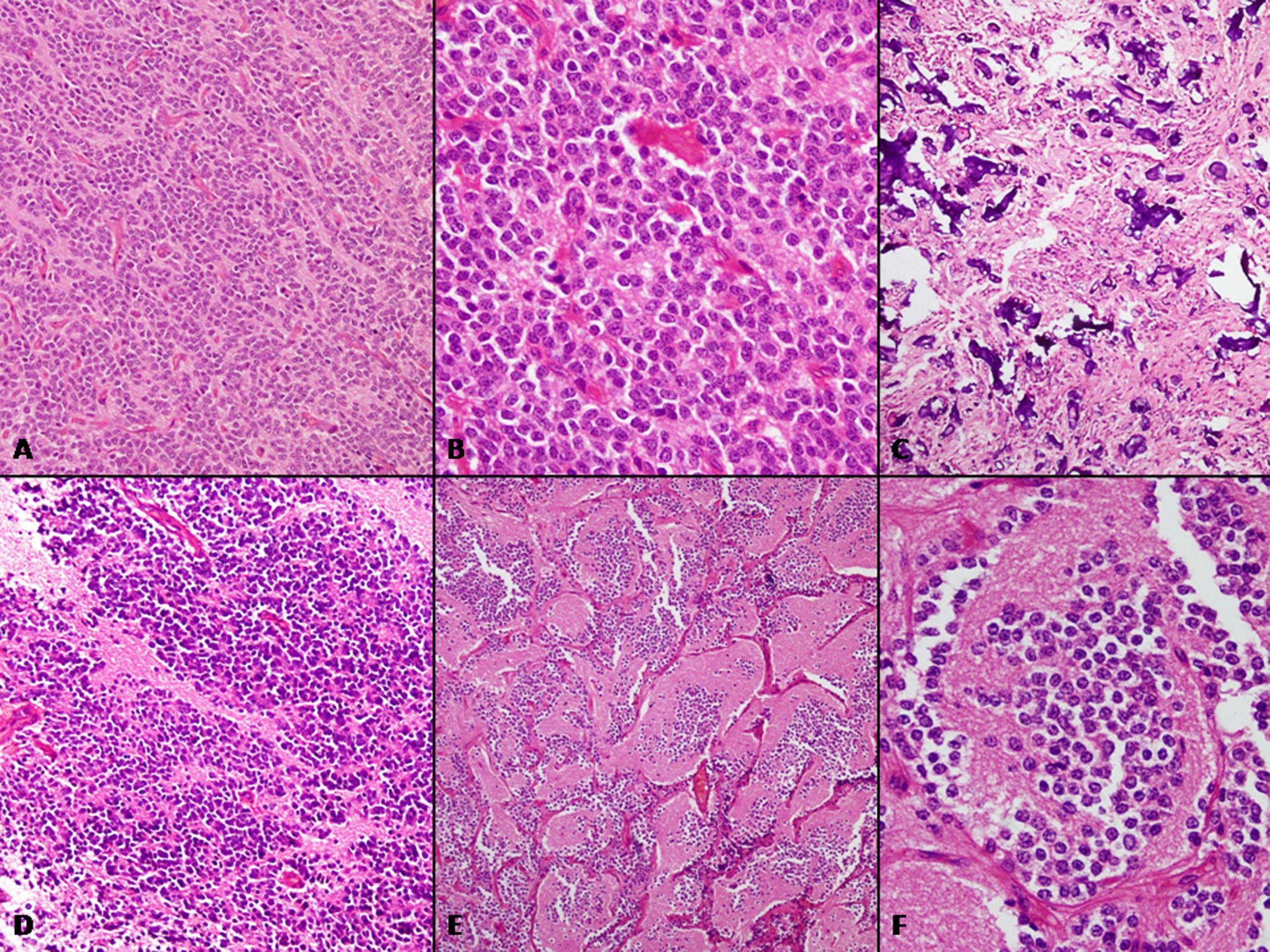
(A) Diffuse sheets of monomorphic round to oval cells supported by rich network of delicate capillaries (H&E, ×200). (B) Uniform nuclei with delicate, speckled chromatin (H&E, ×400). (C) Abundant micro-calcification was identified in one of the tumors (H&E, ×200). (D) Cellular islands alternating with acellular neuropil (H&E, ×200). (E, F) Large islands of neuropil organized as nodular aggregates with central collection of neurocytes (H&E, ×200, ×400). H&E, hematoxylin-eosin.
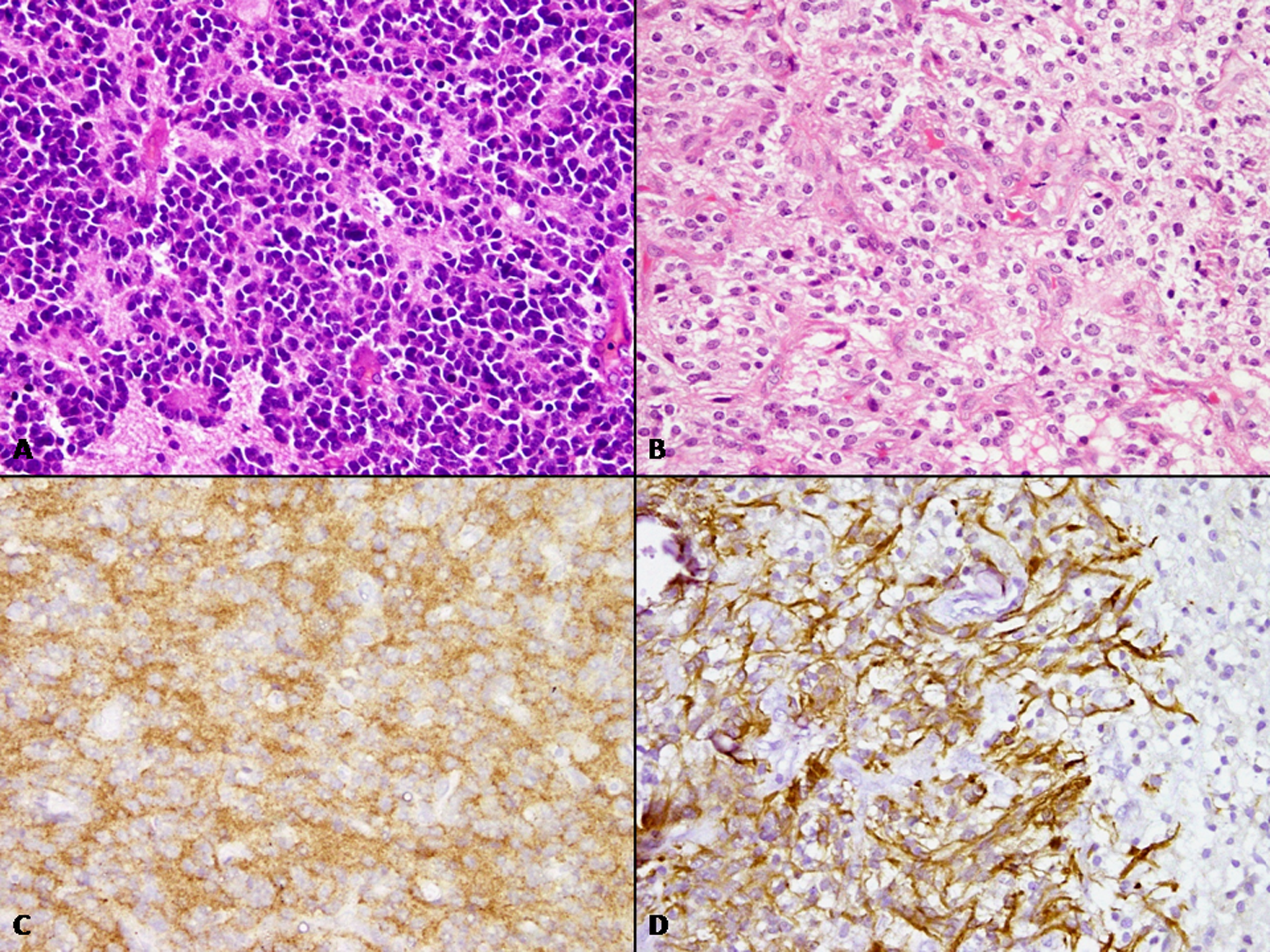
(A, B) Example of “atypical” neurocytoma with frequent mitoses and endovascular proliferation (H&E, ×400). (C) Tumor cells strongly expressing synaptophysin (immunoperoxidase, ×400). (D) Focal glial differentiation identified in example of extraventricular neurocytoma (immunoperoxidase ×400). H&E, hematoxylin-eosin.
Discussion
The current analysis was performed to present our experience with this rare tumor arising in children. It is a rare tumor comprising 0.25% to 0.5% of all intracranial tumors 5 and usually arises in young adults. Examples of this tumor in the pediatric age group are infrequent, being documented in literature as small series and case reports. 6,7 The median age in affected children observed has been 16 years, 6 whereas in our series it was 12.5 years. These are typically located supratentorially in the lateral and/or third ventricle. Attachment to septum pellucidum is a common feature of this tumor. On imaging, heterogenous enhancement is observed and contrast tomography depicts calcification. Children usually present with raised intracranial pressure symptoms. On histology, monomorphism of cells is a characteristic feature with round to oval cells arranged in diffuse sheets in a background of delicate neurofibrillary matrix. Sometimes, large nuclear-free neuropil islands are encountered, and the circular arrangement of cells around these resembles vague rosettes. Homer-Wright rosettes have also been reported. 8 In rare instances, anaplastic features have been described, including brisk mitotic activity (usually ≥3/10 hpf), necrosis, and endovascular proliferation; however, the consensus of grading central neurocytoma as grade III based on these features is not yet established. 5,9,10 However, these are not associated with poor prognosis. 9 -11 MIB-1 labeling index of >2% or 3% have been found to be associated with a significantly shorter recurrence-free survival in earlier studies. 4,12 A higher threshold for MIB-1 labeling index of >4% predictive of poor outcome has been suggested in recent studies. 13,14 Although the number of patients in our analysis is too small and the follow-up period not long enough, it is worthwhile to note the short survival in the 4-year-old girl with tumor demonstrating brisk mitoses. Intriguingly, the recurrence of tumor in Case 6 remains unexplained despite its typical histologic features and near-total excision. The genetic landscape of central neurocytoma is much quiet which perhaps explains its benign course, trisomy 7; chromosome 17 deletion; and gains in 2p, 10q, 13q, and 18q has been reported in various studies. 15
Parenchymal tumors with neurocytoma features located in the cerebral hemispheres are designated as extraventricular neurocytoma. 1 These were introduced as a separate entity in 2007 classification because of a parenchymal site and a more aggressive biologic behavior. 1 While these have been often reported in adults, examples of such tumors in pediatric population are extremely uncommon. 16,17 Neurocytoma arising in 10-year-old male in this series is an example of extraventricular neurocytoma on account of its dominant location in the frontal lobe. Frequent mitoses were observed on histology. Although ganglionic differentiation is more commonly observed, reported in 66% of cases, 18 the tumor in this example included focal areas of glial differentiation highlighted on glial fibrillary acidic protein.
Comparing the central and extraventricular neurocytomas, Xiong and colleagues 19 have observed a higher recurrence rate with aggressive behavior, shorter time to recurrence, and higher nuclear grade and atypia with extraventricular than central neurocytoma. Total surgical resection is the treatment of choice for both central neurocytomas and extraventricular neurocytomas.
In general, central neurocytoma have good prognosis. Surgical excision is the mainstay of treatment for central neurocytoma. It is clear that complete tumor resection offers better outcome both in terms of local control and survival as compared to incomplete resection. 20 -22 As far as the role of adjuvant radiotherapy is concerned, there is lack of consensus because of uncommon occurrence of this disease. Studies that have evaluated the role of adjuvant radiotherapy in central neurocytoma are mainly of adults or a mixed group that consist of both adult and pediatric patients. These show that radiotherapy does not add to the clinical outcome of patients undergoing complete resection of central neurocytoma. 20,22 Although radiotherapy helps in achieving local control after incomplete resection, its role in improving survival, particularly in children, is questionable. 6,21,22
Adjuvant radiotherapy is a concern in the pediatric age group because of the possible secondary effects of radiation on cognition and radiation-induced secondary malignancies. Very few papers have exclusively focused on the follow-up and treatment of pediatric central neurocytoma. 6,7 As described in this series, case 6 showed no increase in the size of the residual lesion (over 4 years of follow-up) even without radiotherapy. In a literature review of central neurocytoma in children, the authors found that radiotherapy after incomplete resection improves local disease control but not the patient survival. After analyzing about 59 patients of pediatric central neurocytoma from various studies, the authors recommend radiotherapy only for children with incomplete tumor resection associated with an MIB-1 labeling index higher than 3%. 6 A recent study conducted by Imber and colleagues that included both adults and children have suggested improvement in progression-free survival with adjuvant radiotherapy after subtotal resection. The authors further indicated that an MIB-1 labeling index of >4% is predictive of poor outcome. 13
In a recent series that compared the outcome between pediatric central and extraventricular neurocytoma, the former showed better prognosis than extraventricular counterparts (overall survival at 20 years, 100% and 40%, respectively). The aggressive behavior of extraventricular neurocytoma was attributed to their high proliferative index (MIB-1 fraction more than 2%). The authors recommend adjuvant radiotherapy after subtotal tumor excision for local disease control. 23 Limited reports are available addressing the role of chemotherapy in pediatric central neurocytoma. 24,25 Atypical childhood central neurocytoma has been shown to respond to multiple chemotherapeutic regimens. 24 Stereotactic radiosurgery may be an alternative to radiotherapy after incomplete resection of central neurocytoma. However, the literature is scarce, especially with respect to pediatric central neurocytoma. 26,27 Furthermore, stereotactic radiosurgery may be technically more demanding in young children.
Limitations of our analysis is the short duration of follow-up and small number of patients. Additionally, we could not evaluate their mutational profile, an area that also remains relatively underexplored in literature. In conclusion, pediatric neurocytomas in general have a favorable prognosis after total excision. Whenever possible, safe complete tumor resection should be attempted. Adjuvant treatment may not be needed in all children and needs to be individualized. Radiotherapy may be reserved for those with incomplete tumor excision and atypical features. Atypical histologic features are noteworthy, and a high MIB-1 labeling index portends poor clinical behavior.
Footnotes
Declaration of Conflicting Interests
The authors declared no potential conflicts of interest with respect to the research, authorship, and/or publication of this article.
Funding
The authors received no financial support for the research, authorship, and/or publication of this article.