





Abstract
Background / Objective
Seizures are a complication for pediatric patients requiring extracorporeal membrane oxygenation (ECMO). There are no standardized guidelines regarding continuous electroencephalography (EEG) monitoring to detect seizures in these patients, and the impact of protocolized monitoring has not been evaluated. Here we examined the effects of continuous EEG protocol implementation in our pediatric ECMO population.
Methods
Retrospective chart reviews were conducted on 57 patients who underwent extracorporeal membrane oxygenation and concurrent continuous EEG out of 165 patients supported on extracorporeal membrane oxygenation. Timing of continuous EEG initiation and seizures detected by continuous EEG was determined for 5 years prior to and 15 months after protocol implementation.
Results
Protocol implementation was associated with increased ECMO–supported patients who were concurrently monitored by continuous EEG. Time from ECMO cannulation to continuous EEG initiation was shorter (median 7 hours after versus 16.2 hours before; P < .001). Patients who had ongoing seizures at the start of continuous EEG recording decreased from 64% preprotocol to 0% postprotocol (P < .001), and there was an associated earlier time to break in status epilepticus postprotocol. Seizures were detected past 48 hours after cannulation in 50% of patients in the postprotocol group.
Conclusions
Protocol implementation resulted in earlier continuous EEG initiation and more EEGs initiated before seizure onset with evidence of altered seizure dynamics. Although current recommendations suggest that continuous EEG duration of 24-48 hours results in seizure detection for >90% of critically ill adults, longer monitoring may be needed to reliably detect seizures in children supported with ECMO, particularly if monitoring is initiated earlier in the post-cannulation period.
Pediatric extracorporeal membrane oxygenation (ECMO) is a life-saving technique that provides temporary support for severe cardiac and pulmonary failure. Because of the severity of the underlying critical illness as well as factors inherent to ECMO, there is a high risk of seizures. 1 Seizure prevalence for children on ECMO using continuous electroencephalographic (EEG) monitoring is estimated at 18% to 23%.2-8 Seizures are associated with worse neurodevelopmental outcome, greater degree of neurologic injury, and increased hospitalization and mortality in several studies.3,4,8,9 Early identification of seizures during extracorporeal membrane oxygenation may provide an opportunity for intervention as well as inform a better understanding of the underlying causes of seizures in this population.
Currently, there are no standardized guidelines for the use of continuous EEG in the pediatric ECMO population, specifically. Consensus statements put forth by the American Clinical Neurophysiology Society (ACNS) recommend continuous EEG monitoring in patients at high risk for electrographic seizures. This includes children on ECMO, especially if they undergo neuromuscular blockade resulting in a lack of clinical manifestations to suggest seizures10,11 making seizures impossible to detect clinically. Prior studies have shown that 87% of seizures in critically ill children are observed within the first 24 hours following continuous EEG initiation. 12 Hence, the ACNS recommends continuous EEG placement as soon as possible in critically ill patients and for a minimum time of 24 hours. 10
Given the high seizure prevalence among patients undergoing ECMO treatment and existing ACNS recommendations, several institutions have implemented protocols for the use of continuous EEG in ECMO. Although several have reported on seizure detection with the use of their protocols,2-4,7,8 the impact of continuous EEG protocol implementation on patient care remains unclear.
In 2019, our institution enacted a continuous EEG monitoring protocol for children undergoing ECMO support. This protocol specifies that all children on ECMO be placed on continuous EEG monitoring for 72 hours with continuous EEG initiation as soon as possible following cannulation. This was a change from prior practice when continuous EEG was placed at the discretion of the treating clinicians if they had a clinical concern for seizures.
In the current study, the effect of continuous EEG protocol implementation on clinical workflow and on seizure detection was examined. The goal of this study was to evaluate protocol implementation as a means to improve clinical care for children supported with ECMO.
Methods
Patients
This was a single-center retrospective chart review of patients monitored by continuous EEG and supported on ECMO between April 2014 and June 2020 at the Johns Hopkins Children's Center. The study period spanned 5 years before and 15 months after the initiation of a protocol dictating that all pediatric patients supported with extracorporeal membrane oxygenation be monitored by continuous EEG unless clinically contraindicated. The implementation of the protocol began in April 2019.
Patients were identified using the search term “ECMO” in an institutional review board–approved database of all patients monitored by continuous EEG at the Johns Hopkins Hospital during the study period and limiting the results by patients cared for in the Children's Center. Only patients undergoing continuous EEG monitoring concurrent with extracorporeal membrane oxygenation treatment were included in the study. Patients with continuous EEG recordings that could not be interpreted because of technical factors were excluded. In clinical practice, a patient undergoing a prolonged period of ECMO support may have more than 1 instance of continuous EEG recordings in the same extracorporeal membrane oxygenation run at the discretion of the primary team. To more accurately identify the effectiveness of protocol implementation, only the first continuous EEG instance for each extracorporeal membrane oxygenation run was included, and subsequent records, separated by more than 24 hours from the first recording period, were excluded (Figure 1). The total number of patients supported on extracorporeal membrane oxygenation during the study period was determined from the institutional administrative ECMO database.
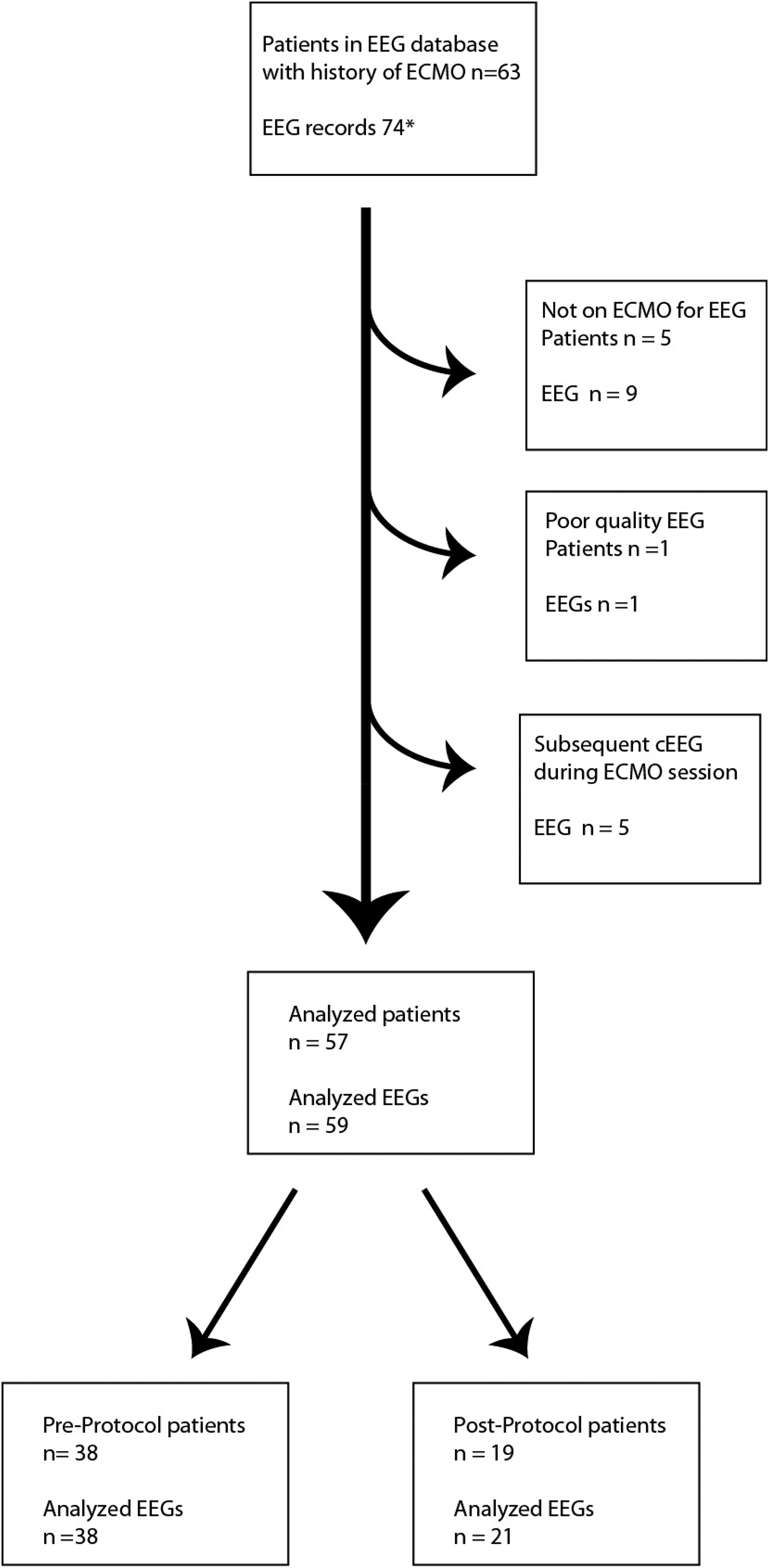
Overview of identification of patients monitored by continuous electroencephalography (EEG) concurrent with extracorporeal membrane oxygen (ECMO) support. Patients and corresponding EEGs were identified from an institutional continuous EEG database. Individual continuous EEG records were excluded if not recorded concurrent with ECMO support, poor quality prohibiting interpretation, or if continuous EEG was not the first continuous EEG recorded during a particular extracorporeal membrane oxygenation run. If a patient had more than 1 extracorporeal membrane oxygenation run, the first continuous EEG recording following each cannulation was included. *Patients may have had more than 1 independent continuous EEG recording at different times after ECMO.
Continuous EEG Monitoring Criteria and Protocol Implementation
The ECMO continuous EEG Monitoring Protocol was formalized and implemented in April 2019 as part of a comprehensive neuromonitoring and neuroimaging protocol update for ECMO–supported patients. The electronic health record order set for ECMO initiation was updated to include a prechecked order for continuous EEG monitoring in addition to a preexisting prechecked consult to Pediatric Neurology. The protocol dictates that continuous EEG should be placed as soon as possible after ECMO cannulation for all patients regardless of medications, paralytics, neurologic examination and Glasgow Coma Score and continued for a minimum of 72 hours if clinically tolerated. The continuous EEG duration can be extended depending on clinical concerns, and if necessary for clinical care can be terminated early. In contrast, prior to protocol implementation, candidacy for continuous EEG monitoring of ECMO–supported patients was determined by the patient's physicians based on concern for seizures or an inability to monitor clinically.
EEG recordings were performed using Nihon Kohden digital EEG systems (Nihon Kohden, Tokyo, Japan). All electrodes were CT compatible and were placed according to the 10-20 system with a full 10-20 montage placed on children greater than 44 weeks’ corrected age (CA) when head circumference and clinical circumstances allowed. In children younger than 44 weeks’ corrected age, a neonatal montage was placed whenever possible. When clinical circumstances or head circumference prevented full or neonatal montages, reduced montages, consisting of Fp1, T3, O1, C3, Fp2, T4, O2, C4, and Cz were used. 13
EEG Analysis
Each continuous EEG was read by a board-certified epileptologist (CWH or KSH) who was blinded to prior clinical continuous EEG reports, medical condition, laboratory values, or indications for extracorporeal membrane oxygenation. Epileptologists had access to age, sex, and medication administration as well as any clinical or EEG annotations that were saved in the record by the clinical EEG reader during the period that the patient was monitored. Study epileptologists read raw EEG with the assistance of Persyst computer-assisted EEG reviewing software (Solana Beach, CA, USA). The entire continuous EEG record was read for all patients, regardless of record duration. Criteria for the EEG review were strictly defined in advance. The readers recorded the presence and timing of seizures and electrographic status epilepticus using REDCap secure data collection software (Vanderbilt University, Nashville, TN, USA). Electrographic seizures and status epilepticus were defined according to the American Clinical Neurophysiology Society (ACNS) terminology. 14 Seizure detection in relation to the start of continuous EEG monitoring was analyzed and EEGs were scored based on whether seizures were ongoing at the start of the EEG or not. If seizures occurred within 30 minutes after continuous EEG initiation, they were considered to be present at onset. Ongoing seizure burden after onset was quantified based on number of hours in which seizures were detected per hour of EEG recoding. Seizure burden was determined as a ratio of the number of hours with seizures identified to the total number of hours monitored. Status break was defined as time to first break in seizure activity >2 hours in duration with some background activity present or to onset of terminal suppression or death. The duration of EEG recording and the timing of EEG initiation relative to ECMO cannulation was obtained from the medical record.
Demographic and Clinical Data
Demographics were collected from electronic patient records and compiled in REDCap. Data collected included sex, age, ECMO indication, duration of ECMO support, requirement of extracorporeal cardiopulmonary resuscitation (eCPR), ECMO type (veno-arterial [VA] or veno-venous [VV]), and survival to hospital discharge. Medical conditions leading to EMO, other medical preexisting conditions, and neurologic conditions prior to initiation of ECMO were also recorded.
Statistical Analysis
Statistical analysis was performed by using GraphPad Prism 9 (La Jolla, CA) and Excel software. Categorical values were reported with counts and frequencies and continuous variables with medians and interquartile ranges (IQRs). Fisher exact test or chi-square test was used to compare frequencies. For continuous variables, the Mann-Whitney U test was used. All hypothesis tests were 2-sided unless otherwise indicated, with a significance level of P < .05.
Results
EEG Monitoring and Study Populations
In this retrospective chart review spanning from April 2014 to June 2020, 74 continuous EEG recordings from 63 individual patients were identified from a continuous EEG database using the search term “ECMO” (Figure 1). Further chart review resulted in the exclusion of 15 EEGs from 6 patients secondary to not being on ECMO during continuous EEG monitoring (n = 9 EEGs and n = 5 patients), poor quality of EEG records (n = 1 EEG and n = 1 patient), and follow-up EEGs during the same ECMO run (n = 5 EEGs). This resulted in 59 analyzed continuous EEG studies from 57 patients as 2 patients had 2 separate ECMO, each with a corresponding continuous EEG record. All continuous EEGs from patients captured prior to continuous EEG protocol implementation on April 6, 2019, were considered part of the preprotocol group (n = 38) and after this date were considered part of the postprotocol group (n = 19) (Figure 1, Table 1).
Proportion of Patients Monitored With Continuous EEG and Seizure Prevalence.

Abbreviations: cEEG, continuous electroencephalography; ECMO, extracorporeal membrane oxygenation.
There were 165 patients supported on ECMO during the study period. Of these, 134 patients underwent ECMO cannulation prior to protocol implementation, with 38 (28%) of these monitored by continuous EEG. After protocol implementation, a total of 31 patients underwent ECMO cannulation with 19 (61%) of these monitored by continuous EEG. Further chart review of the postprotocol group indicated that of the 12 patients supported by ECMO but not monitored by continuous EEG, 6 patients had a complicated course and died within 24 hours of cannulation, 3 patients were awake with intact mental status examinations and therefore the continuous EEG protocol was bypassed in favor of clinical assessments and 1 patient had EEG monitoring initiated but immediately discontinued because of surgical intervention. The 2 remaining patients were not included in the monitored group because although they were monitored on continuous EEG concurrent with ECMO support, they were not identified in the continuous EEG database because of screening failure. Importantly, despite these exclusions, there was a significant increase in the percentage of ECMO–treated patients who were monitored by continuous EEG from 28% in the preprotocol group to 61% postprotocol (Fisher exact, P = .001, OR 4.00, 95% CI 1.7-9.0) (Table 1).
Seizure Prevalence and Patient Demographics
During the study period, seizures were detected by continuous EEG in 17 patients, 10% of the 165 patients treated with ECMO. There were 11 of 134 patients (8%) with seizures detected in the preprotocol group and 6 of 31 patients (19%) with seizures detected in the postprotocol group (P = .10, OR 2.7, 95% CI 0.9-7.3). Of the ECMO patients who were monitored by continuous EEG, there was not a significant difference in the number of patients with seizures detected by continuous EEG between the pre- and postprotocol groups, with seizures in 11 of 38 continuous EEG monitored patients (29%) in the preprotocol group and 6 of 19 continuous EEG–monitored patients (32%) in the postprotocol group (P > .99, OR 1.1, 95% CI 0.3-4.0) (Table 1).
The similar seizure prevalence between the pre- and postprotocol groups is consistent with the similar demographics and severity of illness of the patients included in the groups (Table 2). There was no significant difference between groups with respect to sex, age, diagnosis prior to ECMO, ECMO indication, venoarterial versus venovenous mode and mortality. The study population in both groups was predominantly male and <1 year of age. The indication for ECMO as categorized into the 6 most common reasons for initiation was not different between the pre- and postprotocol implementation periods. Indications for ECMO included congenital heart disease, extracorporeal cardiopulmonary resuscitation (ECPR) / cardiopulmonary resuscitation (CPR), congenital diaphragmatic hernia, persistent pulmonary hypertension and acute respiratory distress syndrome (ARDS) / pneumonia. Some patients had more than 1 indication. The majority of patients underwent venoarterial ECMO in both groups, and there was no difference in hospital mortality between groups.
Clinical Data in Preprotocol vs Postprotocol Children With Continuous EEG Monitoring During extracorporeal Membrane Oxygenation Support.

Abbreviations: ARDS, acute respiratory distress syndrome; cEEG, continuous electroencephalography; ECMO, extracorporeal membrane oxygenation; ECPR, extracorporeal cardiopulmonary resuscitation; IQR, interquartile range; VA, veno-arterial; VV, veno-venous.
Patients may have had more than 1 diagnosis.
“Cardiac other” included cardiac etiologies leading to ECMO that did not fit into the other major cardiac etiologies and included dilated cardiomyopathy, myocarditis, endocarditis, or drug overdose leading to bradycardic episodes or arrythmia.
Extracorporeal Membrane Oxygenation and Continuous EEG Timing and Duration
ECMO and continuous EEG timing and duration was compared between the pre- and postprotocol groups. The median duration of time on ECMO was similar in the preprotocol and postprotocol groups, 4.9 days (IQR, 3-9) vs 4.4 days (IQR, 3-10). The longest continuous EEG recording was 153 hours. There were no cases where EEG was continued throughout the entire ECMO. Median continuous EEG monitoring duration was 45 hours (IQR, 22-91) in the preprotocol group vs 64 hours (IQR, 52-90) in the postprotocol group. There was no statistically significant difference between the 2 groups for median ECMO duration (P = .87) or continuous EEG monitoring duration (P = .06) (Table 2). However, the time from ECMO cannulation to continuous EEG initiation was significantly shorter in the post- vs preprotocol group, median 7 hours vs 16.2 hours (P < .001) (Figure 2).
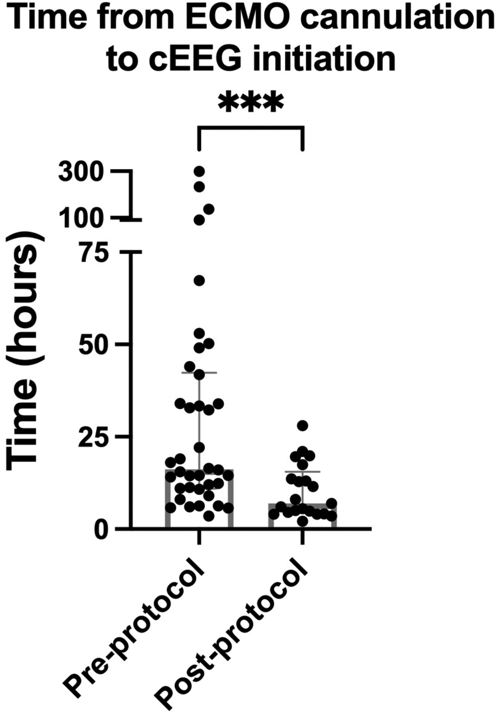
Time interval from extracorporeal membrane oxygenation (ECMO) cannulation to continuous electroencephalography (cEEG) initiation. The timing of ECMO cannulation was determined from the extracorporeal membrane oxygenation patient database and chart review. The initiation of cEEG monitoring was recorded based on the first timestamp of the first continuous EEG record after ECMO cannulation. The interval was significantly shorter in the postprotocol group at 7 hours compared to the preprotocol group at 16.2 hours (P < .001).
Time to Seizure Detection
The percentage of patients with seizures that were ongoing at the start of continuous EEG declined from 64% in the preprotocol group to 0% in the postprotocol group (P < .001) (Figure 3). Further, the duration of time from continuous EEG initiation to seizure detection was significantly shorter in the preprotocol group, median 0 hours (IQR, 0-26) compared to the postprotocol group, median 49 hours (IQR, 23-70) (P = .005) (Figure 4).
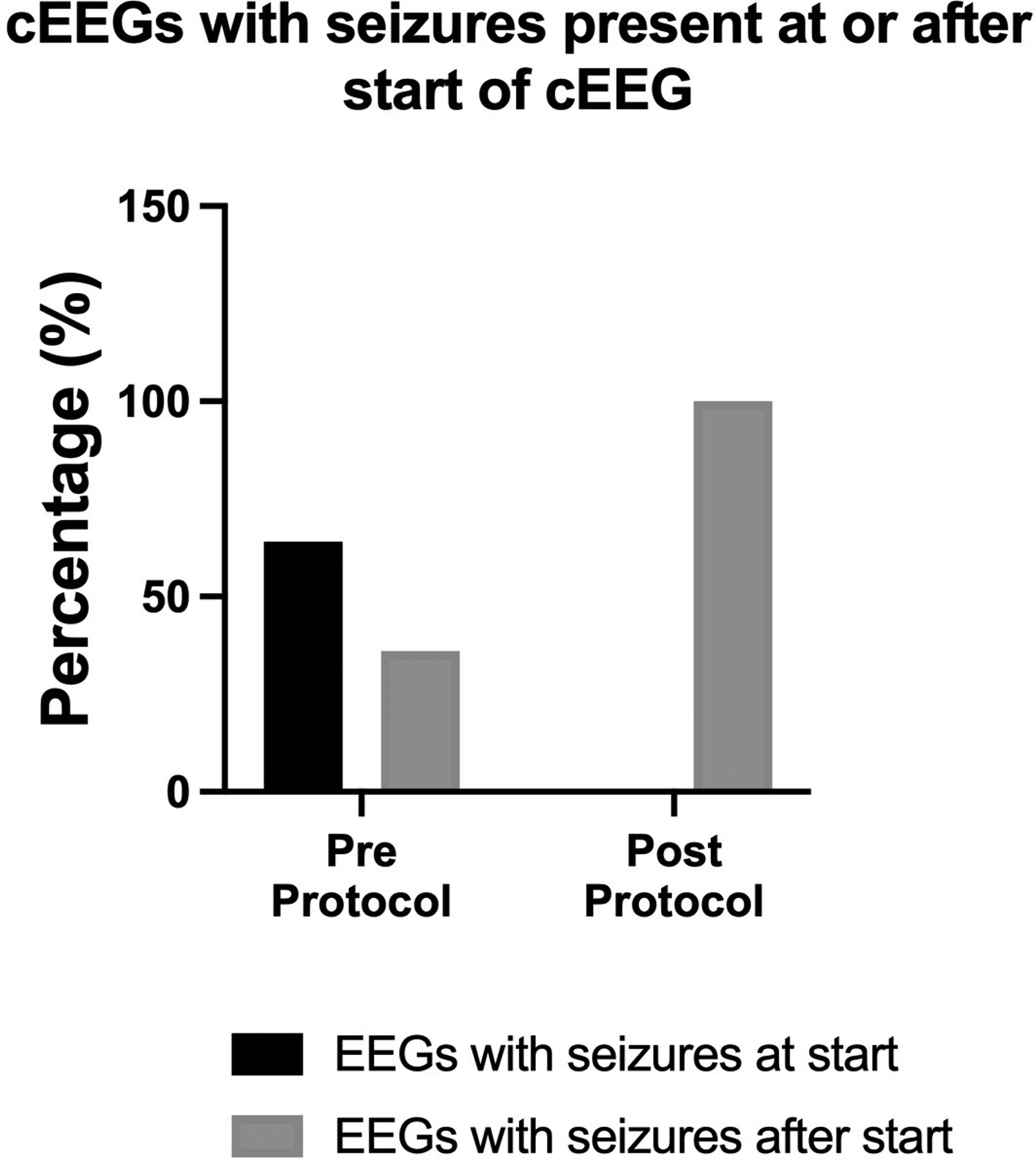
Percentage of patients with ongoing seizures at the start of continuous electroencephalography (cEEG) initiation. The percentage of patients who were already seizing at the start of the EEG decreased from 64% preprotocol to 0% after protocol implementation (P < .001).
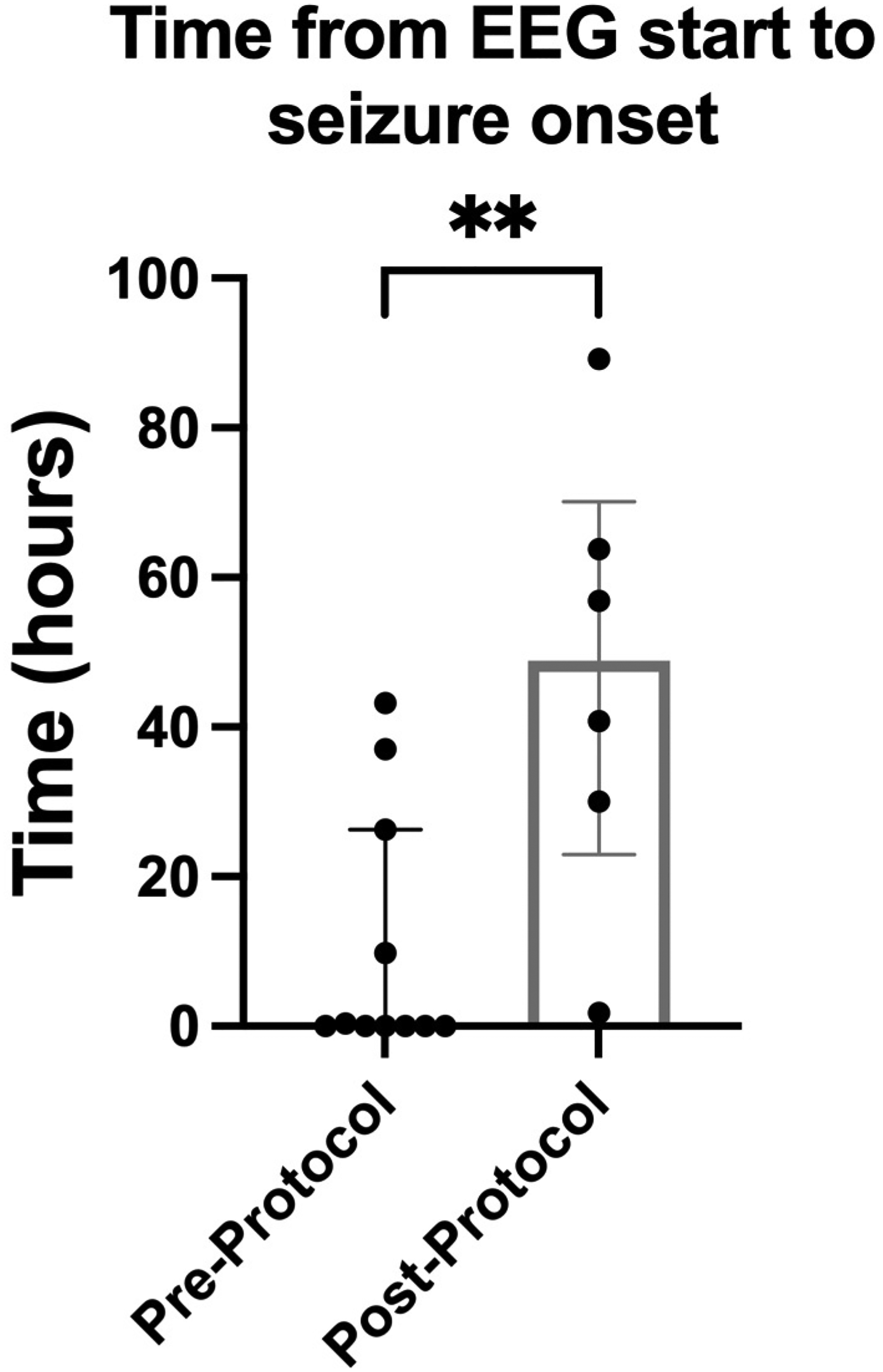
Interval from continuous electroencephalography (cEEG) initiation to seizure detection. The interval was significantly greater in the postprotocol group with a median of 49 hours (interquartile range [IQR] 25%-75% 23-70 hours) compared to the preprotocol group with a median of 0 hours (IQR 25%-75% 0-26 hours) (P = .005).
In order to better understand when seizures occurred in relation to extracorporeal membrane oxygenation cannulation in this study, timing of seizure detection was examined in the postprotocol group where continuous EEG monitoring occurred without the bias of clinical indication. Of the 6 patients who had seizures in the postprotocol group, 1 patient had seizure onset within 24 hours, 2 patients between 24 and 48 hours, and 3 patients 48 hours or longer from the time of cannulation (range 61-102 hours after cannulation). The median time from extracorporeal membrane oxygenation cannulation to seizure onset overall was 54 hours (IQR 30-76 hours).
Although patients in the preprotocol time period were typically connected to continuous EEG based on clinical suspicion for seizures, the actual presence of seizures can only be verified by EEG. As already stated, 64% of preprotocol patients with seizures showed electrographic seizure activity immediately on connection to continuous EEG. To evaluate whether our data for time to seizure onset were biased by delays in initiation of monitoring in the preprotocol group, the median duration of time from extracorporeal membrane oxygenation cannulation to seizure detection was determined for both the pre- and postprotocol groups. Time from ECMO cannulation to seizure detection was not significantly different between the groups, median 37 hours (IQR, 0-37) in the preprotocol vs 54 hours (IQR, 30-76) in the postprotocol group (P = .15), suggesting that the temporal distribution of seizure detection was an accurate reflection of the temporal distribution of actual seizure onset post ECMO cannulation.
Seizure Burden Post Seizure Detection
To begin to address the question of whether the earlier continuous EEG initiation and increased number of hours of monitoring prior to seizure onset in the postprotocol group affected seizure dynamics, post hoc analysis was performed to examine seizure burden, status epilepticus and death in the two groups. Of the 11 patients diagnosed with seizures in the preprotocol group, 8 were determined to be in status and 7 were deceased at discharge. All of these 7 patients with seizures who died were part of the group of 8 patients determined to be in status. Of the 6 patients with seizures in the postprotocol group, 5 were determined to be in status and 3 of these were deceased at discharge. Consistent with similar populations of critically ill patients in the pre- and postprotocol groups (Table 2), there was not a significant difference in the proportion of patients who died (P = .644) or in those who developed status epilepticus (P = .600). In both groups all patients who developed seizures but not status epilepticus survived.
The number of hours in which seizures were identified from time of first seizure detection to out to 72 hours post detection, death, disconnection, or terminal suppression was quantified for all patients in which status epilepticus was diagnosed. There was a trend toward a decrease in seizure burden in the postprotocol group (seizure burden ratio 0.24, IQR 0.085-0.601) compared to the preprotocol group (seizure burden ratio 0.420, IQR 0.3-0.83) (1-tailed P = .080). To evaluate the dynamics of status epilepticus, the time to first status break after seizure detection was determined (Figure 5). Importantly, the number of hours to first status break in the postprotocol period (10 hours, IQR 6.5-12) was significantly less than in the preprotocol group (29 hours, IQR 13.5-62) (P = .031). The total raw number of hours recorded that demonstrated status or terminal suppression were less in the postprotocol group (17 hours, IQR 6.5-32.5) compared to the preprotocol group (58 hours, IQR 21-71.5) (P = .0487). Status resolved within 24 hours in 1 of 8 preprotocol and 3 of 5 of the postprotocol patients. Status recurred, was ongoing, or replaced by EEG suppression at the time of termination of continuous EEG or 72 hours post initial seizure detection in 6 of 8 preprotocol (terminal EEG suppression in 3 and persistent seizures in 3) and 2 of 5 postprotocol patients (1 patient in status until CPR initiated, 1 in persistent status at 72 hours). Across both groups, 100% of patients with ongoing status or terminal EEG suppression at the end of analysis died prior to hospital discharge, whereas only 40% of those in which status resolved died prior to discharge (P = .035).
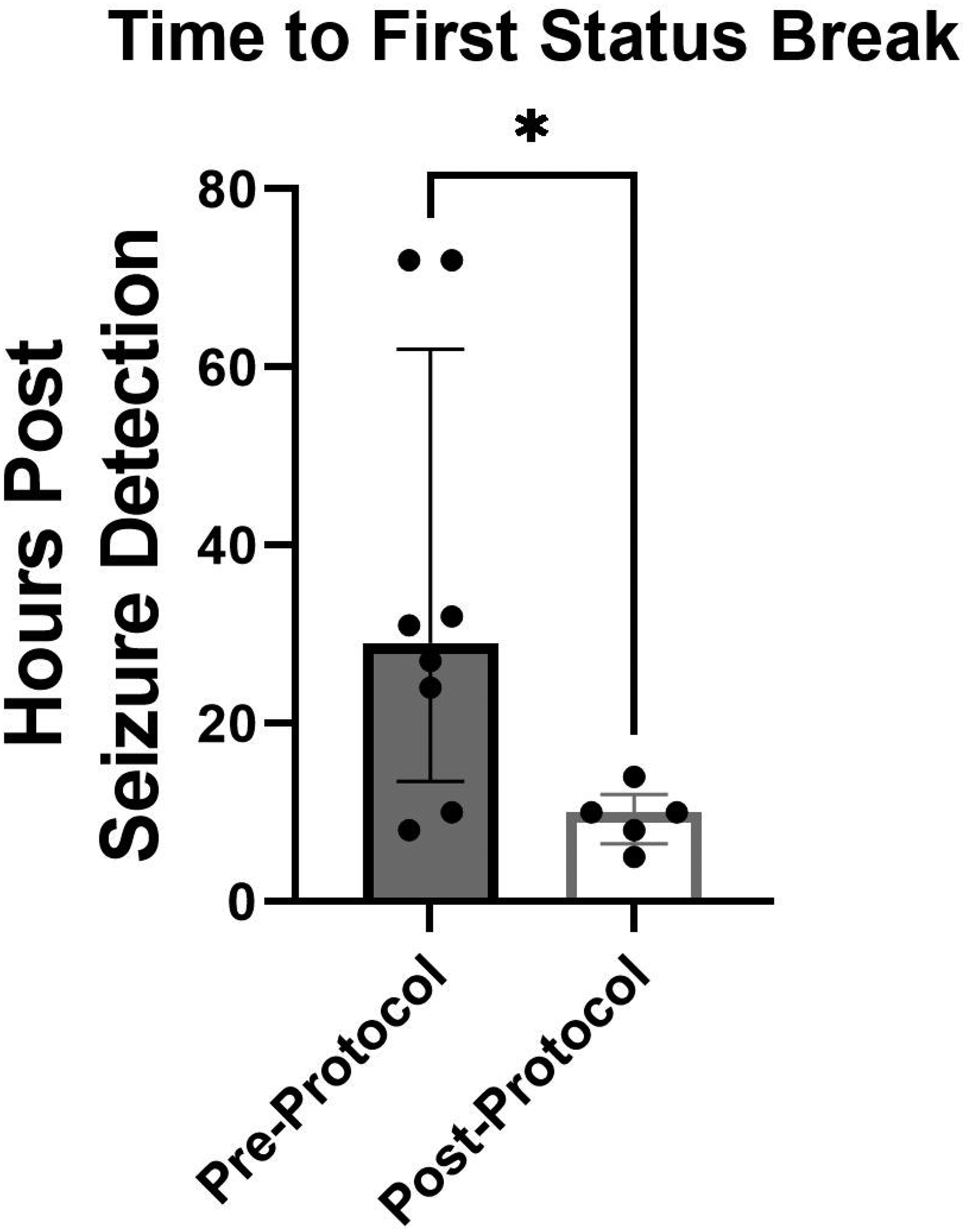
Time to first break in status epilepticus. The period of time between the detection of status epilepticus and the first 2-hour seizure-free period of time in which some background activity was present was determined as the first break in status epilepticus. There was a significantly shorter time to break in status epilepticus in the postprotocol group (10 hours, interquartile range [IQR] 6.5-12) compared with the preprotocol group (29 hours, IQR 13.5-62) (P = .0311).
Discussion
In this study, we examined how continuous EEG protocol implementation in pediatric extracorporeal membrane oxygenation (ECMO) patients changed practice at our institution, and how this affected our understanding of seizures in pediatric patients on extracorporeal membrane oxygenation. Although several institutions have reported on the use of a protocol for continuous EEG monitoring in children undergoing eECMO,2-4,7,8 there have been no studies evaluating the effects of this implementation. This is relevant, as there are no standardized guidelines for continuous EEG use in children undergoing ECMO, whereas seizure prevalence is high in this population.2-8 Our study showed that (1) protocol implementation increased the proportion of patients monitored, (2) reduced the time from ECMO cannulation to continuous EEG monitoring, and (3) resulted in more patients in which the actual seizure onset could be captured and accurately timed in relation to ECMO. In the short years between protocol implementation and analysis, there was a decrease in the number of hours between seizure detection and the first break in status epilepticus, suggesting that earlier detection has the potential to change seizure dynamics. Further, we identified that 50% of our post protocol patients had seizure onset more than 48 hours after the time of extracorporeal membrane oxygenation cannulation. Importantly, this suggests that a significant number of seizures may not be captured with the current minimum recommendation of 24 hours of monitoring when monitoring is begun early in the post cannulation period.
Following protocol implementation, the percentage of ECMO patients monitored on continuous EEG increased significantly. Demographic data, ECMO duration, and duration of continuous EEG monitoring indicate that in both the pre- and postprotocol period, similar patients with similar ECMO exposure were monitored on continuous EEG for similar periods of time. The percentage of seizures captured per continuous EEG in monitored patient was not different between preprotocol (29%) and postprotocol (32%) groups. However, the percentage of continuous EEG–monitored patients out of the total ECMO–supported cohort increased significantly after protocol implementation. Therefore, the number of patients with detected seizures of all patients monitored on ECMO may be a more important metric when considering clinical impact. Protocol implementation resulted in an increase from 8% to 19% seizure detection in all ECMO–supported patients regardless of whether monitored by continuous EEG, creating an opportunity to impact patient care. According to pediatric literature, the percentage of patients on ECMO who develop seizures ranges from 18% to 23%.2-8 Although our post protocol percentage is in keeping with the literature, a seizure prevalence of 8% in the preprotocol period suggests that subclinical seizures in patients not placed on continuous EEG will be missed. Because seizures contribute to morbidity and worse outcomes,3,4,8,9,15,16 initiation of a continuous EEG protocol is the first step toward better-quality care in this patient population.
Implementation of our ECMO continuous EEG protocol decreased time from ECMO cannulation to continuous EEG initiation, but the time from ECMO cannulation to first seizure detection was the same. As a result, the more rapid continuous EEG initiation time in the postprotocol period was accompanied by a prolonged monitoring period prior to the first detected seizure (median 49 hours) compared to the preprotocol period where the majority of patients were already seizing at the onset of monitoring (median 0 hours). Although it is possible that there were seizures that occurred transiently and then stopped prior to continuous EEG initiation, the combination of rapid initiation and the prolonged seizure-free period prior to first seizure detection is most likely consistent with continuous EEG monitoring being initiated before seizure onset in all patients in the postprotocol group (Figures 3 and 4). Accurate timing of seizure onset is important as it is more likely to lead to an enhanced understanding of the events precipitating the seizure and facilitates immediate intervention. Prompt treatment, in turn, provides an opportunity to positively affect clinical outcome, as seizures are more likely to be effectively treated when treatment is started closer to onset. 17 Although the numbers in this study are small, preliminary analysis suggests that in the postprotocol group there was an earlier break in status epilepticus and more hours of status or terminal suppression captured. Additionally, in both the pre and post protocol group, all patients who developed seizures but not status epilepticus survived to hospital discharge, whereas the majority of those patients who developed status epilepticus did not survive. Further, ongoing status or terminal EEG suppression was associated with 100% mortality. Larger patient numbers that take into account mechanism of injury and differences in treatment are needed to determine how earlier detection affects the overall outcomes and will be the subject of future studies. Nonetheless, the current findings emphasize that developing status epilepticus, and in particular, status epilepticus that does not resolve with return of background has devastating consequences and argues for the use of protocolized continuous EEG for prompt detection and aggressive management.
Given the known association between seizures and ischemic infarct or intracranial hemorrhage in pediatric patients in general,15,18 as well as the overall increased risk of these injuries in the ECMO,19-21 identification of factors causing seizures may improve clinical care in other ways. Risk assessment scores for seizures like the ones existing for the adult population 22 may stratify clinical protocols, and early seizure detection may lead to immediate decisions regarding imaging to identify vascular injuries and ultimately provide time-limited treatment such as thrombectomy in the case of stroke or work to expediate decannulation in the case of intracranial hemorrhage.
Our study also identified 3 of 6 patients in the postprotocol group in whom seizures occurred more than 48 hours after ECMO. This contrasts with prior studies reporting that most seizures occur within 24-48 hours, with the median time from continuous EEG initiation to seizure onset being within 48 hours (ranging from 3.2 hours to <48 hours).2-5,8 Although future studies with larger populations are needed to confirm our finding, it argues for longer continuous EEG monitoring periods post extracorporeal membrane oxygenation cannulation. If confirmed, our finding will necessitate a change in clinical practice and practice guidelines, as current recommendations suggest only a minimum time of continuous EEG monitoring duration of 24 hours. 10 These recommendations are based on studying critically ill patients and are not specific to the ECMO population. 10 However, they are currently followed by most centers using continuous EEG during extracorporeal membrane oxygenation support.2-8
Seizure prevalence in postprotocol patients monitored on continuous EEG was 32%. This prevalence is higher than in prior reports where prevalence ranged between 18% and 23%.2-8 It is possible that this may reflect our particular patient population, the majority of which were infants who are known to have an increased risk of seizures while on ECMO and while critically ill in general4,12 or be related to sample size. However, this difference may also reflect the longer continuous EEG monitoring period resulting in the capture of both early- and late-onset seizures. Data collection with strict adherence to our continuous EEG protocol while on ECMO is ongoing at our institutions and may provide a better measure of seizure prevalence in our pediatric extracorporeal membrane oxygenation population.
A limitation to our study is that we were only able to report on a small number of patients, particularly in the postprotocol group. It is likely that the effect of protocol implementation will become clearer once the protocol has been in place for a longer period. Additionally, small numbers limited our ability to identify potential risk factors associated with increased seizure risk.
Conclusion
In summary, this study demonstrates that protocol implementation results in rapid initiation of continuous EEG monitoring following ECMO cannulation, increased numbers of patients monitored, and fewer patients having already ongoing electrographic seizures at the start of continuous EEG initiation and an earlier break in status epilepticus when it develops. Continuous EEG placement prior to seizure onset offers the possibility of creating a window of time for early detection of underlying injuries causing seizures and targeting specific interventions. Lastly, the analysis of predictable patterns of continuous EEG changes may enhance our understanding of the neurologic disease processes associated with ECMO. Future studies should focus on timing and duration of continuous EEG monitoring as well as the impact of early seizure management on outcome as measured by neurologic injury, mortality, and neurodevelopmental outcome.
Footnotes
Author Contributions
DD performed data collection, study design, and data analysis and wrote the first draft of paper. CWH, EKR, and KSH contributed to data collection, data analysis, study design, and writing. MMB contributed to data collection and the writing of the manuscript. CWH and EKR supervised the project.
Declaration of Conflicting Interests
The authors declared no potential conflicts of interest with respect to the research, authorship, and/or publication of this article.
Ethical Approval
The study was approved by the Johns Hopkins Institutional Review Board (IRB NA_0077096) with a waiver of consent.
Funding
The authors disclosed receipt of the following financial support for the research, authorship, and/or publication of this article: Research reported in this publication was supported by the 2020 Doris Duke Charitable Foundation Clinical Scientist Career Development Award and the NIH/NINDS under award number K08NS102526 (CWH) in the form of salary support, and by the NIH/NINDS under award number R01NS106292 (MMB). The content is solely the responsibility of the authors and does not necessarily represent the official views of the National Institutes of Health.
