






Abstract
Lyme disease is the most common vector-borne disease in the United States and has been associated with secondary intracranial hypertension. We reviewed 11 pediatric patients with Lyme-associated secondary intracranial hypertension. All patients presented with headache, ten had papilledema, 7 with a rash, and 5 with a cranial nerve palsy. All patients were treated with acetazolamide, and 3 received combination therapy with furosemide. Three patients were considered to have fulminant intracranial hypertension because of the severity in their presenting courses. Two of the fulminant intracranial hypertension patients were treated with a temporary lumbar drain in addition to medications, whereas 1 fulminant intracranial hypertension patient was treated exclusively with medical therapy alone. The addition of a lumbar drain decreased the time to resolution of papilledema compared to medical management alone. Final visual acuity was 20/20 in each eye of all patients, suggesting that a titrated approach to therapy depending on the severity of presentation can result in good visual outcomes in these cases. Additionally, symptoms can recur after medication wean, so patients should be monitored closely with any discontinuation of intracranial pressure lowering medications.
Introduction
Lyme disease is the most common vector-borne disease in the United States. 1 In the United States, it is most often caused by the spirochete Borrelia burgdorferi and less commonly by Borrelia mayonii. 2 Symptoms include rash, flulike symptoms, and headache. Facial palsy is the most common neurologic manifestation, occurring in 8.4% of cases. 3 Although rare in children, late neurologic symptoms have been described such as encephalitis and polyneuropathy. 2 Previous case studies have reported on Lyme-associated secondary intracranial hypertension and papilledema associated with previously listed symptoms. 4
Primary intracranial hypertension (PIH) is an increase in the pressure of the cerebral spinal fluid around the brain resulting in headaches, nausea, vomiting, and vision problems including diplopia and vision loss not caused by an identifiable condition. 5 Once thought to be a condition predominantly among overweight adult women, the frequency of diagnosis has been on the rise in pediatric populations in part from clearer diagnostic guidelines. 6 Secondary intracranial hypertension is diagnosed when the increased pressure is the result of an identifiable cause, such as medications, sinus venous thrombosis, or infection. 7 In our institution, PIH is diagnosed in pediatric populations by the presence of normal cerebrospinal fluid constituents, a lumbar puncture opening pressure greater than 18 cm H2O if under 8 years of age (25 cm H2O in absence of papilledema) or greater than 25 cm H2O if over 8 years of age, a normal neurologic examination (excluding nerve palsies), and neuroimaging that does not show a clear cause such as mass or hydrocephalus. 8 If a clear antecedent such as meningitis, or known medications, are found, the patients are labeled as having secondary intracranial hypertension. Although uncommon, fulminant intracranial hypertension can be considered the extreme end of intracranial hypertension, seen in either PIH or secondary intracranial hypertension and is considered an emergency because of the risk of permanent visual compromise. It is defined as the acute onset or rapid progression of increased intracranial pressure with evidence of severe vision loss. 9
We performed a review of Lyme meningitis cases seen in the Nationwide Children's Hospital institution's pediatric intracranial hypertension clinic over a 12-year period to look at initial presentation, neurologic symptoms, presence of papilledema, and management and treatment of intracranial hypertension secondary to Lyme disease.
Methods
An institutional review board–approved institutional database of patients 18 years and younger who were followed in the institution's pediatric intracranial hypertension clinic was queried to identify patients with a diagnosis of Lyme disease. Chart review was completed to collect clinical data including patient demographics, body mass index, opening pressure, presence and grade of optic disc edema, visual acuity and visual fields, symptoms leading to diagnosis, workup, including lumbar puncture, medical and surgical interventions, time to medication wean, and disease recurrence.
Inclusion criteria included ≤18 years of age at the time of diagnosis, confirmed serum Lyme infection, and symptoms consistent with intracranial hypertension. Patients without optic edema were still included if they had symptoms suggestive of intracranial hypertension such as cranial nerve palsy, pulsatile tinnitus, or positional headache. Patients were excluded if serum testing did not confirm Lyme infection or were inconclusive.
Resolution was defined as no papilledema on direct funduscopic examination by an ophthalmologist and improvement in headache frequency to none or infrequent. Recurrence was defined as return of headache symptoms at presentation or presence of papilledema following resolution.
Results
We identified 11 patients with Lyme associated secondary intracranial hypertension over the 12-year period. Patients ranged from 4.7 to 18 years of age with an average of 9.5 years at diagnosis (see Table 1). All 11 patients presented with headache, 7 (63.6%) presented with a rash, and 4 (36.4%) reported visual changes. The most common symptoms reported with headache were photophobia and nausea/emesis each seen in 63.6% (7/11) of patients. The average duration of symptoms was 18.7 days at the time of diagnosis. Average body mass index was 19.9 and at the 49th percentile. The average time to first ophthalmology consultation was 1 day compared to 0.5 days for neurology consultation. Five patients presented with a cranial nerve palsy—4 involving cranial nerve VI and 1 cranial nerve VII.
Patient Demographics.
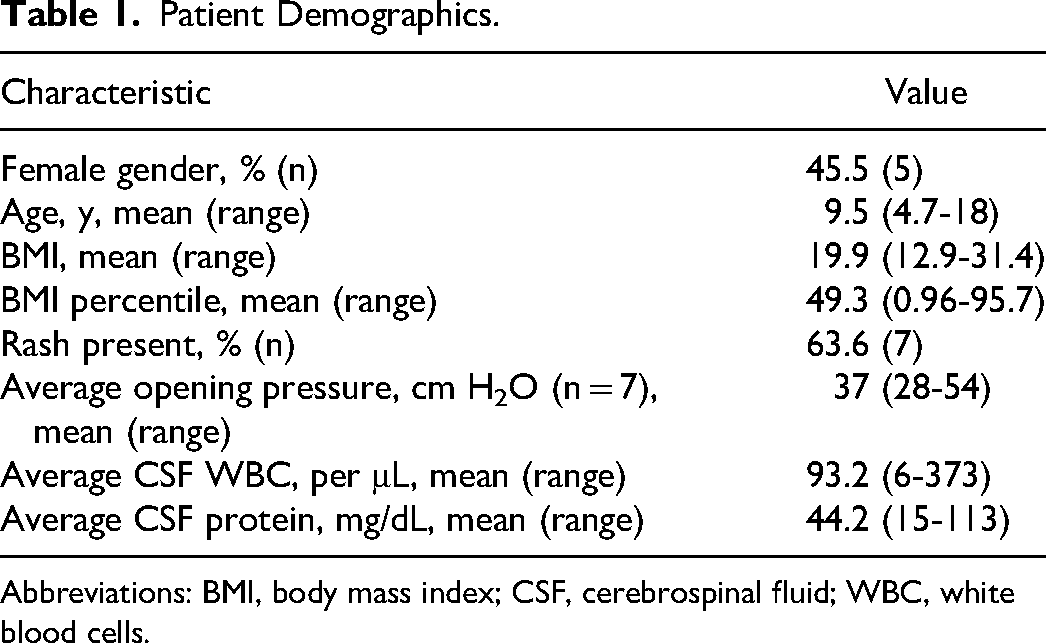
Abbreviations: BMI, body mass index; CSF, cerebrospinal fluid; WBC, white blood cells.
Of the 4 patients who reported visual changes, 3 reported diplopia, 2 of which had bilateral cranial nerve VI palsies. There was an additional patient with bilateral cranial nerve VI palsy and 1 unilateral right nerve VI palsy that did not report visual disturbances. Ten of the 11 patients (91%) presented with papilledema ranging from grade 1 to grade 4 on the Frisen Scale (see Table 2). A higher grade of edema corresponded with a longer time to clinical resolution. Two patients had visual field data available at time of diagnosis; the remainder were too young or too sick to perform visual field testing. Both of these patients had bilateral enlarged blind spots. One patient had a persistent enlargement of the physiologic blind spot after resolution of their papilledema. Visual acuity was largely unaffected in our cohort. Eight of 11 patients (72.7%) had initial visual acuity of 20/20 bilaterally and all patients had a final visual acuity of 20/20 bilaterally. Patient 8, a fulminant intracranial hypertension case, did have a decrease in visual acuity to 20/60 bilaterally over the first few days following diagnosis. However, over the course of treatment her vision improved to 20/20 (see Table 3).
Individual Patients.

Abbreviations: BMI, body mass index; CSF, cerebrospinal fluid; HA, headache; WBC, white blood cells.
Fulminant intracranial hypertension patient.
Patient without optic edema, time to resolution unknown.
Individual Patient Visual Outcomes.
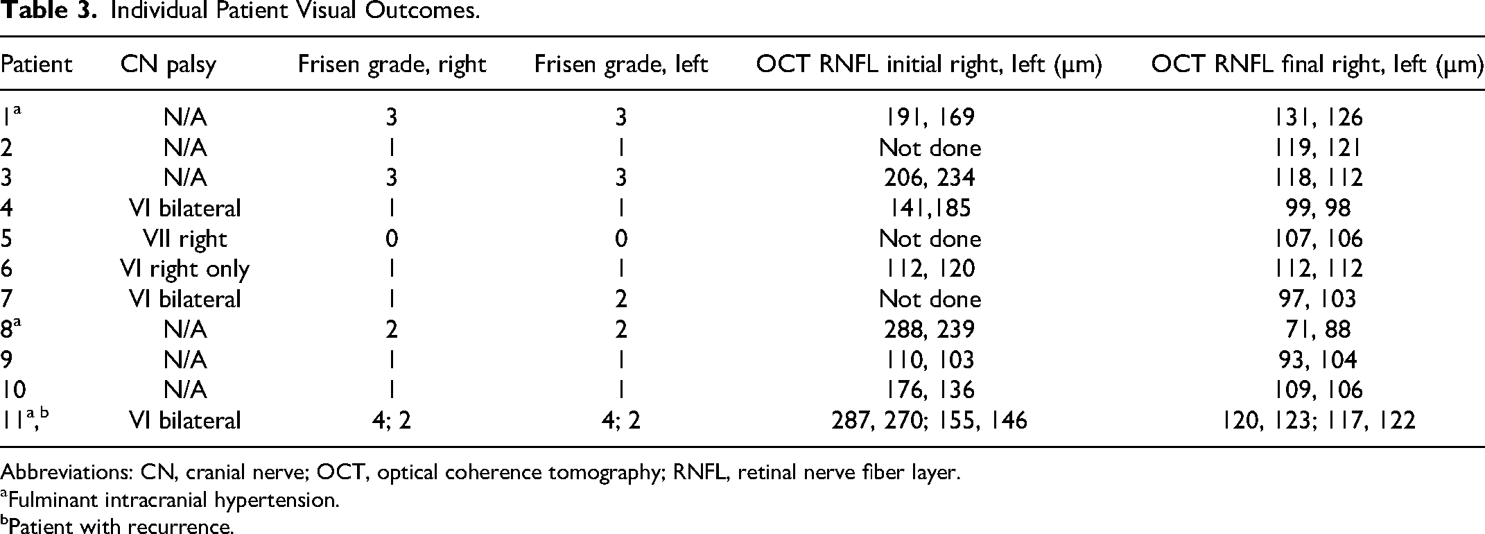
Abbreviations: CN, cranial nerve; OCT, optical coherence tomography; RNFL, retinal nerve fiber layer.
aFulminant intracranial hypertension.
bPatient with recurrence.
All patients had lumbar punctures, 8 had an opening pressure recorded, with 1 exceeding the capability of the manometer at >55 cm H2O. Two patients who did not have an opening pressure recorded and 1 patient had the opening pressure measured while seated. Opening pressures of the remaining 7 ranged from 28 cm H2O to 54 cm H2O, with an average of 37 cm H2O. All patients showed evidence of inflammation with an average cerebrospinal fluid white blood cell count of 93.2/μL (range 6-373/μL), although some patients received their lumbar puncture after starting antibiotics as an outpatient. Eighty-two percent of patients (9/11) had an elevated cerebrospinal fluid protein for age.
All patients were treated with acetazolamide, with maximal doses ranging from 100 to 1000 mg per dose twice a day with an average of 21.3 mg/kg/d (range 8.8-43.3 mg/kg/d). Three patients also took furosemide with doses from 10 to 20 mg twice a day with an average of 1.1 mg/kg/d (range 0.5-1.7 mg/kg/d). All patients received antibiotics for treatment of their Lyme infection, and 5 started antibiotics prior to admission and diagnosis of secondary intracranial hypertension. Antibiotics taken at any point included ceftriaxone, azithromycin, cefalexin, cefepime, doxycycline, and amoxicillin (see Table 2 for specifics). Nine patients received ceftriaxone during their treatment, 6 of whom transitioned to oral doxycycline to complete treatment. Two patients received only doxycycline, 1 initiated prior to the diagnosis of secondary intracranial hypertension and 1 initiated when confirmatory testing returned after discharge. One patient started with doxycycline prior to diagnosis of secondary intracranial hypertension and transitioned to ceftriaxone to finish treatment while inpatient.
The average time to resolution for the 10 patients with optic edema was 67.9 days with a range of 12-207 days. Once clinical resolution was obtained, patients were weaned off medications for their secondary intracranial hypertension per clinic protocol. All patients were weaned off acetazolamide at an average of 65.3 days with a range of 23-116 days. Three patients who additionally received furosemide had this medication weaned first after an average of 35.3 days with a range of 26-46 days. When repeat examination following wean noted continued resolution, acetazolamide was then weaned. Two patients self-discontinued acetazolamide in between visits and were not included in treatment duration calculations.
Three patients were considered fulminant intracranial hypertension. The first fulminant intracranial hypertension patient, patient 1, was seen in 2014 prior to our institutional protocol for fulminant intracranial hypertension and was medically managed with acetazolamide. The other 2 received high-dose acetazolamide, furosemide, and a temporary lumbar drain per institutional protocol. Patient 11 also received intravenous methylprednisolone for 5 days followed by a 2-week oral prednisolone taper. Time to resolution of their secondary intracranial hypertension was 207, 55, and 117 days in patients 1, 8, and 11, respectively.
Patients 1 and 11 were considered fulminant intracranial hypertension at the time of presentation. Patient 1 presented with grade 3 edema plus enlarged blind spots, and patient 11 presented with grade 4 edema plus splinter hemorrhages at the disc margins. Patient 8 initially presented with bilateral grade 2 disc edema and 20/30 visual acuity with an opening pressure of >55 cm H2O. Her edema quickly worsened to bilateral grade 3 edema and visual acuity worsened to 20/60 on maximum therapy prompting a diagnosis of fulminant intracranial hypertension and placement of a temporary lumbar drain. After treatment with the lumbar drain and continued medical therapy, her edema resolved and visual acuity improved to 20/20. Patient 8 had final retinal nerve fiber layer thickness of 71 and 88 µm in the right and left eye respectively, indicating nerve atrophy. Mean normal pediatric retinal nerve fiber layer thickness is approximately 107 ±11.1 µm. 10 She was the only patient with final retinal nerve fiber layer readings indicative of nerve atrophy.
Patient 11 was the only case of recurrence documented. He presented with bilateral grade 4 disc edema and received antibiotic treatment including cefepime, ceftriaxone, and finally doxycycline. After 61 days, the disc edema was declared resolved and he was weaned off medications. At the subsequent follow-up visit off medications, his papilledema had recurred and was grade 2 bilaterally. Oral acetazolamide treatment was reinitiated, with resolution noted at 56 days. He was successfully weaned off medication and has not had further evidence of recurrence. It is likely that the edema had not fully resolved when the patient was initially weaned as retinal nerve fiber layer measurements showed further improvement past those at the first wean attempt after reinitiating treatment.
Discussion
Meningitis is a common presentation of Lyme infection affecting 3 of every hundred patients. 1 It is also a known cause of secondary intracranial hypertension. Previous Lyme secondary intracranial hypertension case studies have shown similar presentations to our cohort, including headaches, papilledema, cranial nerve palsy with associated diplopia, and erythema migrans.4,11 The only presenting symptom shared by all patients in our cohort was headaches, secondary symptoms included cranial nerve palsy in 45.5% (5/11) and erythema migrans in 63.6% (7/11). All patients with valid opening pressure measurements had elevated opening pressures, and 10 of 11 had papilledema. These presentations are consistent with previous case studies on Lyme secondary intracranial hypertension save the inclusion of 1 patient without papilledema who met secondary intracranial hypertension diagnosis from institutional standards. 8
This study does point out the need for inclusion of the opening pressure with lumbar punctures and the need for bedside funduscopic exams. Intracranial hypertension is often not considered when patients present with concerns for headache and meningitis symptoms and thus the opening pressure is not measured. This likely results in underrecognition of secondary intracranial hypertension in the presence of meningitis. This was evident in the 2 patients who did not have an opening pressure recorded, as the procedure notes stated opening pressure was not clinically indicated. The third patient had the opening pressure measured while seated. The average time to first ophthalmology consultation was 1 day compared to 0.5 days for neurology consultation. Headaches are a common complaint in meningitis, whether viral or bacterial. Thus, it is important for practitioners to have a low threshold to examine for other signs of secondary intracranial hypertension. This includes a bedside funduscopic examination (or consult to ophthalmology) looking for optic edema, and examination for presence of a cranial nerve palsy. In patients without these clinical findings, other signs or symptoms that may suggest concurrent secondary intracranial hypertension include headaches that are the most severe in the morning or after prolonged horizontal positioning or exacerbated by maneuvers such as coughing, Valsalva, or bending over. 7 Often not asked, but the presence of pulsatile tinnitus, or a whooshing sound, is highly suggestive of intracranial hypertension. Patients with meningitis often receive magnetic resonance imaging (MRI) and it is important for providers to carefully review the MRI scans for findings associated with intracranial hypertension. These include an empty/partially empty sella, optic nerve sheath distension, flattening of the posterior aspect of the globe, the target sign, and transverse venous sinus stenosis.12,13 These support the possibility of intracranial hypertension but are not required for diagnosis.
Unique to this study is the inclusion of 3 fulminant cases: patients 1, 8, and 11. These patients presented with severe optic disc edema and 1 that had progressive worsening on medical therapy. The last 2 patients were treated with the current institutional protocol that included high-dose acetazolamide, furosemide, intravenous methylprednisolone (in 1 patient), and a temporary lumbar drain. Patient 1 was only medically managed and had a significantly longer time to resolution at 207 days compared to the other cases at 55 and 117 days. Previous studies have shown that lumbar drains have clinical benefits for fulminant intracranial hypertension patients. Ploof et al 9 found that lumbar drains were a viable first option for fulminant intracranial hypertension in the case of IIH and when secondary to influenza and viral meningitis. They noted that treatments were relatively similar and all patients improved on the lumbar drain in spite of having concerns for vision loss on maximum medical therapy. Proper treatment with inclusion of a lumbar drain appears to have a positive effect on the time to resolution of fulminant intracranial hypertension secondary to Lyme meningitis. This is important as quicker resolution of papilledema lessens the risk of permanent visual deficits. 14
The variety of antibiotic courses used did not appear to have an effect on clinical outcomes; however, it is important to note the risks of doxycycline-induced secondary intracranial hypertension. 15 The tetracycline class of medications has been associated with secondary intracranial hypertension with prolonged use, typically for the treatment of acne. 7 Campetti et al reported a 55-year-old woman using doxycycline to treat a Lyme infection that developed secondary intracranial hypertension. 16 However, our study suggests short-term doxycycline treatment can be a safe and effective tool in the treatment of Lyme infections with concurrent secondary intracranial hypertension without worsening clinical course.
All patients had dates of diagnosis between the months of June and August that correlate with CDC data on Lyme seasonality. 1 An interesting note is the number of patients in this study who were diagnosed during the early stages of the COVID-19 pandemic following continued closure of indoor fitness facilities and a mild winter. Only 3 patients had diagnosis prior to January 2020. Six patients were diagnosed in 2020 (beginning of the pandemic), 1 in 2021, and 1 in 2022. This spike in cases contradicts a suspected decrease in the report of Lyme cases but correlated well with an increase in outdoor activity and possible exposure to Lyme-infected tics. 17
This study is not without its limitations, the first being that it is a retrospective analysis. Retrospective analyses are known to have limitations from the accuracy of record keeping and access to important statistics; within this study, there were 3 patients who did not have available opening pressures. Additionally, there were only 11 patients in this study. Although not a large sample size, among Lyme secondary intracranial hypertension studies, this will be the largest study at the time of publishing. Many other Lyme secondary intracranial hypertension articles were case studies or only had up to 4 patients. Having access to the database at Nationwide Children's Hospital allowed access to 400 cases to review, resulting in these 11 patients for this study, making it the largest study of its kind.
Our findings stress the importance of neurologic and ophthalmic evaluations for patients diagnosed with not only Lyme disease but also meningitis, the effects that a lumbar drain can have on the time to resolution for fulminant cases, and the positive outcomes of appropriate doses of doxycycline for patients with Lyme secondary intracranial hypertension.
Footnotes
Author Contributions
JDR contributed by data collection, data review and initial manuscript preparation and editing. COJ contributed with data collection, manuscript review. HEI contributed with data collection, manuscript review. SCA contributed with study design, patient identification, data collection and review, manuscript preparation and review.

Declaration of Conflicting Interests
Shawn C. Aylward does receive honoraria as associate editor of Pediatric Neurology. The other authors have no relevant conflicts of interest.
Funding
The authors received no financial support for the research, authorship, and/or publication of this article.
Ethical Approval
The study was approved by Nationwide Children's Hospital Institutional Review Board (IRB13-00867) with a waiver of consent.