
Abstract

A 16-month-old girl, presented to the emergency department with a progressively worsening 10-day history of high fever, malaise, nasal obstruction, cough, and left otorrhea. She had been previously diagnosed with acute otitis media and started on topical ofloxacin and oral amoxicillin, with no improvement. Otoscopy revealed left-sided purulent otorrhea and a bulging, hyperemic tympanic membrane, associated with periauricular erythema. Blood tests showed elevated inflammatory markers.
The patient was admitted for intravenous amoxicillin-clavulanate and corticosteroid treatment. After 72 hours with only slight improvement, a left retroauricular fluctuant swelling was noted. Laboratory workup revealed further elevation of inflammatory markers, prompting a change to ceftriaxone.
Ear computed tomography (CT) revealed left otomastoiditis with an adjacent subperiosteal abscess, which was drained surgically, along with myringotomy and tympanostomy tube placement. Metronidazole was added.
Brain and ear magnetic resonance imaging (MRI) depicted left acute otomastoiditis with dehiscence and erosion of the internal mastoid wall, with purulent content continuity between the sinus lumen and mastoid. In addition, sigmoid sinus occlusion by a purulent collection was noted (Figures 1 and 2), as well as skull base osteomyelitis and pachymeningitis, along with left cavernous sinus involvement, characterized by a thick enhancement, wall bulging, and stenosis of the cavernous carotid artery, the latter possibly due to arteritis (Figure 3).
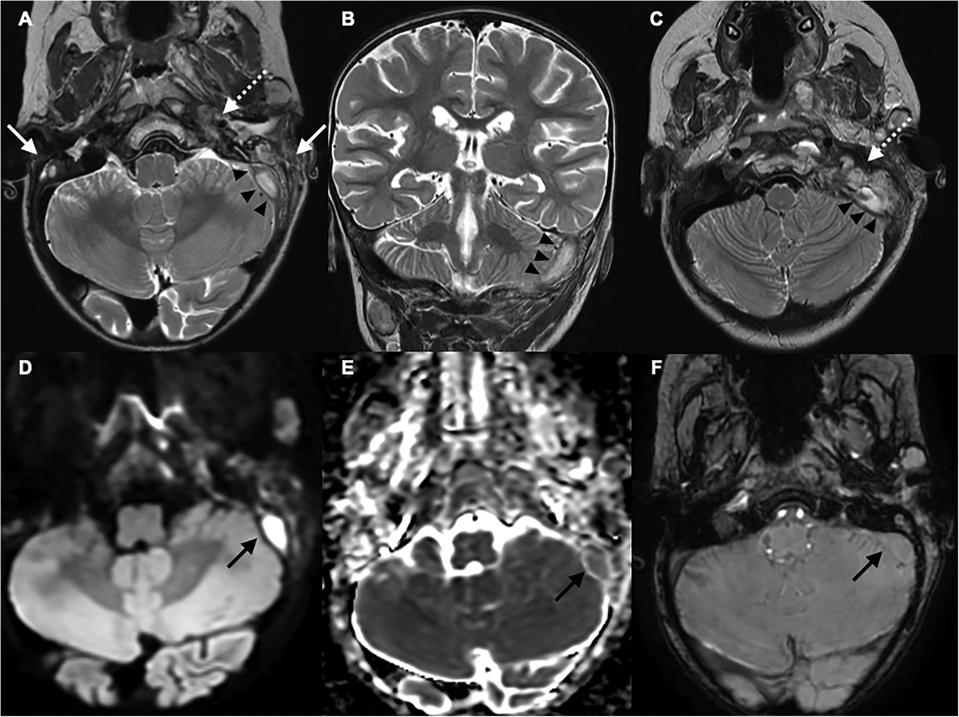
Brain magnetic resonance imaging (MRI) showing bilateral mastoid air cells and middle ears filled with T2-weighted imaging (T2-WI) hyperintense content (A: white arrows), related to bilateral acute otomastoiditis. Note the exuberant high signal intensity on T2-weighted imaging of the skull base at the left side (A and C: white dotted arrow), compared with the right side. Additionally, the sigmoid sinus is filled with pus, with high signal intensity on T2-weighted imaging (A, B, and C: black arrowheads), and marked diffusion restriction (high signal intensity on diffusion weighted imaging [D, black arrow] and low apparent diffusion coefficient [E, black arrow]). Susceptibility-weighted imaging (SWI) (F) showed no low-intensity signal, suggesting no blood thrombus involved (black arrow).
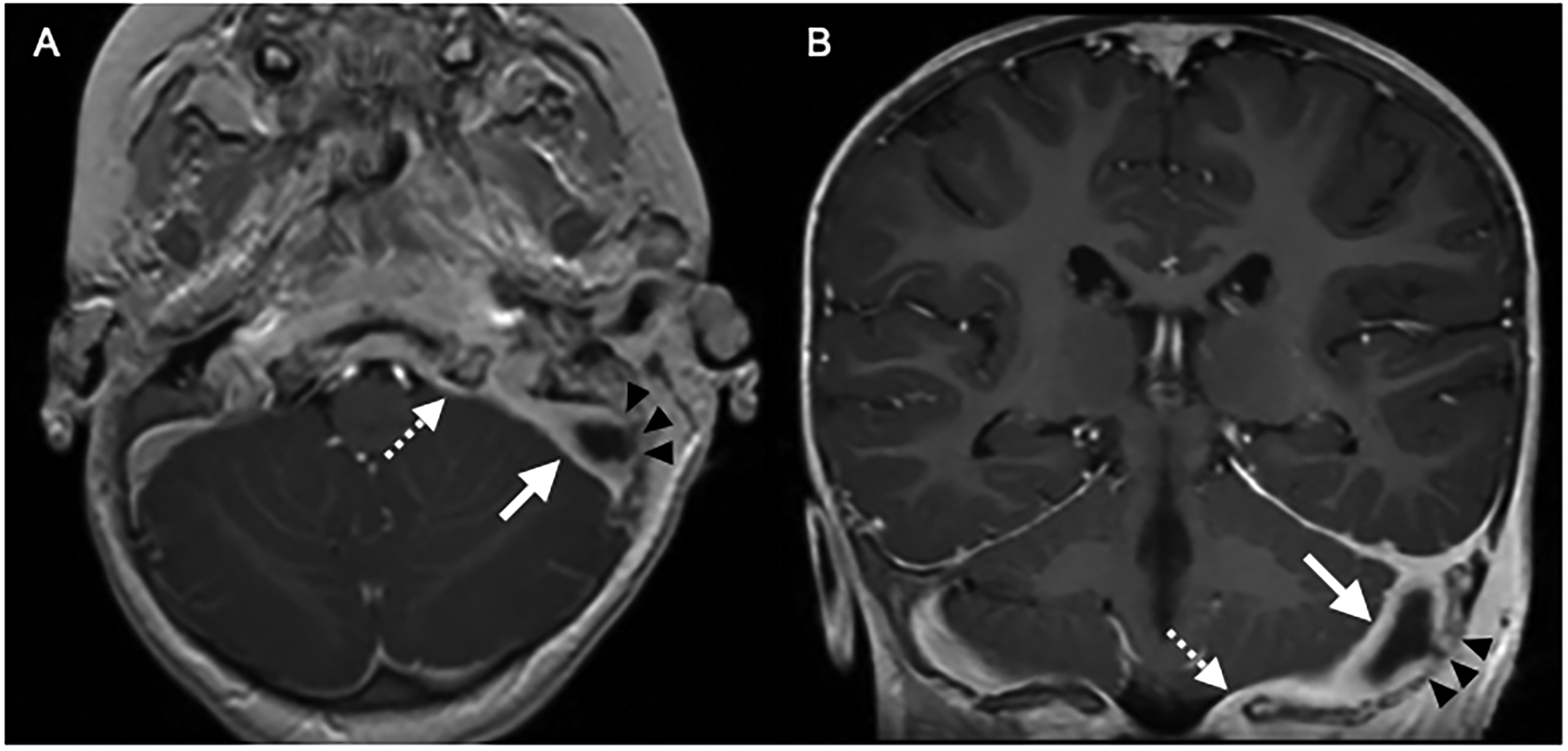
Postcontrast gradient echo T1-weighted imaging (A: axial; B: coronal) confirmed occlusion of the sigmoid sinus associated with thick enhancement of the sigmoid sinus dural walls (white arrows). Communication of the purulent content between the sigmoid sinus and mastoid by a bony defect in the dural sigmoid plate was also evident (black arrowheads), as well as extensive paquimeningitis in the left skull base region (white dotted arrows).
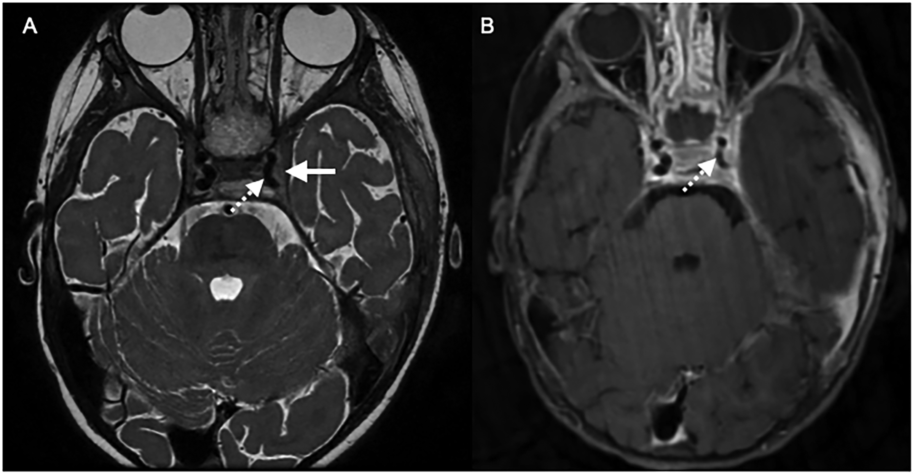
Brain magnetic resonance imaging (MRI) depicting extension of the infectious process to the cavernous sinus, without occlusion, demonstrating high T2-weighted imaging signal intensity and lateral bulging (A: white arrow), associated with moderate stenosis of the cavernous segment of the internal carotid artery (A and B: white dotted arrows).
The patient underwent a simple cortical mastoidectomy with cleaning and debridement of the surgical cavity, which contained necrotic and infected tissue. Dissection extended to the left transverse sinus, where an organized purulent content was found and drained up to the jugular bulb. Local gentamicin was applied and the purulent sample was obtained, which later identified Escherichia coli as the causative agent.
She was discharged on day 28 of hospitalization, after clinical, laboratory, and imaging improvement (Figure 4), maintaining oral levofloxacin for 14 days and multidisciplinary follow-up.
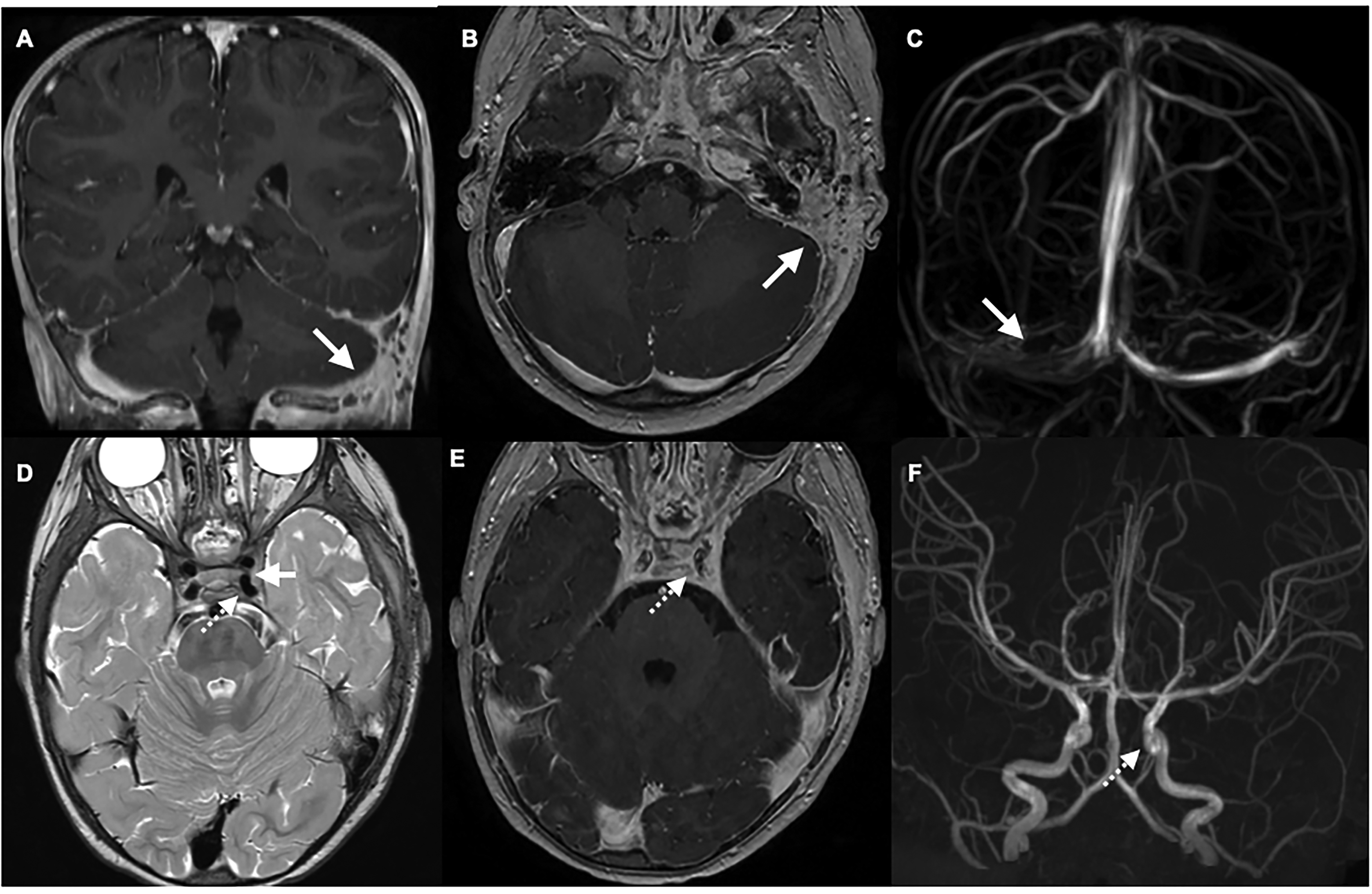
Posttreatment magnetic resonance imaging (MRI) revealed complete resolution of left sigmoid sinus occlusion as depicted by postcontrast T1-weighted imaging (A: coronal; B: axial, white arrows). Magnetic resonance venography confirmed the patency of the left lateral and sigmoid sinus (C: white arrow). Additionally, there was improvement of the left cavernous sinus involvement showed by normal signal intensity on T2- weighted imaging (D: white arrow), and normal diameter of ipsilateral cavernous segment of internal carotid artery (D, E: postcontrast T1-weighted imaging; F: magnetic resonance angiography; white dotted arrows).
Cerebral venous sinus thrombosis is a rare and serious complication of acute otomastoiditis. 1
Because of their proximity to the middle ear and mastoid, the sigmoid/lateral sinuses are vulnerable to thrombophlebitis from otogenic infections. Infection can erode the sinus wall, form granulation tissue or peri-sinus abscesses, and cause pressure necrosis, leading to a mural blood thrombus. 2 Thrombophlebitis may also occur without bony erosion, as pathogens can spread through emissary veins. 3
Classical signs include fever, otalgia, otorrhea, headache, nausea, vomiting, lethargy, and neck stiffness. Early antibiotic use may mask symptoms, leading to a more insidious onset. 4 Cerebral venous sinus thrombosis can impair cerebrospinal fluid absorption, increasing intracranial pressure—a condition known as otitic hydrocephalus. 5 Patients may show papilledema, abducens and facial nerves palsy, vertigo, ataxia, and seizures. MRI should be performed promptly in such cases to rule out cerebral venous sinus thrombosis and other intracranial complications.
Our patient had a sigmoid sinus occlusion by pure purulent content without blood clot involvement, mimicking an abscess on imaging, confirmed by surgery. This rare and life-threatening condition has, to our knowledge, not been elsewhere reported.
Intravenous antibiotics are not sufficient for treatment. A more invasive surgical intervention with sinus drainage, mastoidectomy, and debridement is required. Anticoagulation was not used, as no blood thrombus was present.
Footnotes
Ethical Approval
This case report is based on clinical and patient data collected in accordance with ethical standards and institutional guidelines. The patient's identity has been anonymized to protect confidentiality. The data included in this report were obtained with informed consent from the legal guardian. All clinical and diagnostic information, including medical history, treatment protocols, and outcomes are available upon request.
Author Contributions
FSB - conceptualization and design; drafting of the manuscript and interpretation of the data; DVP - collection of the neuroradiological imaging data; AMM and JD - clinical data collection and initial data analysis; LN - imaging analysis and interpretation of findings; DT - conceptualization and design and revision of manuscript.
Funding
The authors received no financial support for the research, authorship, and/or publication of this article.
Declaration of conflicting interests
The authors declared no potential conflicts of interest with respect to the research, authorship, and/or publication of this article. This manuscript is not under consideration for publication elsewhere and has not been published previously. Additionally, there is no redundant or duplicate publication involved.