
Abstract
In this study, the effect of pharmaceutical-grade alginates on the cell viability of human mesenchymal stem cells derived from umbilical cord was examined and their use in tissue engineering applications was evaluated. The effects of the ratio of the copolymer building blocks (guluronic and mannuronic acids) and their interactions with divalent calcium, the purity of alginates (proteins and polyphenol content), and gelation factors (calcium concentration and sol content) were examined. The high guluronic acid content in the alginates improved the viability of the human mesenchymal stem cells derived from umbilical cord and supported cell growth significantly. It was confirmed that the sol fraction of alginate reduced cell viability. Cells in the presence of alginate beads cross-linked with 50 and 100 mM calcium chloride showed maximum viability; the protein and polyphenol content of the alginates did not affect the viability of the human mesenchymal stem cells derived from umbilical cord, while the monomer ratio did have an obvious effect.
Introduction
The use of alginate as a biomaterial has been studied in many biomedical and pharmaceutical applications, such as dental impressions, wound dressing, and controlled drug delivery1–3 and is approved by the Food and Drug Administration (FDA) as a safe biomaterial for use in the human body.
4
Alginate is a linear polysaccharide, mainly consisting of α-
Alginate polymers containing G residues undergo ionotropic gelation at room temperature in an aqueous medium containing divalent or trivalent cations such as Ca2+, Al3+, or Fe3+.6,7 These gels are reversible, inert in aqueous environments, and dissolve or degrade under physiological conditions.8,9 The physical property of alginate polymers is specifically dependent on the ratio of its two monomers. For instance, G-rich alginates are more susceptible to ionotropic gelation and provide better strength and stability to the final hydrogel product.10,11 Various alginates differing in G/M ratio are commercially available and provide different solution viscosity, mechanical strength, and stability properties. In some studies, the viability of cells has been ascribed to the composition of the alginate polymer and that better bioavailability can be achieved using a G-rich alginate,12,13 while M-rich alginates have been claimed in other studies. 14 Apart from G/M composition, the cell viability in the presence of alginate polymers can also be dependent on the type and amounts of contaminants such as proteins and polyphenols.15–17 Therefore, for tissue engineering scaffolds, these polymers require a comprehensive evaluation of the contaminants and their role on cell viability. In this study, we evaluated cell growth and viability of human mesenchymal stem cells from umbilical cord (hUMSCs) in the presence of two pharmaceutical-grade alginate polymers having equivalent solution viscosity but different G/M composition. In this regard, gelation factors, including calcium concentration and the sol content of the alginate gels, were studied. We used hUMSCs since mesenchymal stem cells (MSCs) have the ability to self-renew and trans-differentiate into all type of tissues.18,19 Moreover, the hUMSCs contain an inexhaustible source of primitive MSCs that can serve as an ideal source for various tissue engineering applications. 20
Materials and methods
Materials
Pharmaceutical-grade sodium alginates (FMC biopolymers, Philadelphia, PA; Protanal LF200S and Protanal LF120M); sodium chloride and calcium chloride dihydrate (Fisher scientific, Pittsburgh, PA, USA); 2,5-diphenyl tetrazolium bromide (MTT) and antibiotic and antimicotic solution as well as phosphate buffered saline (PBS; Sigma, St. Louis, MO); low-glucose Dulbecco’s modified Eagle’s medium (DMEM) and trypsin (Invitrogen, Carlsbad, CA); fetal bovine serum (Atlanta Biologicals, Lawrenceville, GA, USA); and hUMSCs (Sciencell, Carlsbad, CA).
Bead preparation
Two percent w/v solutions of G-rich and M-rich alginate polymers were prepared in physiological saline solution and passed through a 25-µm syringe filter and then extruded dropwise through a 25-gauge needle into a calcium chloride (CaCl2) solution under magnetic stirring. The beads were left in the gelling medium for 15 min and then washed twice in sterile double distilled water, followed by a rinse with sterile PBS.
Cell culture
The hUMSCs were cultured in monolayers in T75 flasks with low-glucose DMEM supplemented with 10% fetal bovine serum and 1% antibiotic and antimicotic solution. The cells were fed with a fresh DMEM every 2–3 days. Cultures were propagated at 37°C under humidified conditions using 5% CO2. The cells grown up to 70%–80% confluency were treated with trypsin to dislodge them from the flask. Suspended cells were plated either in 12- or 24-well plates, according to the experimental design, where series of experiments were conducted to investigate the effects of alginates and calcium chloride on the hUMSC viability.
Cell viability in the presence of alginate beads
A hUMSC suspension (density of 30 × 104) in a growth medium, containing DMEM, 10% fetal bovine serum, and 1% antibiotic and antimicotic solutions, was placed in a flat-bottom, 12-well plate. After the cells reached 30% confluency, freshly prepared alginate beads (~30, 60, and 120 mg) were added and incubated at 37°C in a 5% CO2 incubator for 48 hours. The cell viability was evaluated by a standard colorimetric MTT assay. The same procedure was used to prepare alginate beads cross-linked at different calcium chloride solutions (50, 100, 150, and 200 mM) and used to evaluate their effect on cell viability.
Cell viability in alginate solutions
Monolayer cells in a 24-well plate with 30%–40% confluency were exposed to ~30 mg w/v of LF200S and LF120M alginate solutions. Cell viability was estimated by MTT assay (described below) after 48 hours of incubation period.
Cell viability in calcium chloride solution
The hUMSCs plated in a 24-well plate with a density of 3 × 10⁴ were exposed to 30 mg w/v of CaCl2 (10, 50, 100, and 200 mM) solutions. Cell viability was estimated after 48 hours of incubation period.
MTT assay
Viable hUMSCs were counted using MTT colorimetric assay after being cultured in the presence of 2 wt% alginate beads for 48 hours. Following Mosmann et al., 21 100 µL of 2 mg/mL of MTT solution in a PBS was added to the wells after the designated incubation times and was further incubated for 4 hours at 37°C in a CO2 incubator. The medium containing the free dye was removed, the cell layer was briefly rinsed with PBS, and the formazan crystals were dissolved in 200 µL of dimethyl sulfoxide (DMSO), where its absorbance was measured at 570 nm. The values for each of the treatments represent the average from duplicate wells.
Protein content
The protein content of the alginates was determined utilizing bicinchoninic acid (BCA) method. To a total of 100 µL protein sample and 2 mL of BCA reagent was added and incubated for 30 min at 37°C.
Total phenols content
The total phenol content in all alginate samples was measured using Folin–Ciocalteu (FC) method. 22 Serially diluted quercetin (standard phenolic compound) was mixed with 400 µL of FC reagent followed by 800 µL of 20% sodium carbonate. The mixture was allowed to stand for 15 min and total phenol was determined by a spectrophotometer at 765 nm. The standard curve was prepared using 0, 2.5, 5, 10, 20, and 40 µg solutions of quercetin in ethanol:water mixture (50:50 v/v). Total phenol content was expressed in terms of quercetin equivalent.
Statistical Analysis
A Student’s t-test (two-tailed, paired) was used to evaluate the data, and a p < 0.05 was considered to be statistically significant.
Results and discussion
Sodium alginate is composed of two acid residues, G and M. The biopolymer, through its guluronic acid residues, can interact with metal ions, in particular calcium, to form a gel. 23 The “egg box” model has been suggested to explain the cation–G acid complexation in alginate polymers. 11 As with other biopolymers with natural origin, commercial alginate polymers contain different ratios of the two acids. Apparently, due to the gelling reactivity of the G residues toward cations, an alginate with higher G content would extensively interact with calcium cation to make a stronger gel after complexation. 24 However, a softer gel is formed if the alginate polymer contains more M residues. Since G and M residues vary with the alginate type, different physical and chemical properties are expected from these polymers. The gel and the sol fractions of an alginate polymer are critically dependent on its G and M content. In the presence of adequate calcium cations, an alginate polymer at presumably 100% G content would produce a network with no sol fraction. However, no gel would be formed if an alginate polymer is composed entirely of M residues, even in high calcium concentrations. Consequently, to determine the biocompatibility of ionogelling polymers, the cell viability was evaluated in the presence of each individual components of the gelling system, that is, calcium–cross-linked alginate (gel fraction), calcium chloride (gelling agent), and the non-cross-linked alginate (sol fraction). Due to different levels of porosity, stability, and structural integrity in the gel state, the composition of an alginate polymer is assumed to have a strong role in the rate and extent of cell growth and survival.24,25 The effects on cell viability of common impurities of alginates, such as proteins and polyphenols were also investigated.
Cell viability in the presence of alginate beads cross-linked with calcium
Cells grown in monolayer were exposed to three different quantities of alginate beads (Figure 1), and their viability was assessed (mean ± SD, n = 3) against control (cells in the absence of beads). As shown in Figure 2, the cells were comparatively viable in the presence of LF200S, especially at low and medium doses of 30 and 60 mg, respectively, while the cells were 23% less viable at the maximum gel dose of 120 mg. However, at all gel doses, the viability of the hUMSCs was significantly decreased up to 34% in the presence of LF120M alginate gels at the maximum gel dose. Regardless of the gel type, the cells showed less viability at the maximum gel dose, even the rate of survival was less in the presence of LF120M gel beads. Between the two grades of alginate, the hUMSCs were generally shown to be less viable in the presence of LF120M. This may be accounted for in terms of the high M content of this polymer or high G content of the LF200S, which affects the gel strength after complexation.23,26 Wang et al. 27 observed that higher proportions of guluronic acid provided better colonization and differentiation of bone marrow cells in rats.
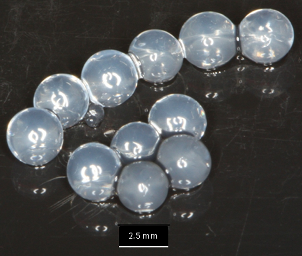
Alginate gel beads.
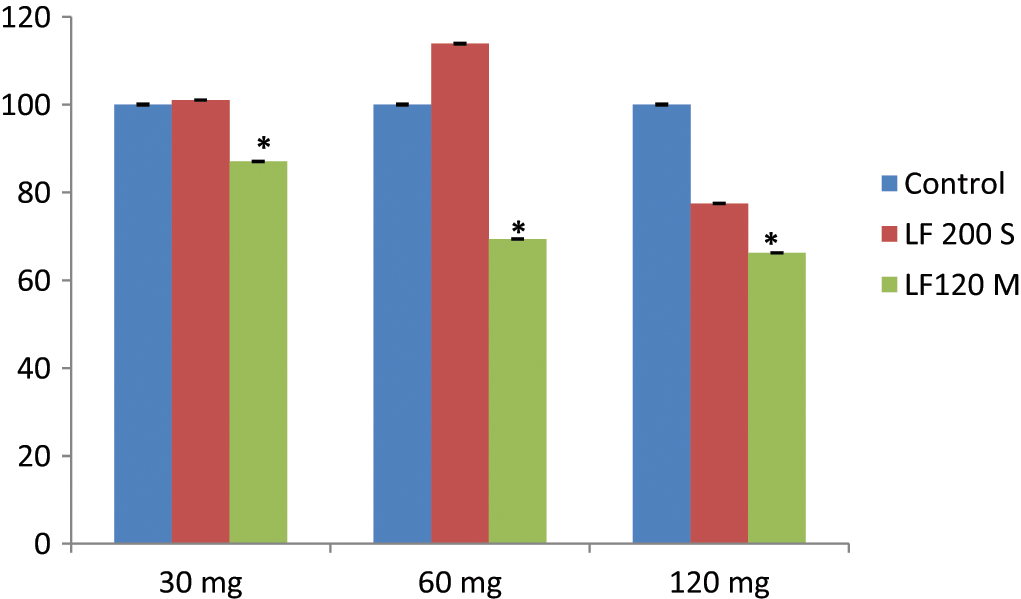
hUMSC percentage of viability in the presence of various doses of alginates (mean ± SD, n = 4).
Since the biocompatibility of alginates depends on various factors such as alginate concentration, molecular weight,28–31 solution viscosities,32,33 guluronic acid content29,34, and purity levels,35–37 our studies revealed that the viability of hUMSCs depends on the alginate composition as well as its optimum concentration and dose. To elucidate the effect on cell viability of the G content of the polymer, another series of experiments were conducted in which the concentration of the gelling agent ranged between 50 and 200 mM.
Cell viability in the presence of alginate beads cross-linked with different calcium concentrations
At the lower CaCl2 concentrations (50 and 100 mM), the hUMSC viability was comparable to that of the control in the presence of LF200S gel beads (Figure 3). However, cells were found to be 15% less viable at the higher CaCl2 concentration range of 150 to 200 mM. However, the viability at all CaCl2 concentrations was significantly less for the cells in the presence of LF120M. The minimum viability of ~75% was observed at the lowest concentration and ~15% less viability was observed in the range of 100–200 mM. Interestingly, cell viability was ~15% less for both alginate gels over a concentration range of 150–200 mM. The minimum viability of 74% for the cells in the presence of LF120M gel beads can be accounted for in terms of the weak physicomechanical properties of the gel due to the lower cross-link density. However, there is an effective CaCl2 concentration range (50–100 mM), where comparable hUMSC viability was observed with LF200S gel beads.
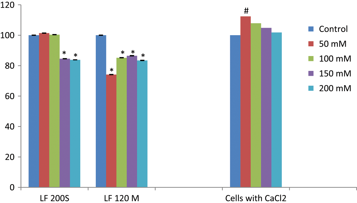
hUMSC % viability in the presence of alginate gel beads (LF200S and LF120M) cross-linked with different calcium chloride concentrations and calcium chloride (CaCl2) alone (mean ± SD, n = 4); bars from left to right: control, 50, 100, 150, and 200 mM.
When gel beads are in contact with the cells, two components of the gel can potentially be extracted into the medium over time; these are CaCl2 residues and the water soluble (non-cross-linked fraction or sol fraction) alginate polymer, which might affect the cell viability at high concentrations. To monitor the hUMSC viability in the presence of non-cross-linked alginate polymers, experiments were conducted in the absence of calcium and in pure CaCl2 solutions at different concentrations.
Cell viability in alginate solutions
Experiments were conducted at 30-mg gel dose using two grades of alginates. The cell viability was dramatically less in the presence of LF120M (33%) compared to LF200S (18%) (Table 1). Since the only obvious difference between the two grades is their G/M ratio building blocks, the viability is apparently related to the G or M contents of the alginate polymer.
hUMSC viability in the presence of non-cross-linked 2% alginate solutions.
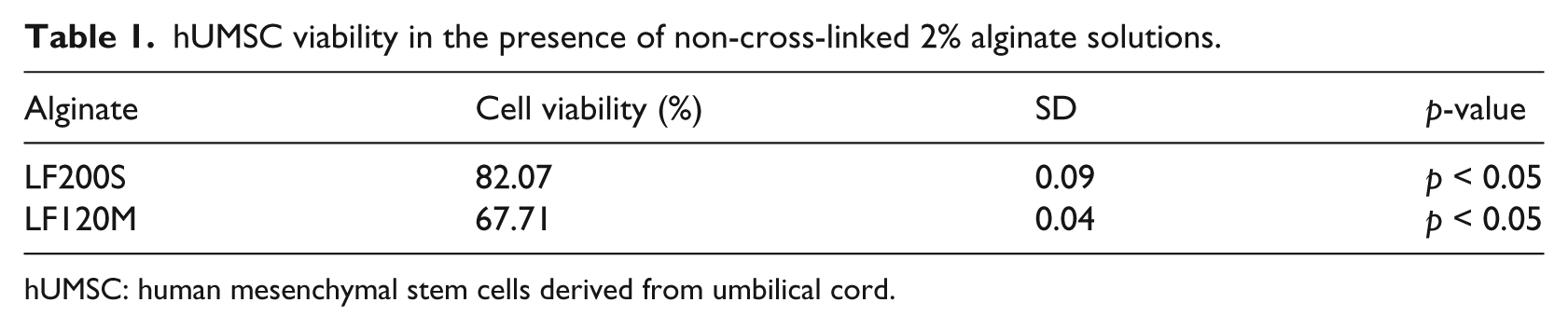
hUMSC: human mesenchymal stem cells derived from umbilical cord.
Cells viability in calcium chloride solutions
The hUMSC viability was comparable to that of control over the CaCl2 concentration ranging from 50 to 200 mM and significant at the minimum concentration of 50 mM (Figure 2). Physiologically, calcium ions seem to regulate a wide range of intracellular functions, including cell attachment, motility, and signal transductions 38 as well as that of a second messenger. The intracellular free calcium concentration is ~100 nM in the cytoplasm and ~100 µM in the endoplasmic reticulum, while extracellular calcium concentration is ~1.2 mM. 39 However, an excess of calcium provokes cell toxicity, leading to apoptosis followed by cell death.38–40 In this study, cells treated with calcium chloride over the range of 50 to 200 mM did not show any significant decrease in cell viability. The hUMSC viability was decreased at high calcium concentration of 150 to 200 mM (Table 2), but the lower cell viability was apparently not due to higher calcium residues based on the viability data at high calcium concentration.
G/M ratio, protein, and polyphenol contents of the two alginates.
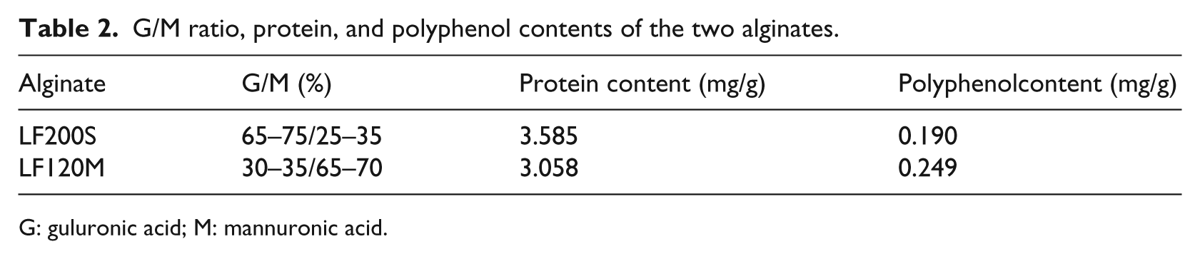
G: guluronic acid; M: mannuronic acid.
Alginate protein and polyphenol content
The major contaminants of sodium alginate, due to its natural origin, are proteins, polyphenols, and endotoxins. These contaminants cause inflammatory reactions in vivo. Since there was no purity data for the commercial alginates used in this study, the impurity level was measured to see if they may affect hUMSC viability. Although both alginates’ grades differed considerably in G and M content, the amounts of protein and polyphenol were similar (Table 2).
Conclusions
With systematic evaluation of alginate composition and gelation factors, it was found that hUMSC survival is superior in the presence of G-rich alginate, especially at low calcium concentrations. In addition, the sol content of the alginate gel seems to play a significant role on cell viability. High hUMSC survival can be achieved, however, if the alginate polymer contains a high proportion of guluronic acid, used at low concentration, and is cross-linked using the lowest feasible calcium concentration.
Footnotes
This research work was supported by Chancellor’s Faculty Research and Development Grant from Nova Southeastern University.