






Abstract
Biomimetic nanocomposite scaffolds were fabricated by electrospinning poly(
Keywords
Introduction
The treatment of bone loss or damage arising from trauma, tumor, and disease conditions is a challenging situation. Surgical interventions of bone grafts are the most common modality to treat most skeletal deficiencies. 1 Autogenic and allogenic bone grafts are currently being employed, but the problems include limited availability and associated morbidity due to second surgery. Hence, there is a need to substitute natural grafts with scaffolds, the best of which mimics the extracellular matrix (ECM) of the host tissue and supports adhesion and proliferation of the host cells. Electrospinning was selected in this study to fabricate highly porous nanofibrous scaffolds that resemble the ECM of the host tissue. Interconnected pores enhance the mineral deposition rate as well as the depth of the infiltration, which provide space for blood vessels to nourish growing bone. 2
Several polymeric materials have been electrospun for the fabrication of bone tissue–engineered scaffolds. However, due to lack of cellular recognition sites on the molecular architecture of synthetic polymers, these have been blended with natural polymers, such as collagen.
3
The main practical problems with collagen are its antigenicity, high cost, and difficult processing.4,5 Therefore, in this study, gelatin (Gel) was chosen as the natural polymer and mixed with poly(
Hydroxyapatite (HA) is the major inorganic mineral (60%) in natural bone, which is embedded into collagen matrix. Several polymer–ceramic composites have been studied, including blends of collagen/HA, 6 PLLA/Ca-deficient HA (dHA), 7 chitosan/HA, 8 poly(lactic-co-glycolide)(PLGA)/HA, 9 PLGA/multiwalled carbon nanotubes (MWNTs)/HA, 10 polycaprolactone (PCL)/collagen/HA, 11 and silk/HA. 12 Mineralized polymeric substrates have been fabricated by several methods, such as in situ precipitation,8,11 mixing nano-HA (n-HA) powder into electrospinning solution,6,9,13 biomimetic mineralization by simulated body fluid (SBF) method,14,15 and electrochemical deposition. 16
The blending of HA powders with polymers has several problems, including poor dispersion and agglomeration of HA, which leads to reduction in mechanical properties. 17 The presence of HA on the scaffold surface is minimal, which reduces osteoconductivity by the scaffolds. 6
PCL/HA/gelatin composite fibrous scaffolds prepared by blending HA powders in PCL/gelatin solution showed highly flexible tensile properties and allowed osteoblasts to penetrate into the scaffolds. 18 These results contradict the observations made by Fabbri et al. 19 and Kim et al. 17 who observed reduction in mechanical properties on mixing hydrophilic HA powders in polymer solution.
Mineralized electrospun PLLA scaffolds have been fabricated by the SBF method by Chen et al., 14 but it requires several days to mineralize the scaffolds. Recently, Andric et al. 20 reported the mineralization of PLLA/gelatin scaffold by SBF method and found higher mineralization in the presence of gelatin. However, all these methods are complex and time-consuming. Taguchi et al. 21 developed alternate soaking process to form bone-like apatite with low crystallinity and nanoscale size on polymeric surfaces. This process is simple and efficient than conventional SBF method to mineralize the scaffolds. The presence of n-HA in scaffolds not only mimics the natural bone environment but also significantly amends the mechanical properties, degradation profile, and osteoconductivity and creates a 3D microenvironment for differentiation of stem cells. n-HA particles enhance the interaction and proliferation of human osteoblast–like cells compared to normal HA particles. 22
The differentiation capability of mesenchymal stem cells (MSCs) into adipocytes, chondrocytes, osteoblasts, and myoblasts makes them promising candidates for cell-based regenerative therapy.23,24 Several polymer/composite nanofibrous composite scaffolds have been investigated for osteogenic differentiation of MSCs.25–29 However, the limited sources of MSCs, invasive isolation procedure, and the low cell number upon harvest have led many researchers to look for alternate sources of MSCs. 30 Adipose tissue is an abundant source of stem cells that differentiate toward the osteogenic, adipogenic, myogenic, and chondrogenic lineages in vitro under respective induction. Adipose tissue–derived stem cells (ADSCs) can be isolated by simple surgical procedure from human subcutaneous tissue that has a faster expansion rate. 31 McCullen et al. 32 demonstrated the differentiation of human ADSCs (hADSCs) on PLLA/tricalcium phosphate (TCP) scaffold and showed that electrospun PLLA/20% TCP accelerated the osteogenic differentiation of hADSCs. Haimi et al. 33 observed enhanced proliferation of ADSCs and their osteogenic differentiation on a PLLA/β-TCP composite scaffold compared to other scaffolds (PLLA and PLLA/bioactive glass 10% and 20%).
Keeping these advantages (of alternate soaking, gelatin, and n-HA) in mind, our objective was to prepare PLLA/Gel/n-HA composite scaffold via the alternate soaking process and to evaluate its mechanical properties before and after mineralization process. The biocompatibility of the scaffolds was confirmed with human blood cells and murine epididymal adipose tissue–derived stem cells (mE-ADSCs). Osteoblastic lineage differentiation ability of PLLA/Gel/n-HA scaffold makes it a potential applicant for bone tissue engineering.
Methods and materials
Materials
PLLA, average molecular weight 300,000 dalton, was obtained from Polysciences, Inc., Warrington, FL, USA. Gelatin (Type A) from porcine skin, 1,1,1,3,3,3-hexafluoro-2-propanol (HFP), trypsin-ethylenediaminetetraacetic acid (EDTA), penicillin–streptomycin, triton-X, dimethyl sulfoxide (DMSO), and 3-(4,5-dimethylthiazol-2-yl)-2,5-diphenyltetrazolium bromide (MTT) were purchased from Sigma–Aldrich (St Louis, MO, USA). Fetal bovine serum (FBS), Dulbecco’s phosphate-buffered saline (PBS), and Dulbecco’s modified Eagle’s medium (DMEM) were purchased from Gibco-BRL (Grand Island, NY, USA). Calcium chloride (CaCl2), disodium hydrogen phosphate (Na2HPO4), and glutaraldehyde were purchased from Merck, Water was distilled and deionized (deuterium-depleted water (DDW)) using Milli-Q system (Millipore, Bedford, MA, USA).
Preparation of nanofibrous scaffold
PLLA was dissolved in HFP to make 8% (w/v) solution for electrospinning. To fabricate PLLA/Gel scaffolds, the PLLA and gelatin were dissolved in HFP at 3:1 ratio (w/w) to make 8% final solution. The experimental procedure for fabrication of nanofibers by electrospinning process is as follows. Polymer solution was kept overnight for stirring at room temperature before electrospinning. Polymer solution was filled in a 5-mL plastic syringe with blunt-ended metallic needle (24 gauge; BD, Delhi, India) taking care to avoid air bubble entrapment. The syringe filled with the polymer solution was loaded into a syringe pump, and the syringe needle tip was connected to the positive output of a high-voltage power supply (Gamma High Voltage Ormond, Beach, Florida, USA), while the aluminum foil–covered collector was grounded. The needle tip to collector distance was 12.5 cm, the voltage was 14 kV, and the flow rate was 0.7 mL h−1. The entire process was conducted in a fume hood at a room temperature of 27°C and a relative humidity of 55%. The electrospun scaffold was kept under vacuum overnight before carrying out mineralization.
PLLA and PLLA/Gel scaffolds were mineralized by previously described alternate Ca-P soaking method. 34 Scaffolds were first immersed in a 0.5 M calcium chloride solution (pH 7.4) for 30 min at 37°C followed by washing with deionized water (DI). Then scaffolds were immersed in 0.3 M disodium hydrogen phosphate for 30 min at 37°C and finally washed with DI water. This consisted of a single cycle of Ca-P treatment. PLLA scaffolds underwent five cycles for mineralization, while PLLA/Gel scaffolds just took three cycles of alternate dipping. After the last cycle, scaffolds were left in DI water for 2 h at 37°C and then lyophilized for 24 h. After mineralization, PLLA and PLLA/Gel scaffolds were named as PLLA/n-HA and PLLA/Gel/n-HA, respectively.
Scaffold characterization using SEM and TEM
The morphology of electrospun polymeric and composite scaffold was examined by scanning electron microscope (SEM; Hitachi S-3400 N, Hitachi, UK) at an accelerating voltage of 15 kV. For SEM, the samples were cut into 5 mm × 5 mm squares, mounted on sample stubs, and coated with gold by sputter coating using SC7640 Sputter Coater (Quorum Technologies Ltd, Hailsham, UK). The average fiber diameter of the scaffolds was determined from the SEM micrographs using image analysis software (ImageJ, National Institutes of Health (NIH), Bethesda, MD, USA). Microstructure of the fibers was studied by transmission electron microscope (TEM; Tecnai 20; Philips FEI, Netherlands) at a voltage of 100 kV. Fibers of PLLA and PLLA/Gel were collected on TEM grids during the process of electrospinning, and mineralization was done in the same way as described above.
Surface topography using atomic force microscopy
Atomic force microscopy (AFM) studies were conducted in tapping mode on nonmineralized and mineralized scaffolds using Nanoscope IV equipped with 6626E scanner (Digital Instruments, Santa Barbara, CA, USA). A silicon nitride probe cantilever was used which had spring constant of 40 N m−1, length of 115–135 nm, and nominal tip radius of curvature of 8–10 nm. The scaffolds were cut in small pieces and stuck on metal disk (a puck, with an outer diameter of 1 cm) by double-sided cellophane tape.
X-ray diffraction and Fourier transform infrared spectroscopy
The mineral phase in the scaffolds was determined by X-ray diffraction (XRD) using a PANalytical X’Pert Pro X-ray diffractometer (Philips analytical B.V. Lelyweg 1, AA Almelo, The Netherland) at 40 kV and 30 mA, and Cu Kα monochromatic radiation was used. The XRD patterns were recorded between 20° and 60° (2θ) in step of 0.010 intervals with 1 s counting time at each step.
Fourier transform infrared (FT-IR) spectra were recorded for unmineralized and mineralized scaffolds using Nicolet Magna-IR FT-IR 550 (Ontario, Canada) spectrophotometer. The spectra were obtained with 30 scans per sample ranging from 4000 to 400 cm−1.
Wettability of the scaffold
The effect of mineralization on scaffold’s wettability was determined by water contact angle measurement using Digidrop GBX (GBX Instruments, Romans, France) at ambient temperature. Samples were placed on sample stage, single drop (20 µL) of distilled water was dropped at three different places, and the contact angle was measured immediately.
Mechanical testing of the scaffold
The tensile properties of rectangular specimens of 10 mm × 50 mm were measured using a tabletop tensile tester (Material Testing Machines, Tinius Olsen H1KT, Tinius Olsen, UK) with a load cell of 50 N.8,15,35–37 Test samples were vertically mounted on two mechanical gripping units of the tensile tester at its ends, leaving a 30-mm gauge length for mechanical loading. Load deformation data were recorded at crosshead speed of 5 mm min−1 and ultimate tensile strength, Young’s modulus, and percentage elongation at break were calculated from the stress–strain curve.
Quantification of n-HA by thermal gravimetric analysis
The amount of n-HA on PLLA/n-HA and PLLA/Gel/n-HA scaffold was quantified by thermal gravimetric analysis (TGA) using NETZSCH-STA 409 PC (Selb, Germany). Samples (n = 3) were loaded within the measurement chamber and heated up to 650°C with an ascending rate of 10°C min−1. PLLA and HA samples were also analyzed as control. The residual mass remaining in PLLA/n-HA and PLLA/Gel/n-HA samples was considered as amount of n-HA present in the scaffolds.
Biological evaluation of scaffold
Hemocompatibility of scaffold
Interaction of erythrocytes with scaffold is essential to study the release of hemoglobin (called hemolysis). The hemocompatibility of unmineralized and mineralized scaffolds was evaluated by hemolysis assay described elsewhere. 38 Briefly, 20 mL of whole blood was collected from a healthy individual, as per institutional ethical guidelines in place, into two 10-mL BD Vacutainer® Plus plastic plasma tubes containing 150 United States Pharmacopeial Convention (USP) units sodium heparin (spray coated) anticoagulant. The heparinized whole blood was immediately centrifuged for 15 min at 1200 r min−1 to pack erythrocytes. Scaffolds were equilibrated in normal saline for 30 min at 37°C, and all samples were incubated with 1 mL of blood for 1 h at 37°C in water bath. Thereafter, blood was centrifuged (1000 r min−1, 10 min), and the hemolytic percentage was determined by photometric analysis of supernatant at 545 nm. The hemolysis ratio (HR) was obtained by the equation
where AS, AP, and AN are absorbance of sample supernatant, positive control, and the negative control, respectively.
Platelet adhesion assay
Platelet adhesion study was done on scaffolds as described previously. 39 In brief, platelet-rich plasma (PRP) was obtained by centrifuging the fresh human blood (1500 r min−1, 20 min). Scaffolds of 1 cm × 1 cm size were kept in 12-well tissue culture plate, equilibrated with PBS for 30 min at 37°C, and incubated with 1 mL of PRP for 2 h at 37°C. After incubation, scaffolds were washed with PBS to remove unattached platelets and dehydrated with graded ethanol from 10% to 100%. Samples were dried overnight and sputter coated with gold and observed under SEM.
Culture of mE-ADSCs
mE-ADSCs were isolated at National Centre for Cell Science (NCCS), Pune, India.
30
mE-ADSCs were cultured in DMEM containing 2 mM
Cell proliferation
The cell proliferation on nanofibrous scaffolds was determined by MTT assay. The mE-ADSCs (1 × 10⁴) were seeded on PLLA, PLLA/n-HA, PLLA/Gel, and PLLA/Gel/n-HA scaffolds materials for different time periods (days 1, 7, and 11). The MTT solution was prepared at 1 mg/mL in PBS and was filtered through 0.2-µm filter. MTT (100 µL) and 400 µL of DMEM without phenol red were added into each well of 24 well plates. After the cells were incubated for 4 h at 37°C in a humidified CO2 incubator; the MTT solution was removed and replaced with 200 µL of DMSO. The plate was further incubated for 5 min at room temperature, and the absorbance was determined using a plate reader (Dynatech MR 700, Dynatech Laboratories Inc., Chantilly, Virginia, USA) at a test wavelength of 570 nm and a reference wavelength of 630 nm.
Cell attachment
Sterilized scaffolds (1 cm × 1 cm) were kept in a 12-well plate, and 1 × 10⁴ mE-ADSCs were seeded on unmineralized and mineralized scaffolds. On day 5, the medium was discarded, and the cell attachment on the scaffolds was analyzed by fluorescence microscopy and SEM. At the end of day 5, the scaffolds containing cells were harvested and washed with PBS; the cells were then fixed with 3.7% formaldehyde for 5 min at room temperature. The cells were permeabilized with 0.1% triton-X solution in PBS. To determine the morphology and integrity, the cells were stained with fluorescein isothiocyanate-phalloidin (FITC-ph) at 1:100 dilutions for 30 min in the dark at room temperature, to expose the F-actin filaments of the cells. 4′,6-Diamidoino-2-phenylindole (DAPI) was added to stain and visualize the nuclei. After staining, the samples were washed with PBS and mounted on glass-slides with VECTASHIELD mounting medium (Vector laboratories, Burlingame, CA, USA). Images were captured using a Zeiss-LSM 510 (Carl Zeiss, Jena, Germany) laser scanning confocal microscope using 63×/1.3 oil objectives.
For SEM, the scaffolds containing cells were washed with PBS. The cells were fixed in 2.5% glutaraldehyde for 2 h at room temperature, washed with PBS, and dehydrated with graded ethanol from 10% to 100%. The samples were dried overnight, sputter coated with gold, and observed under SEM at 15 kV.
Differentiation of mE-ADSCs
Expression of osteocalcin by cells is a late osteogenic differentiation marker. Gene expression of osteocalcin from mE-ADSCs seeded on nonmineralized and mineralized scaffolds was determined by reverse transcription polymerase chain reaction (RT-PCR). 40 The total RNA was extracted from each scaffold construct on day 14- of culture using tri-reagent (Invitrogen, Carlsbad, CA, USA). RNA pellets were dissolved in 20 µL of RNase- and DNase-free water, and RNA yields were estimated based on A260. The RNA was reverse transcribed into cDNA, and 150 ng of this cDNA was used for PCR reaction. PCR reaction conditions were 94°C for 30 s, 55°C for 30 s, and 72°C for 60 s. The extension was allowed to run for 5 min at 72°C. PCR products were separated by gel electrophoresis using 1% agarose gel and stained with ethidium bromide. Bands were visualized using UV illumination and recorded using UV Tech Gel documentation system (model UVIsave, Cambridge, UK). Image analysis software ImageJ (Version 1.6, NIH) was used for gel band quantitative analysis. Selected bands were quantified based on their relative integrated density. 41
Mineralization analysis
Mineralization of the mE-ADSCs on the scaffolds was assessed using alizarin red-S (ARS) dye, which binds to calcium salt selectively. 36 ARS staining was performed on day 14 of culture. The cell–scaffold constructs were washed with PBS thrice and fixed in 70% ice-cold ethanol for 1 h and stained with 40 mM alizarin for 30 min. Qualitative examination was done by imaging of stained cell–scaffold construct using Olympus BX51 (Olympus, Tokyo, Japan) microscope. Image analysis software ImageJ (Version 1.6, NIH) was used for quantitative analysis. Images were thresholded in the blue channel whose complimentary correspond to ARS stain. The common threshold of 69 was used for all images.
Statistical analysis
Quantitative data were collected in triplicate (n = 3) and reported as mean ± standard deviation where indicated. Statistical significance of the differences was evaluated by a one-way analysis of variance (ANOVA, Tukey’s method), using Origin pro 8 software. The p values of less than 0.05 were considered as significant.
Results and discussion
The SEM morphology analysis of the electrospun nanofibers of PLLA and PLLA/Gel revealed uniform, smooth nanofibers without beads (Figure 1). Both scaffolds exhibited random, nonwoven architecture with interconnected pores, which mimic the ECM of bone. The average fiber diameters of PLLA and PLLA/Gel were observed at 615 nm and 506 nm, respectively. Both kinds of scaffolds were successfully mineralized with n-HA by the alternate soaking method.
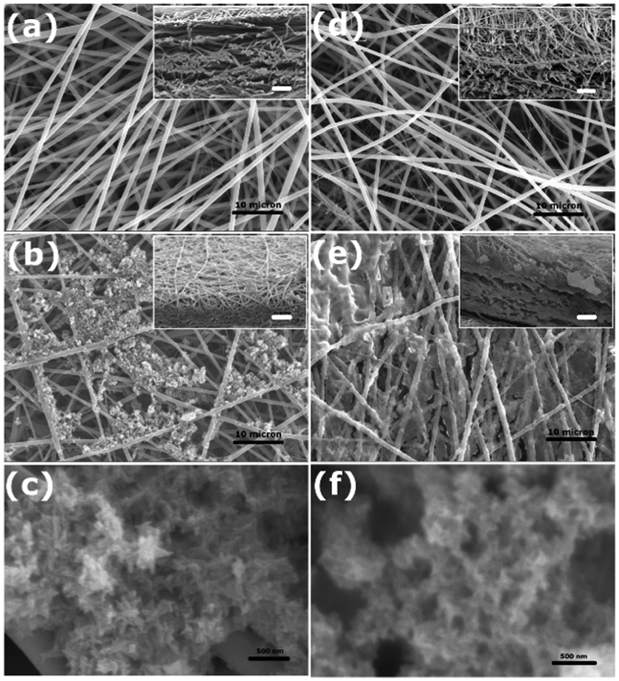
Morphology of electrospun (a) PLLA, (b) PLLA/n-HA, (d) PLLA/Gel nanofibers, and (e) PLLA/Gel/n-HA nanofibers (scale bar = 10 µm). (c) and (f) showing n-HA particle on PLLA/n-HA and PLLA/Gel/n-HA nanofibers, respectively (scale bar = 500 nm). Inset showing cross-sectional SEM micrographs of respective nanofibers (scale bar = 10 µm).
The n-HA was deposited on scaffold surface using multiple cycles of the alternate soaking method, until the surface of the scaffolds showed complete mineralization by SEM. This took five cycles for the PLLA scaffold due to hydrophobic and inert nature of the PLLA and three cycles for the PLLA/Gel scaffold due to the presence of anionic carboxyl (COO−) groups of gelatin in PLLA/Gel, which accelerated the chelation of Ca+2 ions and nucleated more PO4−3 ions.42–45 Unlike the PLLA scaffolds, the deposition of n-HA on the PLLA/Gel scaffolds was uniform (Figure 1(b) and (e)). Deposition of n-HA on PLLA and PLLA/Gel scaffold was observed throughout the scaffolds, that is, the surfaces as well as between layers (Figure 1, inset).
The TEM micrographs are shown in Figure 2(a) and (b) for the PLLA and PLLA/Gel fibers that had smoother edges compared to the rougher mineralized fibers (Figure 2(c) and (d)). The TEM images of the n-HA particles are shown in Figure 2(e), and the size range of HA nanocrystals in the native bone was 50–100 nm. 46 Electron diffraction pattern of the n-HA showed the polycrystalline nature of the n-HA particles.
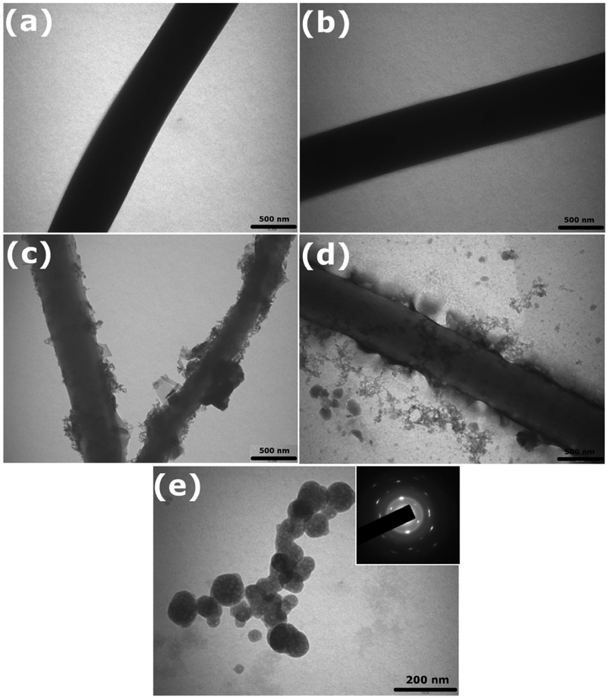
TEM images of electrospun nanofiber of (a) PLLA, (b) PLLA/Gel, (c) PLLA/n-HA nanofiber, (d) PLLA/Gel/n-HA (scale bar = 250 nm), and (e) HA nanoparticles (scale bar = 200 nm). Inset showing electron diffraction pattern of n-HA particle.
The topography of scaffold surface plays an important role in cell attachment and proliferation. 47 AFM images exhibited changes in topography of scaffold after incorporation of gelatin in PLLA scaffold (Figure 3 is 3D); small granular patterns are depicted on the surface of PLLA/Gel scaffold (Figure 3(b)), whereas the PLLA scaffold appeared flat (Figure 3(a)). Also clearly indicated in the AFM images (Figure 3(c) and (d)) are the changes in the surface topography after mineralization of n-HA on PLLA and PLLA/Gel scaffold. Spiky ridges were observed on the PLLA/n-HA scaffolds, whereas the PLLA/Gel/n-HA surface showed larger granular structures. This was attributed to the efficient nucleation of n-HA in the presence of gelatin in the scaffold.
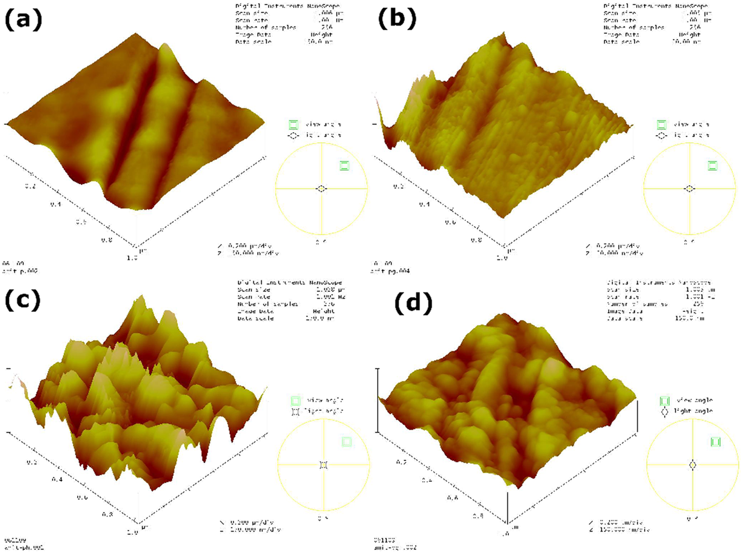
A 3D surface topography of nanofibrous scaffold using atomic force microscopy: (a) PLLA, (b) PLLA/Gel, (c) PLLA/n-HA, and (d) PLLA/Gel/n-HA.
The n-HA deposit on the scaffold was confirmed by XRD and FT-IR spectra. The XRD peaks at 26.1 [022] and 31.7 [211] observed in PLLA/n-HA and PLLA/Gel/n-HA are characteristic peaks of HA 3 and were absent in PLLA and PLLA/Gel scaffold (Figure 4(a)). The peaks at 26.1 [022] and 31.7 [211] are broader for the PLLA/Gel/n-HA due to fine grain size of the HA crystals. 31 The XRD data were analyzed using the X’pert Highscore software to obtain the crystallite size via inbuilt Scherrer calculator using (211) reflection plane. The crystallite size of n-HA was 33.6 and 27.5 nm in PLLA/n-HA and in PLLA/Gel/n-HA, respectively. The difference in n-HA particle size was determined by TEM, and crystallite size was determined by XRD. All the diffraction patterns were compared to International Centre for Diffraction Data (ICDD) database (ICDD Powder Diffraction File No. 00-009-0432).
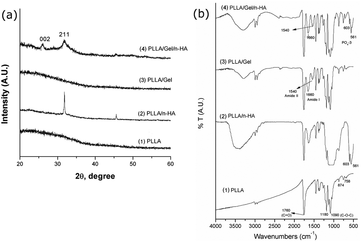
(a) XRD pattern of nanofibrous scaffold showing HA peaks and (b) FT-IR spectra of (1) PLLA nanofibers, (2) PLLA/n-HA nanofibers, (3) PLLA/Gel nanofibers, and (4) PLLA/Gel/n-HA nanofibers.
The FT-IR spectra of the scaffolds are shown in Figure 4(b); the carbonyl stretch was at 1760 cm−1 due to the PLLA (C=O) in all the scaffolds. Other major PLLA peaks were at 1090 cm−1 (C–O–C) and at 1180 cm−1 (A-type C–O–C stretch). 46 Peaks at 603 and 561 cm−1 corresponding to PO4−3 from n-HA 48 were in both PLLA/n-HA and PLLA/Gel/n-HA spectra but absent in the unmineralized PLLA and PLLA/Gel scaffold. Peaks at 1660 and 1540 cm−1 in PLLA/Gel and PLLA/Gel/n-HA attributed to the amide I and amide II peaks of gelatin, 49 and these peaks were absent in the PLLA and PLLA/n-HA scaffold.
The hydrophilicity of the surface of scaffolds, measured by water contact angle (Figure 5(a)) demonstrated the hydrophobic nature of PLLA surface (124.4°). Presence of gelatin in the PLLA/Gel scaffold failed to make the surface of PLLA hydrophilic, resulting in a similar contact angle (126.5°) as that of the PLLA scaffold. This does not rule out the presence of gelatin on the surface because contact angle measurements are of macroscopic nature as compared to the size of electrospun fibers. Notably, deposition of n-HA on top of the surface of both PLLA and PLLA/Gel made them completely hydrophilic (contact angle 0°). This result is in accord with Lee et al., 25 where the addition of HA made the poly(lactide-co-glycolide) (PLGA) surface hydrophilic. However, nucleation of mineral occurred at submicroscopic chemical regions of the surface; hence, the presence of gelatin enhanced the mineralization without affecting wettability. The wettability, as measured by sessile drop technique, is governed largely by the surface topography as in case of lotus leaf. 50
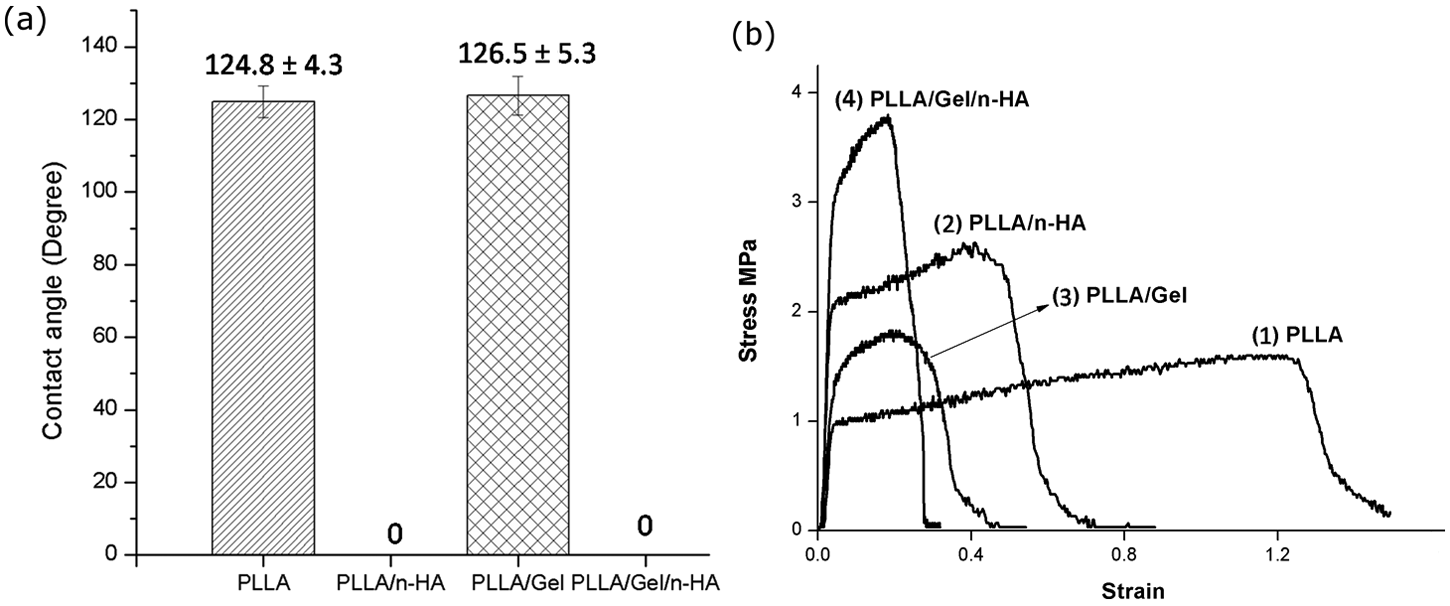
(a) Water contact angles of PLLA, PLLA/n-HA, PLLA/Gel, and PLLA/Gel/n-HA nanofibers and (b) stress–strain curve of (1) PLLA, (2) PLLA/n-HA, (3) PLLA/Gel, and (4) PLLA/Gel/n-HA scaffold.
The mechanical properties of electrospun scaffold were measured as ultimate tensile stress, Young’s modulus, and percentage elongation at break. Typical stress–strain curves for PLLA, PLLA/n-HA, PLLA/Gel, and PLLA/Gel/n-HA scaffolds were obtained (Figure 5(b)). The Young’s modulus and ultimate tensile stress of PLLA were 53 and 1.59 MPa, respectively, which increased to 100.0 and 2.72 MPa, respectively, after mineralization (Table 1). Likewise, the Young’s modulus and ultimate tensile stress of PLLA/Gel was 44.6 and 1.68 MPa, respectively, which also increased to 157.2 and 3.88 MPa, respectively, after mineralization. This indicates that n-HA provides strength to the resultant nanofiber. However, the percentage elongation at break of both PLLA and PLLA/Gel (140% and 46.19%) decreased (85.4% and 22.9%) after mineralization of the scaffolds. Increase in the Young’s modulus and ultimate tensile stress after incorporation of n-HA is in accordance to the results of Jeong et al., 51 where the strength of the electrospun PLLA nanofibers increased after addition of HA. In another study, the strength of poly(ϵ-caprolactone) was increased nearly fourfold when mixed with 20% HA. 52
Young’s modulus, tensile stress, and percentage elongation of nanofibrous scaffolds
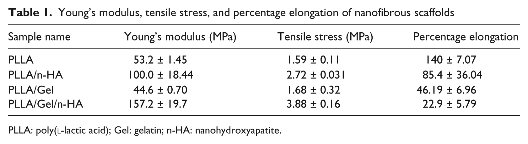
PLLA: poly(
The TGA was done to determine the amount of n-HA in PLLA/n-HA and PLLA/Gel/n-HA scaffold with PLLA and HA as controls. It was observed that PLLA and gelatin both degraded at around 360°C, but HA remained stable till 650°C. Thus, the residual mass remaining in PLLA/n-HA and PLLA/Gel/n-HA was taken as amount of n-HA present in the scaffolds. 53 The amount of n-HA in mineralized PLLA and PLLA/Gel was found to be 50.5% and 66.8%, respectively. The amount of n-HA on the PLLA/Gel was comparable to the PLLA scaffold. This showed the beneficial effects of gelatin in the formulation by efficiently nucleating n-HA. The amount of HA in natural human bone was approximately 60%, 46 so with the alternate soaking method and addition of gelatin to the formulation, it was possible to achieve the n-HA content similar to natural human bone, in contrast to Niagm et al. 54 who reported merely 37.8% and 30.2% HA content after 5 and 3 cycles of alternate soaking, respectively, in case of PLLA and PLLA/Col nanofibers.
Biological evaluation of scaffold
Platelet adhesion assay
Adhesion, aggregation, and activation of platelets are crucial steps in the process of thrombus formation. 55 SEM micrographs were employed to assess platelet adhesion study on surface of all types of scaffolds, which are shown in Figure 6. The platelets that adhered on the PLLA and PLLA/Gel scaffolds had round morphology with a few filopodia. On PLLA/Gel/n-HA and PLLA/n-HA scaffold, platelets were aggregated with flat morphology and elongated filopodia. The mineralized scaffolds promoted the activation of platelets more effectively than unmineralized scaffold, thereby, implying that mineralized scaffolds lead to faster bone regeneration.
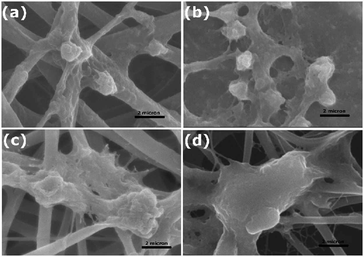
SEM micrographs of platelet adhesion on (a) PLLA, (b) PLLA/Gel, (c) PLLA/n-HA, and (d) PLLA/Gel/n-HA scaffold (scale bar = 2 µm).
Hemocompatibility of scaffold
The percentage hemolysis of nonmineralized and mineralized scaffolds is shown in Figure 7(a). The PLLA and PLLA/n-HA scaffolds had 2.4% and 2.7% hemolysis, respectively, while the PLLA/Gel and PLLA/Gel/n-HA had 2.8% and 2.7%, respectively. The percentage hemolysis of all scaffolds was under permissible limit of 5%. 38 These results confirm that mineralization of n-HA does not compromise the hemocompatibility of scaffold.
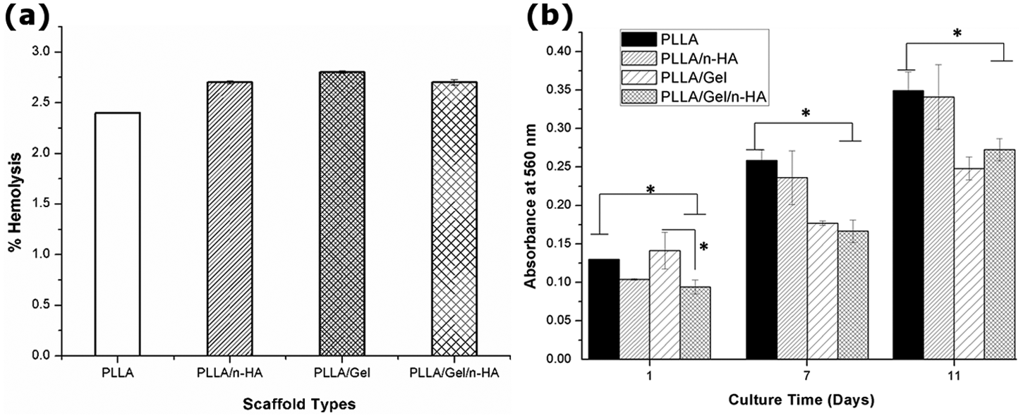
(a) Bar graph showing percentage hemolysis for PLLA, PLLA/n-HA, PLLA/Gel, and PLLA/Gel/n-HA nanofibers and (b) proliferation of mE-ADSCs on PLLA, PLLA/n-HA, PLLA/Gel, and PLLA/Gel-nHA after 1, 7, and 11 days of culture.
Cell proliferation
The proliferation of mE-ADSCs was assayed by MTT on days 1, 7, and 11 after cell seeding on different scaffolds (Figure 7(b)). Cell proliferation occurred on all of the scaffolds that increased with culture time. Increase in optical density with time clearly indicated that the mineralized scaffolds did not have any cytotoxic effects on cells. On day 1, there was less absorbance (less adhesion) on the PLLA/n-HA and PLLA/Gel/n-HA scaffolds compared to the PLLA and PLLA/Gel. This may be due to the pore structure and increased wettability of the scaffolds after mineralization. This could have led to more rapid spreading of the cells on to the surrounding culture plates and, consequently, decreased cell seeding efficiency. Cell proliferation on PLLA/Gel/n-HA was significantly lower than on the PLLA scaffold on days 7 and 11. The lower absorbance on PLLA/Gel/n-HA scaffold may be due to the number of cells triggered to differentiate. Similarly, low cell proliferation on composite scaffold was reported for the proliferation of human mandible–derived hMSCs on poly(l-lactide) (PLA) and PLA/demineralized bone powder (DBP) nanofibers as well as osteoblast cell proliferation on PCL/HA and PCL scaffolds, respectively.23,56
Ngiam et al. 57 also found significantly lower cell proliferation on both mineralized PLGA and PLGA/collagen (PLGA/Col) compared to unmineralized scaffold at all culture time points. In our studies, the PLLA/Gel/n-HA scaffold has a different dynamic in the growth of cells initially, but within 7 days, scaffold had good cell proliferation.
SEM of cell attachment
Adhesion of mE-ADSCs to scaffolds was analyzed by SEM on the fifth day of culture (Figure 8). The SEM micrographs of mE-ADSCs cultured on PLLA, PLLA/Gel, PLLA/n-HA, and PLLA/Gel/n-HA exhibited good cell adherence on all of the scaffolds. The presence of n-HA on the scaffolds favored cell aggregation, indicating the interaction among cells promoted cellular activity and cell differentiation. While in nonmineralized scaffolds, cells were observed as single cell with flattened morphology. Ramires et al. 58 also reported cell aggregation on HA-deposited titanium surfaces.
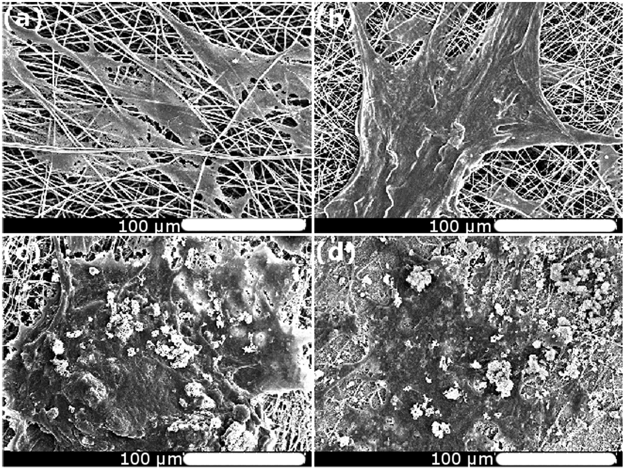
SEM micrograph of mE-ADSCs attached on (a) PLLA, (b) PLLA/Gel, (c) PLLA/n-HA, and (d) PLLA/Gel/n-HA scaffold after 5 days of culture.
Confocal microscopy of cell attachment
On the fifth day of culture, cell attachment of mE-ADSCs and development of actin filaments were observed by fluorescent microscopy (Figure 9). The mE-ADSCs on PLLA and PLLA/Gel scaffolds were single cells with well-stretched filopodia (Figure 9(a) and (b)); however, on the PLLA/n-HA and PLLA/Gel/n-HA scaffolds (Figure 9(c) and (d)), they formed cell aggregates. These results are in accord with the SEM results.
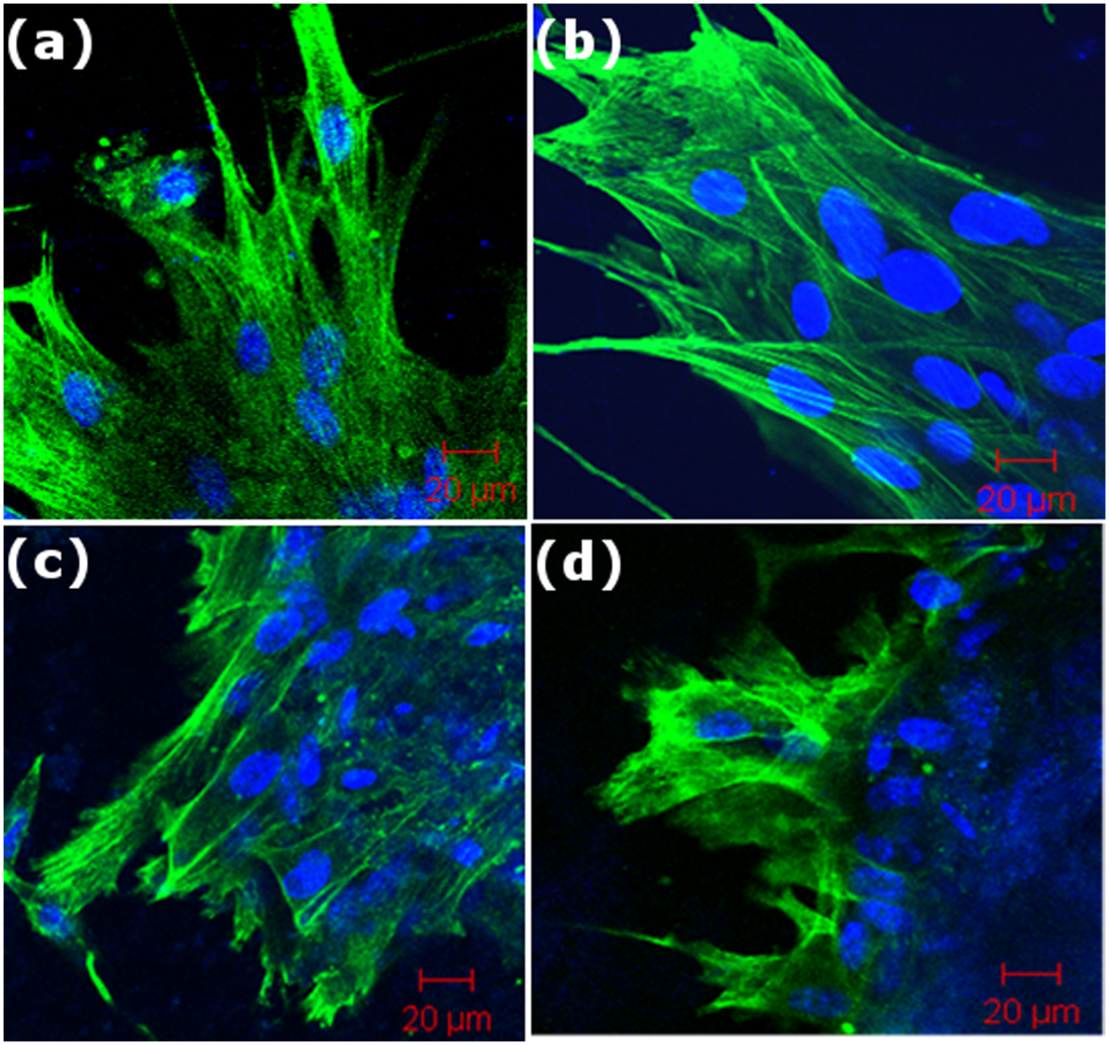
CLSM image of mE-ADSCs attached on (a) PLLA, (b) PLLA/Gel, (c) mineralized PLLA, and (d) mineralized PLLA/Gel scaffold after 5 days of culture. Actin filament (green) and nucleus (blue) were stained with FITC and DAPI are shown, respectively (scale bar = 20 µm).
Differentiation of mE-ADSCs
The expression of osteocalcin mRNA by mE-ADSCs cultured on the PLLA, PLLA/n-HA, PLLA/Gel, and PLLA/Gel/n-HA on day 14 is shown in Figure 10. The expression levels of mRNA in the cultured cells were semiquantified by band intensities. Osteocalcin levels were 8.89% less in the PLLA scaffolds than the PLLA/n-HA scaffolds. Similarly, the intensity of the osteocalcin band was 56.5% less for the PLLA/Gel scaffold than for the PLLA/Gel/n-HA scaffold. The band intensities of expression of housekeeping gene glyceraldehyde 3-phosphate dehydrogenase (GAPDH) were almost similar for all of the scaffolds. The osteocalcin expression was maximum in the case of PLLA/Gel/n-HA. It appears that gelatin and n-HA in the scaffold synergistically stimulated osteocalcin expression. This is in good agreement with a previous report, where differentiation of hMSCs was upregulated after 7 days of culture on PLGA/HA nanofibrous scaffold. 22 Hence, mineralized scaffolds, specially PLLA/Gel/n-HA, support intrinsic properties of mE-ADSCs differentiation. This preliminary analysis shows the significant potential of this scaffold and points the way for further studies of bone sialoprotein (BSP), osteopontin, and osteonectine in order to understand mE-ADSCs differentiation toward osteodifferentiation.
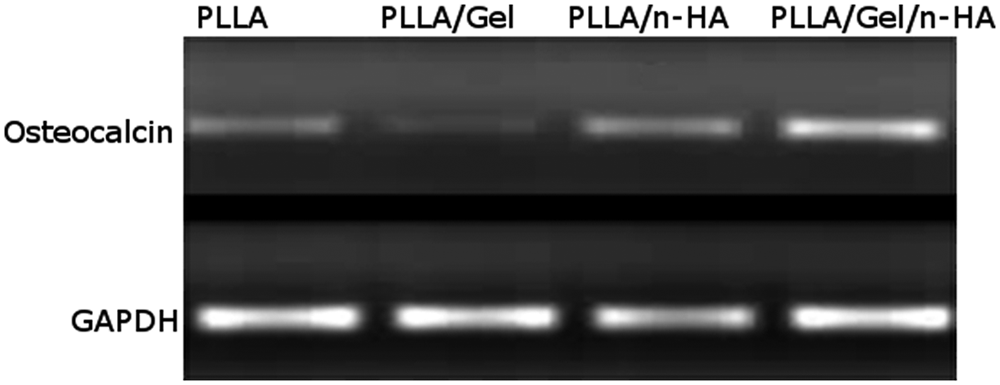
Expression of osteocalcin mRNA by mE-ADSCs cultured on electrospun PLLA, PLLA/Gel, PLLA/n-HA, and PLLA/Gel/n-HA scaffold on day 14.
Mineralization analysis
Mineralization of mE-ADSCs cultured on different scaffolds was assessed by ARS staining. Optical microscopy images of cell–scaffold construct with ARS staining on day 14 is shown in Figure 11. The bright red spots (ARS–calcium chelate) present on PLLA, PLLA/n-HA, and PLLA/Gel/n-HA scaffold correspond to the presence of calcium. PLLA/Gel scaffold showed comparatively lower mineralization than PLLA scaffold. A much higher intensity of the mineral stains was observed for the mineralized nanofibrous scaffold compared to polymeric nanofibrous scaffold. The PLLA/n-HA scaffold had 32.7% more mineralization than the PLLA scaffold. Similarly, PLLA/Gel/n-HA had 99.9% more mineralization compared to PLLA/Gel (Figure 11). Thus, based on our results, gelatin alone with PLLA did not show any positive effect in mineralization process, while an increase in the mineralization was observed with n-HA with PLLA. However, the combination of gelatin and n-HA with PLLA markedly increased the differentiation of mE-ADSCs toward the osteoblastic lineage. This indicated that gelatin, along with n-HA, in PLLA-Gel/n-HA scaffold synergistically stimulated differentiation of the mE-ADSCs.

Light microscopy images of alizarin red staining of mineralization of mE-ADSCs on day 14 on (a) PLLA, (b) PLLA/Gel, (c) PLLA/n-HA, and (d) PLLA/Gel/n-HA scaffold (original magnification 10×).
Conclusion
PLLA and PLLA/Gel nanofibrous scaffolds were fabricated via electrospinning and their surface was composited with n-HA by alternate soaking in Ca-P solutions. Gelatin blended with PLLA offered several advantages, first in terms of fewer soaking cycles to achieve sufficient mineralization by alternate soakings, which proved to be a rapid and efficient method for the mineralization of the scaffolds. SEM micrographs revealed smooth, uniform fibers of PLLA, while the PLLA/Gel and the mineralized nanofibers had better mechanical strength than the unmineralized nanofibers. The deposition of n-HA made the surface of both PLLA and PLLA/Gel completely hydrophilic with contact angles of 0°. The expression of osteocalcin, a bone marker gene, was significantly enhanced for PLLA/Gel/n-HA scaffolds, which supported the significance of n-HA and gelatin in the differentiation of mE-ADSCs. Consequently, the PLLA/Gel/n-HA scaffold is a very promising entity for stem cell regenerative therapy of bone defects.
Footnotes
Acknowledgements
We thank Centre for Research in Nanotechnology and Science (CRNTS), Department of Metallurgical Engineering and Materials Science (MEMS), and Department of Physics of Indian Institute of Technology Bombay, Mumbai, for characterization facility. We thank DST for support through FIST, Nanomission, IRPHA, SERC, and Indo-Spain schemes and for access to EM. We thank Ashwini Atre of NCCS, Pune, for assistance with confocal microscopy. We thank Dr Sachin Kadam and Mr Ganpat Dahe for all the suggestions and Hemlata Chhabra for critically reading the manuscript.
Funding
Amit K. Jaiswal is supported by a fellowship from the Council of Scientific and Industrial Research (CSIR), Government of India.