
Abstract
A new class of pH-responsive multivalent host–guest interactions to manipulate polypeptide-based nano-vehicles was developed. Poly(
Keywords
Introduction
In recent decades, molecular design studies aimed at developing artificial autonomous nano-vehicles have attracted significant attention from biomedical researchers, following findings on a new class of nano-vehicles that have provided insights into breakthrough tumor therapies.1–14 For example, the molecular design of nano-sized molecular motors, which perform mechanical work on a nano-sized scale when given a specific biological signal, is a challenging task. The successful design of such motors will provide multifunctional nano-vehicles with great potential as diagnostic and therapeutic systems.1–14 Currently, the construction of these nano-vehicles typically involves adjusting the intermolecular force (such as electrostatic and hydrophobic interactions) using ionic or amphiphilic polymers.1,15 However, these technological attempts have not always resulted in precisely constructed nano-vehicles with a smart molecular motor, primarily due to the limited availability of nano-vehicle accessories (polymer parts). 16 Additionally, the lack of functionality of the nano-vehicles accessories used to produce drug-carrying nanosystems generally results in a relatively low effectiveness. 16
A host–guest chemical approach provides a new tool for fabricating nano-vehicles. 17 For example, cyclodextrin has gained considerable attention in molecular design, because it has a special pocket with a hydrophobic interior and hydrophilic exterior. Thus, an external molecule 17 can fit into this pocket in a manner similar to nut and bolt fastenings. In this respect, hybridizing a cyclodextrin component into a stimuli-responsive nano-vehicle will offer a unique opportunity for developing a multifunctional nano-vehicle for drug delivery.
In this study, self-zipping/self-unzipping nano-vehicles using polypeptides grafted with β-cyclodextrin (CD) and 2,3-dimethylmaleic acid (DMA) (Figure 1(a)) were developed. This system includes a built-in molecular motor, whereby the molecular configuration changes with pH between the inclusion (i.e. intra-chain zipping for formation of nano-vehicles) state of the polypeptide chains with CD or DMA to the bond-scissoring (i.e. intra-chain unzipping for disintegration of nano-vehicles) state of DMA and the polypeptide (Figure 1(b)). The self-zipping/self-unzipping behavior of these nano-vehicles was evaluated using physicochemical studies and in vitro/in vivo experiments.
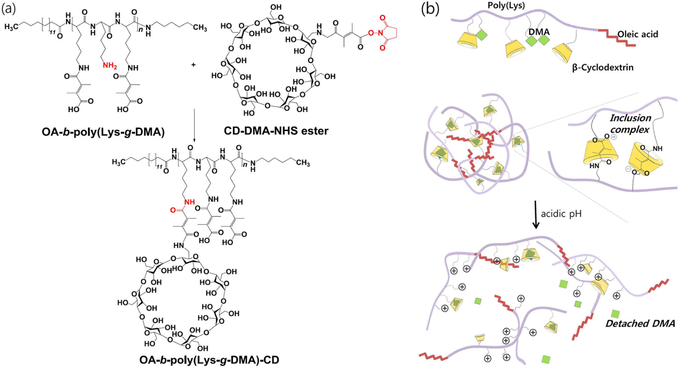
(a) Synthesis scheme of OA-b-poly(Lys-g-DMA)-CD and (b) schematic concept for a proposed a self-zipping/unzipping nano-vehicles. See the text for more details.
Materials and methods
Materials
Nε-benzyloxycarbonyl-
Synthesis of OA-b-poly(Lys-g-DMA)-CD
Poly(Nε-benzyloxycarbonyl-
Acid–base titration
Polymers or NaCl (as a control) dissolved in deionized water (30 mM) were adjusted to a pH of 10 using 1 M NaOH or HCl. The solutions were titrated via the stepwise addition of 1 M HCl solution to obtain the pH titration profile. 19
Nano-vehicles preparation
Polymers (10 mg) dissolved in DMSO (2 mL) were transferred to a pre-swollen dialysis membrane tube (Spectra/Por MWCO 15K) and dialyzed against a phosphate buffered saline (PBS; 150 mM, pH 7.4) solution for 24 h. The outer phase was replaced three times with fresh PBS solution. The solution was subsequently lyophilized by freeze-drying for 2 days. 10
Zeta-potential analysis
The zeta-potential change over time of each sample (1 mg/mL, PBS 150 mM) at different pH values (pH 7.4–5.0) was measured with a Zetasizer 3000 (Malvern Instruments, USA). Before the test, each sample was stabilized at room temperature for 4 h. 13
Particle size distribution
Particle size analysis was conducted using a Zetasizer 3000 (Malvern Instruments) equipped with a He-Ne laser beam at a wavelength of 633 nm and a fixed scattering angle of 90°. 19 The samples (0.1 mg/mL) were exposed to different pH conditions (PBS 150 mM, pH 7.4–5.0) for 4 h before measuring the particle size and the particle size distribution. To evaluate the morphology of each nano-vehicle, the samples were prepared by casting a dilute nano-vehicle solution (0.05 mg/mL, pH 7.4 or 6.8) on a slide glass, which was then dried. The nano-vehicles were imaged via field-emission scanning electron microscopy (FE-SEM; S-4800, Hitachi, Japan) to examine their morphologies. 19
Circular dichroism analysis
The circular dichroism profile of each sample (0.1 mg/mL, PBS 150 mM) at different pH values (pH 7.4–6.0) was measured with a J-815 CD spectrometer (Jasco International, UK).
DOX loading
Before DOX loading into the nano-vehicles, the HCl salt of DOX·HCl was detached by reacting DOX·HCl with a 2-mole ratio of TEA in DMSO overnight. 11 Subsequently, the polymer (10 mg) and DOX (2 mg) were dissolved in DMSO (1 mL) and dialyzed against PBS (150 mM, pH 7.4) for 24 h. The outer phase of the dialysis bag containing DOX and the polymer was replaced three times to remove non-encapsulated free DOX. The DOX concentration in the nano-vehicles was determined by measuring the ultraviolet (UV) absorbance of the DOX-loaded nano-vehicles dissolved in a DMSO/PBS mixture solution (95:5 vol%) at 481 nm using a UV/visible spectrophotometer. The DOX loading efficiency was 82–87 wt%, as calculated by dividing the loading DOX content by the feeding DOX content.11,19
DOX release test
A DOX-loaded nano-vehicle solution (1 mL) was added to a dialysis membrane bag (Spectra/Por MWCO 15K). The dialysis membrane bag was sealed and then immersed in fresh PBS (20 mL, 150 mM) at different pH values (pH 7.4–5.0). The release test of DOX from the nano-vehicles was performed using mechanical shaking (100 rpm) at 37°C. The outer phase of the dialysis membrane bag was extracted and replaced with fresh buffer solution at predetermined time intervals to maintain a sink for the DOX. The DOX concentration was measured at 481 nm using a UV/visible spectrophotometer.11,19
In vitro cellular uptake test
Human nasopharyngeal epidermal carcinoma KB cells (from Korean Cell Line Bank, a model tumor cell) were maintained in RPMI-1640 medium with 1% penicillin–streptomycin and 10% FBS in a humidified standard incubator at 37°C with a 5% CO2 atmosphere. Prior to testing, cells (1 × 10⁵ cells/mL) that were grown as a monolayer were harvested via trypsinization using a 0.25% (wt/vol) trypsin/0.03% (wt/vol) ethylenediaminetetraacetic acid (EDTA) solution. KB cells suspended in RPMI-1640 medium were seeded onto each well plate and cultured for 24 h prior to the in vitro cell testing. 5 The tumor cellular uptake of each sample (equivalent DOX 1–10 µg/mL, 4 h treatment) was monitored at pH 7.4, 6.8 or 6.0 using an Axio Imager D2 fluorescence microscope (Carl Zeiss, USA) and a FACSCalibur™ Flow Cytometer (Becton Dickinson, USA).10–14
In vitro cell cytotoxicity test
KB tumor cells incubated with DOX-loaded nano-vehicles (equivalent DOX 0.1–10 µg/mL) or free DOX (0.1–10 µg/mL) at pH 7.4 or 6.8 for 12 h were washed three times with fresh cell culture medium (without the nano-vehicles or free DOX) and then further incubated for 24 h. The cell viability was measured using a CCK-8.10–14
Animal care
In vivo studies were conducted in 7- to 8-week-old female nude mice (BALB/c nu/nu mice; Institute of Medical Science, Tokyo, Japan). The nude mice were maintained under the guidelines of an approved protocol from the Institutional Animal Care and Use Committee (IACUC) of the Catholic University of Korea (Republic of Korea).
In vivo photoluminescence imaging and tumor inhibition
Fluorescent dye (Cy5.5 NHS ester) (1 mg) was added drop-wise to OA-b-poly(Lys-g-DMA)-CD (10 mg) in pH 7.4 PBS (1 mL) at room temperature and was then stirred for 24 h, allowing the chemical reaction between the NHS ester of Cy5.5 and the residual amine groups of polymer. The solution was dialyzed with a dialysis membrane bag (Spectra/Por MWCO 5K), followed by lyophilization.
For the in vivo animal experiments, KB tumor cells were introduced into female nude mice via a subcutaneous injection of 1 × 10⁶ cells suspended in PBS 7.4 (150 mM) medium. When the tumor volume reached 150 mm3, Cy5.5-labeled OA-b-poly(Lys-g-DMA)-CD nano-vehicles (50 mg/kg body) were injected (only once) intravenously into tumor-bearing nude mice through a tail vein or injected intratumorally to the tumor site of nude mice directly. A 12-bit charge-coupled device (CCD) camera (Image Station 4000 MM; Kodak, USA) equipped with a special C mount lens and a long-wave emission filter (600–700 nm; Omega Optical, USA) were used to measure live fluorescence images of the nude mice. At 24 h post injection, the nude mice were sacrificed, and excised organs (tumor, liver, spleen, lung, kidney, and heart) were also analyzed.10,13,14
For the in vivo tumor inhibition test, DOX-loaded nano-vehicles (equivalent DOX 10 mg/kg body) or free DOX (10 mg/kg body) were injected intravenously through a tail vein of the nude mice (n = 5 per each sample). The tumor volume was monitored over time and calculated using the formula: tumor volume = length × (width)2/2.10,13,14 After 7 days, the nude mice were euthanized.
Statistical analysis
All results were analyzed via Student’s t-test or analysis of variance (ANOVA) at a significance level of p < 0.01 (indicating **). MINITAB® release 14 statistical software was used.10,13
Results and discussion
To establish self-zipping/self-unzipping nanoparticular system, we first designed a functional polypeptide using poly(
As shown in Figure 1(b), the host–guest interaction between CD and DMA and the hydrophobic interaction between the OA molecules are expected to enable a self-assembled nano-vehicle arrangement. A DMA-conjugated primary amine can be changed to free primary amine at a slightly acidic pH (~pH 6.8) due to the detachment of DMA via the rapid hydrolysis of the weak-acid linkage, as we previously demonstrated.13,14 As a result, the inclusion complex nano-vehicles can be destabilized in an acidic milieu (e.g. human tumor extracellular pH: ~6.8)15,20–22 (Figure 1(b)).
We first investigated pH-responsive properties of OA-b-poly(Lys-g-DMA)-CD using the acid–base titration profile (Figure 2(a)). The cleavage of DMA at acidic pH generated protonated free amine groups,13,14 which appeared to be responsible for most of the proton-buffering capacity from pH 6.0 to 7.2, regardless of the presence of CD on polypeptide.
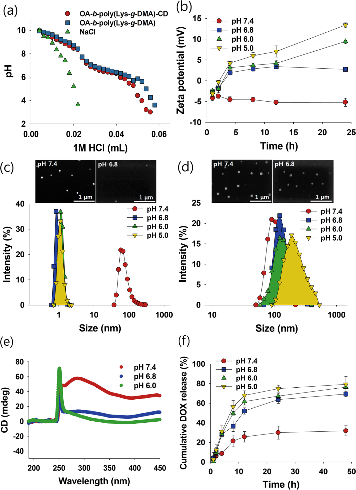
(a) The pH profile of polymers or NaCl measured by acid–base titration; (b) zeta-potential change of OA-b-poly(Lys-g-DMA)-CD nano-vehicles (1 mg/mL, pH 7.4–5.0, 150 mM PBS) with time (n = 3); (c) particle size distribution or FE-SEM images of OA-b-poly(Lys-g-DMA)-CD nano-vehicles (0.1 mg/mL) at various pH values (pH 7.4–5.0); (d) particle size distribution or FE-SEM images of OA-b-poly(Lys-g-DMA) nano-vehicles (0.1 mg/mL) at various pH values (pH 7.4–5.0); (e) Circular dichroism of OA-b-poly(Lys-g-DMA)-CD nano-vehicles (0.1 mg/mL) at various pH values; and (f) the pH-dependent DOX release from DOX-loaded OA-b-poly(Lys-g-DMA)-CD nano-vehicles at various pH values for 0–48 h of incubation (n = 3).
Shown in Figure 2(b) are the changes in zeta-potential of the nano-vehicles that were formed using OA-b-poly(Lys-g-DMA)-CD via a dialysis process. For a 2- to 4-h incubation period, the zeta-potential changed drastically, indicating the rapid detachment of the DMA moieties from the OA-b-poly(Lys-g-DMA)-CD nano-vehicles (Figure 2(b)). After a 24-h incubation, the zeta-potentials of the OA-b-poly(Lys-g-DMA)-CD nano-vehicles changed from −5 to +13 mV upon a pH reduction from 7.4 to 5.0. These data indicate that the negative zeta-potential, which originated from the DMA moieties, was offset by the formation of protonated free amine groups at acidic pH.
Shown in Figure 2c-d and Supporting Figure S2 are the changes in the nano-vehicles size with pH. The size of the OA-b-poly(Lys-g-DMA)-CD nano-vehicles changed considerably between pH 7.4 and 6.8. The images obtained from FE-SEM reveal that the nearly spherical OA-b-poly(Lys-g-DMA)-CD nano-vehicles at pH 7.4 disappeared at pH 6.8. However, the size of the OA-b-poly(Lys-g-DMA) nano-vehicles (without CD) (Figure 2(d)) and OA-b-poly(Lys)-CD nano-vehicles (data not shown) did not change significantly with pH (Figure 2(d)), indicating the formation of pH-insensitive nano-vehicles with hydrophobic core (OA) and ionized hydrophilic shell (poly(Lys-g-DMA)). We also confirmed that poly(Lys-g-DMA)-CD (without OA) did not form self-assembled nano-vehicles: no detection in the particle size analysis and the FE-SEM image (data not shown). On the other hand, the OA-b-poly(Lys-g-DMA)-CD nano-vehicles at pH 7.4 were stable for 2 weeks without precipitation, and the changes in the size of the nanoparticles were not significant (data not shown). From these data, we concluded that both the inclusion interaction between CD and DMA (or possible inclusion interaction between CD and OA) and the hydrophobic interaction between OA molecules are responsible for the nano-vehicles’ formation. Here, it was expected that the cleavage of the DMA moieties would destabilize the nano-vehicles, and the ensuing water diffusion inside the nano-vehicles was expected to lead to dissociation of the supramolecular assembles.
Shown in Figure 2(e) is a circular dichroism spectrum of OA-b-poly(Lys-g-DMA)-CD nano-vehicles as the pH of solution decreased from pH 7.4 to 6.0. The high circular dichroism signals in the near-UV region (250–350 nm) are attributable to the presence of DMA at pH 7.4. However, the low circular dichroism signals in the near-UV region at pH 6.8 are a good indication of DMA cleavage, suggesting a rearrangement of the molecular structure. Consequently, the anticancer drug (DOX) release patterns from DOX-loaded OA-b-poly(Lys-g-DMA)-CD nano-vehicles were pH dependent (Figure 2(f)): highly increased DOX release at pH 6.8 and minimized DOX release at pH 7.4. However, DOX release patterns from DOX-loaded OA-b-poly(Lys-g-DMA) (without CD) nano-vehicles at pH 7.4 and pH 6.8 was not significantly different (Supporting Figure S3).
The fluorescence images and flow cytometry profiles of human nasopharyngeal epidermal carcinoma KB cells treated with DOX-loaded OA-b-poly(Lys-g-DMA)-CD nano-vehicles in cell culture media of various pH values are shown in Figure 3(a) and (b) and Supporting Figure S4. The protonated amine groups resulting from the DMA cleavage contributed to the high cellular uptake at pH 6.8, unlike that at pH 7.4 (Figure 3(a) and (b)), through electrostatic interaction between positively charged nano-vehicles and negatively charged tumor cellular membrane. 21 Furthermore, DOX-loaded OA-b-poly(Lys-g-DMA)-CD nano-vehicles allowed tremendous DOX release at pH 6.8 (Figure 2(f)), increasing the tumor cell death at pH 6.8 relative to that at pH 7.4 (Figure 3(c) and (d)).
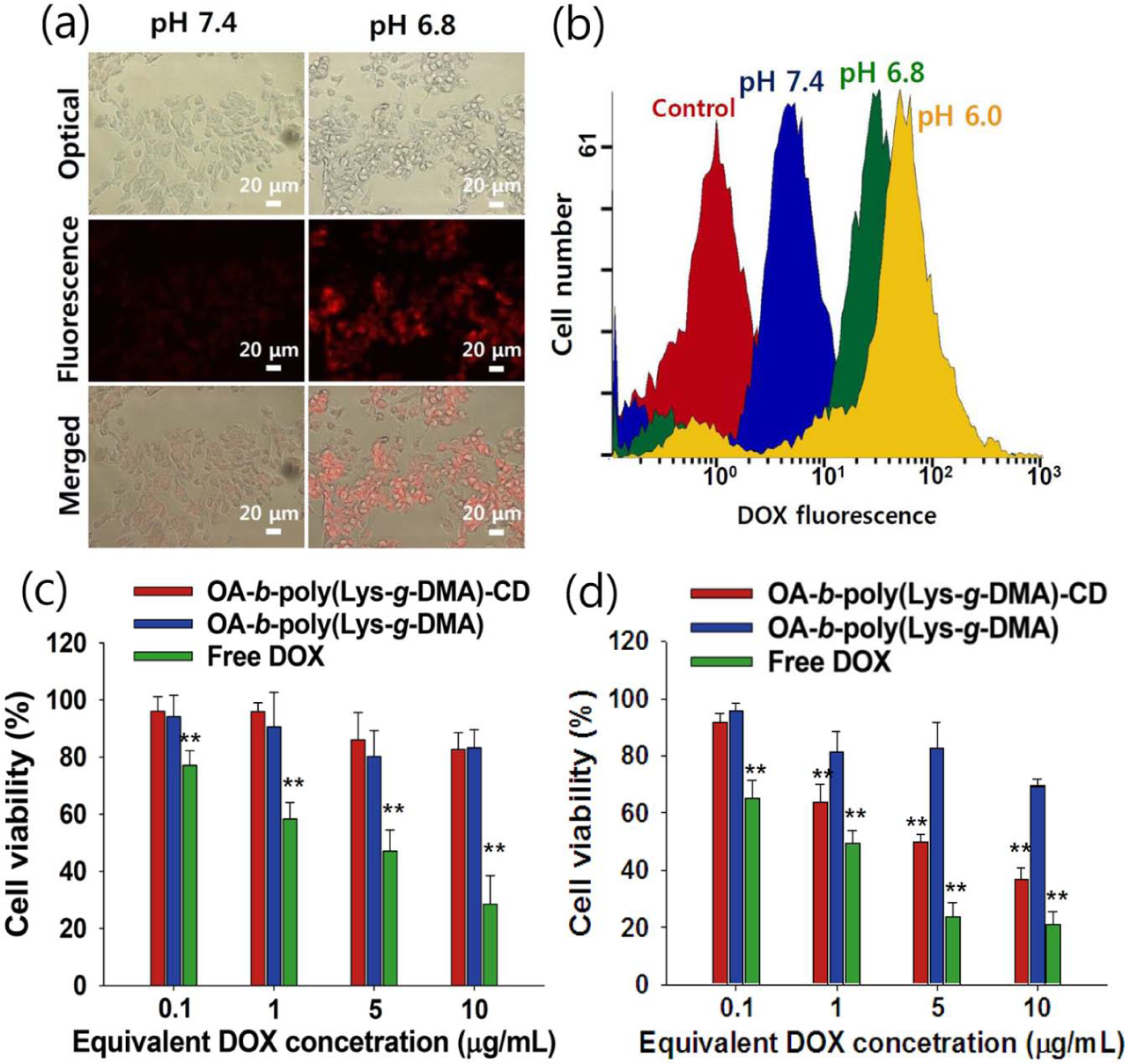
(a) Fluorescence images of KB tumor cells treated with OA-b-poly(Lys-g-DMA)-CD nano-vehicles (equivalent DOX 10 µg/mL) at pH 7.4 or 6.8 (4 h incubation) (red fluorescence: DOX) and (b) cellular uptake study of OA-b-poly(Lys-g-DMA)-CD nano-vehicles (equivalent DOX 1 µg/mL) for KB tumor cells at pH 7.4–6.0 (4 h incubation) using flow cytometry. Cell viabilities of KB tumor cells treated with each nano-vehicles or free DOX at (c) pH 7.4 or (d) pH 6.8 (n = 7) (**p < 0.01 compared with OA-b-poly(Lys-g-DMA) nano-vehicles).
The in vivo photoluminescence images of KB tumor-bearing nude mice are shown in Figure 4(a) and (b) and Supporting Figure S5. The results reveal that intratumorally or intravenously injected OA-b-poly(Lys-g-DMA)-CD nano-vehicles allow for high-resolution photoluminescence at the tumor site, primarily owing to passive tumor penetration via the enhanced permeability and retention (EPR) effect.23–30 The intravenously injected DOX-loaded OA-b-poly(Lys-g-DMA)-CD nano-vehicles are more efficient for tumor regression as shown in Figure 4(c). These results strongly support the possibility of improved, effective tumor treatment using a self-zipping/self-unzipping nano-vehicle.
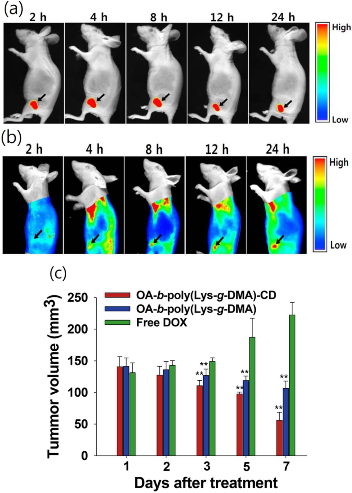
In vivo noninvasive photoluminescence imaging of Cy5.5-labeled OA-b-poly(Lys-g-DMA)-CD nano-vehicles (dose: 50 mg/kg) injected (a) intratumorally or (b) intravenously into KB tumor-bearing nude mice. Fluorescence images were obtained 2–24 h post injection. The tumor site is indicated by an arrow. (c) Tumor volume regression of KB tumor-bearing nude mice by intravenously injected nano-vehicles or free DOX (dose: equivalent DOX 10 mg/kg) (n = 5) (**p < 0.01 compared with free DOX).
Conclusion
We have developed a drug-carrying polypeptide nano-vehicle containing CD and DMA. We utilized advantage of both the pH-activated zipping/unzipping behaviors between polypeptide chains with CD and DMA moieties and the accelerated drug release of polypeptide nano-vehicles at acidic pH. This trial resulted in a significant enhancement of tumor inhibition under in vitro and in vivo conditions. We believe that this multifunctional nano-vehicle provides a promising pathway for tumor therapy.
Footnotes
Appendix 1
Declaration of conflicting interests
The authors declared no potential conflicts of interest with respect to the research, authorship, and/or publication of this article.
Funding
This work was financially supported by the Research Fund, 2014, of the Catholic University of Korea, the GRRC program of Gyeonggi province (GRRC 2013-B01, Development of industrial nano-/micro-sized biomaterials for high-performance drug release control), Basic Science Research Program through the National Research Foundation of Korea (NRF) funded by the Ministry of Education (NRF-2013R1A1A2004375), and by a grant of the Korean Health Technology R&D Project, Ministry of Health & Welfare (No. A111291), Republic of Korea.