
Abstract
Uniform poly(ε-caprolactone) microspheres containing a variety of water-soluble antibiotics, such as tobramycin, vancomycin, and gentamicin, were prepared by a simple fluidic device with a pristine or tapered glass capillary. Each type of antibiotic was dispersed in an organic solvent by ball-milling prior to microsphere preparation. The poly(ε-caprolactone) organic solution containing the powder of each antibiotic was introduced as the discontinuous phase into the fluidic device, where an aqueous phase containing surfactant served as the continuous phase. The poly(ε-caprolactone) microspheres were obtained after solvent evaporation. A tapered glass capillary was tested to produce poly(ε-caprolactone) microspheres, leading to the size reduction of the microspheres from 47.46 ± 0.72 to 25.49 ± 1.05 µm without destroying size uniformity. This size range should be suitable for parenteral injection into the human body. The release analysis revealed that gentamicin and vancomycin were released from the poly(ε-caprolactone) microspheres up to approximately 2 months in a more sustained manner than tobramycin, which is due to the solubility difference in the antibiotics in water. The antimicrobial activities of each type of antibiotic released from the poly(ε-caprolactone) microspheres were evaluated using Staphylococcus aureus and Escherichia coli.
Keywords
Introduction
Depot injections capable of releasing drugs for a prolonged period have been of great interest for pharmaceutical applications because they can minimize discomfort for both patients and medical doctors. Various depot injections for leuprorelin acetate, risperidone, and goserelin have been commercialized.1–5 Depot injections are characterized by the use of biodegradable polymers, such as poly(
A number of methods for the fabrication of biodegradable microspheres for parenteral drug delivery have been developed, including homogenization, ultrasonication, spray-drying, membrane emulsification, and micro-fluidic focusing.7–12 Among them, the micro-fluidic focusing method has been attractive due to its unique feature of producing uniform emulsion droplets. Xu et al. 13 fabricated PLGA microspheres containing bupivacaine using conventional homogenization and micro-fluidic focusing and demonstrated the superior performance of uniform PLGA microspheres prepared by the micro-fluidic focusing method.
Previously, a simple fluidic device consisting of a syringe needle, a glass capillary, and a flexible tube was developed to obtain uniform microspheres. 11 In addition to the production of uniform microspheres, the reliable reproducibility of the fluidic device makes it possible to systematically compare the performance of the microspheres containing drug. Therefore, we used the fluidic device as a model system to fabricate uniform biodegradable microspheres containing a variety of antibiotics and compared their performances as drug delivery carriers for depot injections. Among the various antibiotic drugs, vancomycin, gentamicin, and tobramycin were chosen as model drugs because of their wide clinical use. Staphylococcus aureus and Escherichia coli were selected as Gram-positive and Gram-negative bacteria to evaluate the antibiotic effect of the microspheres.14–17 These results could potentially provide a guideline for the future design of polymeric microspheres for the treatment of infection.
Materials and methods
Materials
PCL (molecular weight (Mw) ≈65,000), dichloromethane (DCM), polyvinyl alcohol (PVA; Mw ≈13,000–23,000), o-phthaldialdehyde, and sodium azide were purchased from Sigma-Aldrich (St Louis, MO, USA). Gentamicin, tobramycin, and vancomycin were kindly provided by Samjin Pharm. Co. Ltd (Seoul, Korea). Nutrient agar (BD Difco; Becton, Dickinson and Co., NJ, USA), S. aureus ATCC 6538, and E. coli ATCC 8739 were supplied by Ecowell Co., Ltd (Seoul, Korea) Sodium borate and 2-hydroxy-ethylmercaptan were purchased from Bio-Rad Laboratories, Inc. (CA, USA) and Junsei Chemical Co. (Tokyo, Japan), respectively.
Fabrication and characterization of uniform PCL microspheres
Tobramycin, vancomycin, and gentamicin (0.3 wt%, solid phase) as obtained were separately added into the oil phase of DCM and then ball-milled (Pulverisette 7; Fritsch, Idar-Oberstein, Germany) at 400 r/min for 6 h. Afterward, the PCL polymer (3 wt%) was dissolved in each antibiotic dispersion, making a solid-in-oil (S/O) phase. To assess the dispersibility of the antibiotic powders, the PCL solution containing antibiotics was cast onto a glass slide and photographs were taken using an optical microscope (IX71; Olympus, Tokyo, Japan) after solvent evaporation. The size of the antibiotics was analyzed by the photographs using ImageJ software (National Institutes of Health, Bethesda, MD, USA).
The simple fluidic device consisted of a syringe needle (30G), a glass capillary (0.5 mm i.d. × 1 mm o.d.), and a Tygon® tube (1/32 in i.d. × 3/32 in o.d.), as previously reported. 11 A tapered glass capillary was fabricated by heating with an alcohol lamp at its middle and elongating to 2 mm in the axial direction. PCL microspheres were prepared using the fluidic devices with pristine or tapered glass capillaries, based on a solid-in-oil-in-water (S/O/W) emulsion system. PCL (3 wt%) was subsequently dissolved in the S/O dispersion containing each antibiotic. The S/O dispersion and an aqueous PVA solution (3 wt%) served as the discontinuous and continuous phases, respectively. Each phase was introduced into the fluidic device using peristaltic pumps, forming S/O droplets in the water phase. The flow rates of the discontinuous and continuous phases were kept at 0.02 and 5.0 mL/min, respectively. The resultant S/O droplets were gently stirred in the collection phase (the aqueous PVA solution) to evaporate solvent for 24 h. The PCL microspheres were washed with water three times to remove PVA and were freeze-dried. Some of the PCL microspheres containing each type of antibiotic were sectioned using a microtome (Cryotome FSE; Thermo Scientific, Loughborough, UK) and scanning electron microscopy (SEM; S-4800; Hitachi, Tokyo, Japan) was used to analyze surface and inner morphology.
Release behaviors of antibiotics
The encapsulation efficiency of the antibiotics in the PCL microspheres was determined by an extraction method.18–20 The obtained PCL microspheres (0.1 g) were dissolved in DCM (10 mL) for 1 h and then phosphate buffered saline (PBS) (10 mL) was added into the organic DCM phase. The mixture was vigorously stirred for 1 h to extract the antibiotic from the organic phase into the aqueous phase. After centrifuging at 3000 r/min for 5 min, the aqueous PBS phase was sampled and analyzed to determine the antibiotic concentration. The concentration of gentamicin and vancomycin was analyzed after slight modification using the o-phthaldialdehyde assay. 21 O-phthaldialdehyde (2.5 g), methanol (62.5 mL), and 2-hydroxyethyl mercaptan (3 mL) were added to 560 mL distilled water with 0.04 M sodium borate, making o-phthaldialdehyde reagent. The sampled PBS (200 µL) containing antibiotic and the o-phthaldialdehyde reagent (200 µL) were added into isopropanol (200 µL) and kept at room temperature under dark conditions for 45 min, followed by measuring with an ultraviolet (UV)–visible (vis) spectrophotometer (Lambda1050; Perkin Elmer, Norwalk, CT, USA) at 333 nm.22,23 The concentration of vancomycin was determined by a UV–vis spectrophotometer at 280 nm without o-phthaldialdehyde reagent.24–26 The encapsulation efficiency was calculated as the ratio of the actual amount to the theoretical amount of antibiotic.
To evaluate the cumulative release of the antibiotics from the PCL microspheres, the PCL microspheres (0.1 g) containing each type of antibiotic were added into PBS buffer (pH 7.4, 5 mL) with 0.02 w/v% sodium azide at 37°C in a shaking water bath. Sodium azide was added to prevent contamination by bacteria. 27 At predetermined times, the PBS phase (4 mL) was sampled for analysis and was replaced with fresh PBS buffer (4 mL). The concentration of antibiotics was determined in the same way as the method for measuring the encapsulation efficiency.
Antimicrobial activity
The agar diffusion assay is a well-known method used to quantify the efficacy of antibiotics inhibiting bacteria growth.28,29 Disks (15.32 ± 0.14 mm in diameter and 1.24 ± 0.02 mm in thickness) were prepared by pelletizing the PCL microspheres (0.1 g) containing antibiotics. After autoclaving, nutrient agar sol (Becton, Dickinson and Co.) at 45°C was dispensed into a Petri dish and kept at room temperature for 2 h. When the agar sol changed into gel, a bacteria strain (S. aureus ATCC 6538 and E. coli ATCC 8739) was administered over the agar gel. Each type of pelletized disk was gently placed on the agar gel. The Petri dishes were maintained in an incubator at 37°C in a humidified atmosphere containing 5% CO2 for 24 h. The inhibition area was photographed and analyzed using ImageJ software (National Institutes of Health). All data were expressed as mean ± standard deviation.
Results and discussion
Previously, we developed a simple fluidic device using a syringe needle, a glass capillary, and a Tygon tube to produce uniform microspheres. 11 Based on an S/O/W emulsion system, uniform PCL microspheres containing water-soluble antibiotics were prepared using the fluidic device for the sustained release of antibiotics in the treatment of infection after surgical operations. Tobramycin, vancomycin, and gentamicin were selected as model antibiotics due to their wide clinical use. The SEM images of the antibiotic powders before and after ball-milling are shown in Figures 1(a) and 2(b), respectively. The average sizes of the tobramycin and gentamicin (Figure 1(a)) before ball-milling were 18.21 ± 7.91 and 30.07 ± 20.03 µm, respectively. The size of vancomycin was hard to measure because of its nonspecific shape. The sizes of tobramycin, vancomycin, and gentamicin were reduced to 0.41 ± 0.37, 0.91 ± 0.45, and 0.53 ± 0.43 µm, respectively (Figure 1(b)). To confirm the dispersibility of the antibiotics treated with the ball-mill process in the organic polymer solution, the S/O dispersion containing the dissolved PCL polymers was cast onto a slide glass and photographs were taken after solvent evaporation (Figure 1(c)). The antibiotic powders were observed to be individually dispersed in the polymer matrices, which might be attributed to the increase in the viscosity of the S/O phase after adding PCL polymer.
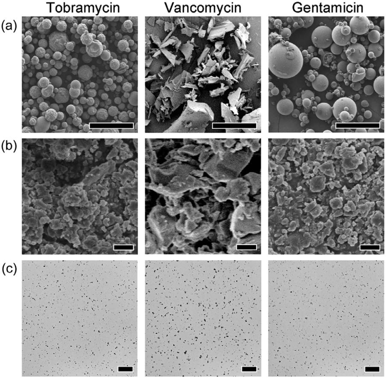
SEM images of antibiotics: (a) before and (b) after ball-milling, and (c) optical microscopy images of antibiotics dispersed in the PCL matrix that were prepared by casting the PCL solution on a slide glass. Scale bars are 100, 1, and 50 µm for (a), (b), and (c), respectively.
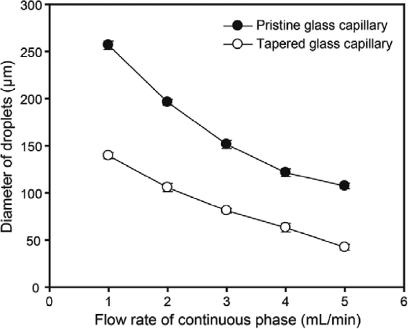
Effects of the flow rate of the continuous phase on the diameter of emulsion droplets prepared using fluidic devices with a pristine or tapered glass capillary. The flow rate of the discontinuous phase was kept at 0.02 mL/min.
The diameters of the microspheres can be tuned by changing the dimensions of the fluidic device, the polymer concentration, and the flow rates of the discontinuous and the continuous phases. Generally, a lower polymer concentration and higher flow rate of the continuous phase lead to the production of smaller microspheres. 11 However, these conditions might cause relatively high polydispersity and a small production amount of microspheres. Our preliminary results revealed that it was difficult to fabricate a large amount of microspheres with less than 50 µm diameter by changing the dimensions of the syringe needle and the flow rate of the continuous phase. Therefore, a glass capillary with a tapered shape in its middle was fabricated by extending a glass capillary using the heat from an alcohol lamp in an effort to produce a large amount of PCL microspheres with less than 30 µm diameter. This diameter range is considered to be suitable for parenteral injection into the human body. 30 As illustrated in Scheme 1, the flow rate of the continuous phase was fast at the end of the needle, imposing high shear force onto the oil droplets, eventually resulting in smaller microspheres even at the same flow rate of the continuous phase as used in the pristine glass capillary.
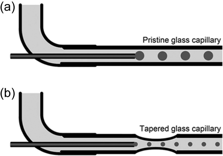
Schematic diagrams of fluidic devices with a (a) pristine or (b) tapered glass capillary.
As shown in Figure 2, the diameter of the PCL microspheres was controlled by changing the flow rate of the continuous phase in the fluidic device with a pristine or tapered glass capillary. An increase in the flow rate of the continuous phase led to a decrease in the diameter of the microspheres. In all cases, the diameter of the PCL microspheres decreased approximately 50% by using the tapered fluidic device, which is attributed to flow focusing at the taped glass capillary.
The SEM images and size distributions of the PCL microspheres prepared using fluidic devices with a pristine or tapered glass capillary at the same flow rates are shown in Figure 3. Both types of the PCL microspheres had a spherical morphology and uniform size. The size of the PCL microspheres was reduced from 47.46 ± 0.72 to 25.49 ± 1.05 µm by employing the tapered glass capillary in the fluidic device. The coefficient of variation (CV) increased from 1.52% to 4.12%, suggesting that uniformity in the diameter of the PCL microspheres prepared using a tapered glass capillary was maintained.
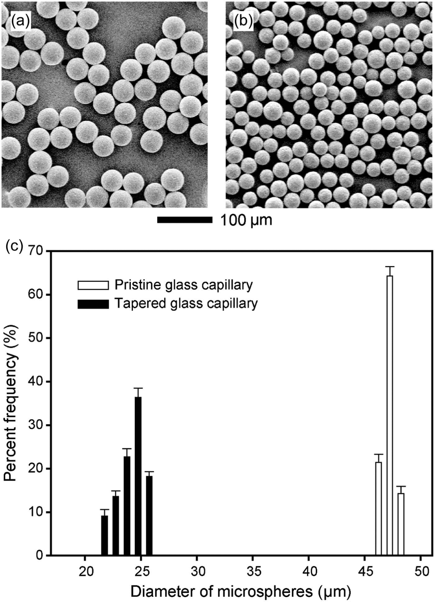
(a) and (b) SEM images and (c) a plot of the diameter distribution of the PCL microspheres prepared using fluidic devices with a (a) pristine or (b) tapered glass capillary, where the flow rates of the discontinuous and continuous phase were 0.02 and 5.0 mL/min, respectively.
The SEM images of surface and cross sections of the PCL microspheres containing antibiotics prepared using the fluidic device with a tapered glass capillary are shown in Figure 4(a) and (b), respectively. Hereafter, the PCL microspheres containing tobramycin, vancomycin, and gentamicin are referred to as Tob-PCL, Van-PCL, and Gen-PCL microspheres, respectively. There was no significant difference in the morphologies of the surface and inside between the microspheres prepared using a pristine or tapered glass capillary, except for their overall diameters. The Tob-PCL microspheres exhibited a rougher surface with more dents compared to the Van-PCL and Gen-PCL microspheres. The Gen-PCL microspheres had a relatively smooth surface. To observe the inner morphology, the PCL microspheres were sectioned with a microtome and washed with water to remove water-soluble antibiotics. The PCL microspheres were observed to have many inner pores of approximately 3 µm in size, which were the spaces that were previously occupied by antibiotic powder before removal with water. The size of the inner pores was slightly larger than that of the antibiotic powder, which is due to the aggregation of antibiotic powder during solvent evaporation in the collection phase. The Tob-PCL microspheres had a relatively less porous structure than the Gen-PCL and Van-PCL microspheres. In addition, the encapsulation efficiencies of tobramycin, vancomycin, and gentamicin in the PCL microspheres were 65.35%, 76.08%, and 78.38%, respectively. These results are attributed to the difference in the water solubility of the antibiotics. From the literature, tobramycin, vancomycin, and gentamicin had 538, higher than 100 and 100 mg/mL solubilities in water, respectively. 31 The higher water solubility of antibiotics resulted in PCL microspheres with the more porous structures, both inside and at the surface, because there was a large chance for antibiotics with a higher water solubility to diffuse into the outer water phase during solvent evaporation.
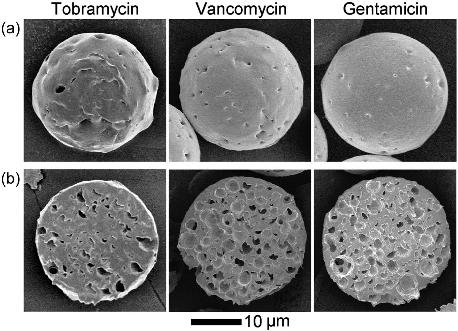
SEM images of (a) surfaces and (b) cross sections of the PCL microspheres containing antibiotics.
The release profiles in percents and amounts of antibiotics, from each type of PCL microsphere, are shown in Figure 5(a) and (b), respectively. A significant amount of tobramycin (39.0%) was released from the Tob-PCL microspheres at 1 day, reaching a plateau after 21 days. In contrast, the Van-PCL and Gen-PCL microspheres showed sustained release profiles with 23.7% and 15.9% of burst release at 1 day, respectively. The Gen-PCL and Van-PCL microspheres exhibited more sustained release patterns than Tob-PCL, which is due to the lower water solubility of gentamicin and vancomycin. Note that the release of gentamicin and vancomycin was prolonged for 60 days. To verify the antimicrobial effect of the released antibiotics from the PCL microspheres, the PCL microspheres were pelletized in the form of disks that were used in an antibiotic sensitivity test. The representative photographic images of the disks on layers of S. aureus (Gram-positive) and E. coli (Gram-negative) after 24-h post administration and a plot of the quantified inhibition zone are shown in Figure 6(a) and (b), respectively. The Gen-PCL and Tob-PCL microspheres exhibited similar efficiency in both Gram-positive and Gram-negative bacteria. The Gen-PCL and Tob-PCL microspheres were fairly effective in the treatment of the Gram-positive strain and the Gram-negative strain due to their broad spectrum. The Van-PCL microspheres had a smaller effect on each strain than the Gen-PCL and Tob-PCL microspheres. The disks with different antibiotics were immersed in PBS for 28 days under the same condition as the release analysis and then the inhibition zone was measured to evaluate the long-term antimicrobial effect. As shown in Figure 6(c), the disks prepared with the Van-PCL and Gen-PCL microspheres had antimicrobial effects even at 28 days, whereas the antimicrobial effect of the disk prepared with Tob-PCL microspheres was greatly reduced. The differences in the antimicrobial effects were attributed to the released amount of antibiotics from each type of disks, as observed in Figure 5.
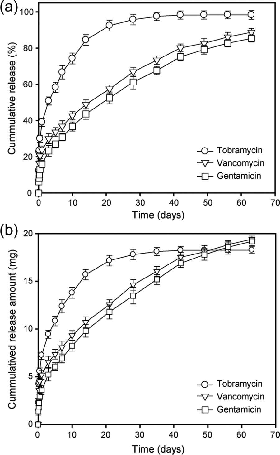
Cumulative release profiles: (a) percent and (b) amount of antibiotics from the PCL microspheres.
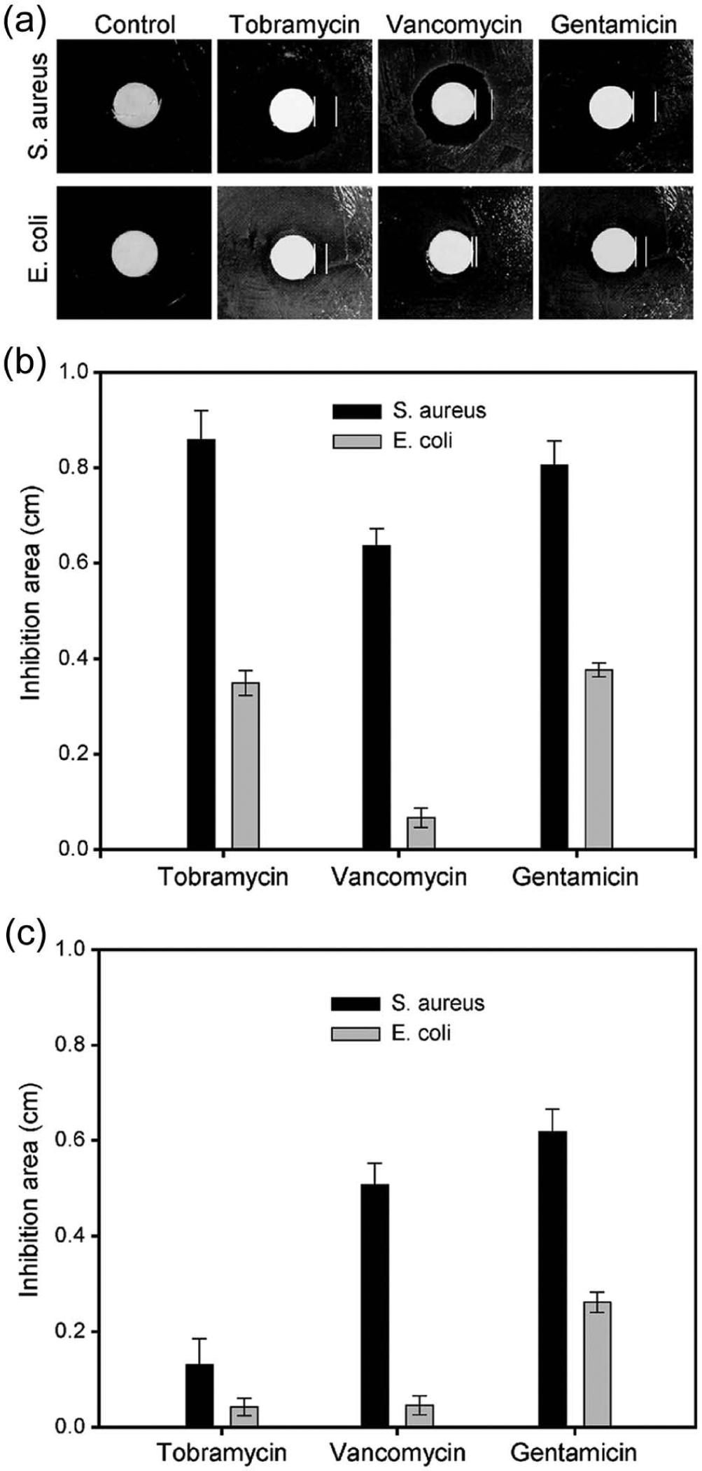
(a) Photographic images for the antimicrobial effects of pelletized disks made from the PCL microspheres containing antibiotics, and (b) and (c) plots for the quantified inhibition zone of (b) the disks after 1 day post administration and (c) the disks that were immersed in PBS for 28 days prior to administration.
Conclusion
Uniform biodegradable microspheres with suitable diameters for parenteral injection were fabricated using a simple fluidic device with a tapered glass capillary. The employment of the tapered glass capillary led to a large production of small PCL microspheres with less than 30 µm in diameter. Using the fluidic device as a model fabrication system, we tested various antibiotics and evaluated their potential application in sustained drug delivery. The solubility of the antibiotic was a crucial factor in determining the encapsulation efficiency and release behaviors. We believe that these uniform microspheres, capable of releasing antibiotics in a sustained manner, can be effectively used for the treatment of intracellular infections. Our next goal is to focus on the practical application of these biodegradable microspheres to reduce infections after implantation of bone substitutes in regenerative medicine.
Footnotes
Declaration of conflicting interests
The authors declared no potential conflicts of interest with respect to the research, authorship, and/or publication of this article.
Funding
This work was supported by the Basic Science Research Program through the National Research Foundation of Korea (NRF) funded by the Ministry of Education, Science, and Technology (2011-0023064); a grant of the Korean Health Technology R&D Project, Ministry of Health & Welfare, Republic of Korea (HI11C0388); and a grant from the “GRRC” Project of Gyeonggi Provincial Government, Korea.