
Abstract
Defected peripheral nerve regeneration is still a challenge in clinical treatment. Conductive polymers show great potential in nerve tissue engineering because of their electrical property based on bioelectricity in vivo. In this study, conductive composite nerve conduit was synthesized with tetra-aniline and poly-dl-lactic acid. Their properties and the differentiation of rat pheochromocytoma 12 (PC12) cells in vitro stimulated with 200 mV for 1 h were investigated. Different amounts of tetra-aniline (0%, 5%, 10%, and 15%) were used to synthesize the conduits with different conductivities (0, 0.00625, 0.01, and 0.025 s/m, respectively), tensile strengths (2.45, 3.40, 4.45, and 5.50 MPa, respectively), and contact angles (80°, 78.5°, 62.5°, and 61.5°, respectively). The percentage of neurite-bearing cells and the median neurite length increased with an obvious raise of the content of tetra-aniline. In addition, the conduit with subcutaneous implantable experiments in vivo showed less inflammatory response. These promising results illustrated that this poly-dl-lactic acid/tetra-aniline conductive composite conduit had potential for nerve tissue engineering.
Introduction
Across the world, nerve injuries 1 are clinical common diseases which puzzle more than 1 million people every year. Nerve injury regeneration is therefore particularly attractive due to difficult treatment. The diseases seriously impact the quality of life of patients, 2 and the repair of peripheral nerve injury is a worldwide problem. In the past few decades, nerve repair experienced four stages including autologous nerve graft, allogenic nerve transplantation, 3 autologous nerve tissue transplantation, and nerve conduit. In recent years, nerve conduits were widely used in nerve tissue engineering. It is a great important scientific problem to develop a promising conduit that can replace the autologous nerve graft to meet the important crucial needs of peripheral nerve injury repair. While it is difficult to prepare a perfect nerve conduit from a single material, synthetic nerve conduit materials include natural biomaterials and synthetic degradable materials, and they can be complementary in performance. So it is crucial and difficult to find the perfect natural and synthetic biological material. So far, the nerve conduit repair has not generated an excessive impact over the autologous nerve graft repair. 4 Meanwhile, endogenous and exogenous nerve growth factors (NGFs) filled in the nerve conduit are necessary to promote synaptic growth. However, the utilization rate of exogenous filler is not high and is easy to lose. Bioelectric stimulation creates a new situation for the preparation of nerve conduits. Conductive material has an effect of promoting tissue growth induction. At the same time, the effect of electrical stimulation is equivalent to multiple NGFs. Conductive polymers could conduct bioelectric signals in the human body and provided local electrical stimulation to local positions, and the multiple functions of cells can be modulated by the electrical stimulation section such as proliferation, migration and differentiation, protein synthesis and secretion of nerve cells. One of the biggest advantages of conductive nerve conduit as a nerve repair material is to restore nerves to electrical stimulation and promote nerve regeneration, so it is essential to study the conductivity of conduit. The electrical stimulation applied by such nerve conduit is mainly concentrated in around the conduit, and it is easy to control spatially the loaded stimulus without using the stimulation to localize the stimulus using an external electromagnetic field. 5
Conductive polymer6,7 is a class of polymers possessing macromolecule and conductive properties. With the invention of conductive poly-acetylene, conductive polymer has received plenty of attention8,9 and its potentiality in conductivity has been evidenced by scientists such as Shirakawa et al. 10 The organic conductive polymers such as poly-pyrrole (PPy), 11 polyaniline (PANi), 12 and polythiophene (PTh)13,14 were intensively studied. PPy is one of the mostly reported conductive polymers due to its characteristic properties such as high conductivity and good biocompatibility. 15 However, it is insoluble and non-degradable. 16 Compared with PPy, the proceeding of the research of PANi 17 is relatively slow in nerve tissue engineering. The use of PANi in biological applications is limited due to its low processability, week flexibility, and non-biodegradability, and chronic inflammation appeared once implanted in the body. 18 Therefore, the researches of PANi mainly focused on how to improve its biocompatibility and biodegradability. PTh was discovered relatively late compared to PPy and PANi. However, it is one of the most important polymers and widely studied because of its excellent electrical property, chemical stability, and reversible doping process.19,20 Nevertheless, its use is also limited because of low processability and dissolvability in an organic solvent.
Compared with PPy, PANi, and PTh, tetra-aniline (TA) bears advantages such as good processability, biodegradability, and solubility. In the past years, TA was mainly used in semiconductor, battery, and coating because of its good conductivity. Recently, researchers have paid more attention to study the electroactivity of TA because of its outstanding performance. The least unit number of electrical conduction is four for PANi. The conductivity of TA is similar to that of PANi. Due to their short polymer units and low molecular weight, degradation of TA is faster than that of PANi, PPy, and so on. Because of the good solubility in all kinds of organic solvents, TA can be easily processed. Molecular design strategy was utilized to synthesize monodisperse TA with different end groups. TA was synthesized by one-step oxidation coupling method. 21 TA with amine end groups was synthesized by the method of end group protection. 22 A polysaccharide cross-linked with TA grafting oxidized sodium alginate was prepared for biomedical applications. 23 A novel copolymer by crosslinking aniline pentamer with chitosan could significantly improve and induce PC12 cell differentiation without external electrostimulation. 24 A novel electroactive compound was prepared by coupling an aniline trimer with an epoxy silsesquioxane. The compound modified with the polypeptide had good biocompatibility with the PC12 cells. 25 Guo et al. 26 synthesized the multi-armed branched poly-l-lactic-acid-bearing aniline trimer segments and the polymer showed good solubility, biodegradability, and redox properties.
However, good tissue engineering scaffolds require the support of the base material. Poly-dl-lactic acid (PDLLA) is a kind of biological plastic. It is non-toxic, non-irritating, and has good mechanical property, biocompatibility, and biodegradable absorption. 27 It is the most widely used nerve tissue engineering material. 28 Our group has studied PDLLA/PPy conductive conduit and produced PDLLA/chondroitin sulfate/chitosan/NGF conduits 29 and injectable chitosan–hyaluronic acid hydrogels for NGF. 30 These results have laid a certain foundation for this work. However, the results of the repair of peripheral nerve need to be improved to meet the requirement of the clinical treatment.
In this study, we developed a new kind of nerve conduit material by the blending of PDLLA and TA to obtain a nerve conduit with electrical stimulation effect. Unlike other traditional scaffold materials, this nerve conduit owns good electrical stimulation and biodegradability. It was reported that conductive scaffolds were able to increase the length of neurites and promote neuronal differentiation as the scaffolds acted as a communication platform for cell interaction. PC12 cells were cultured on the films to assess the biocompatibility and bioactivity of the conduit. In vivo animal experiments were carried out to assess biocompatibility. The aim of this work was to investigate the performance of PDLLA/TA conduit and demonstrate their potential application for nerve tissue engineering.
Materials and methods
Materials
PDLLA with a molecular weight of 100,000 Da was purchased from Jinan Daigang Bioengin Co., Ltd (Wuhan, China). N-phenyl-p-phenylenediamine (PPDA) was purchased from Shenshi Chem Co., Ltd, (Wuhan, China). Dulbecco’s Modified Eagle’s Medium (DMEM), fetal bovine serum (FBS) trypsin, Cell Counting Kit-8 (CCK-8), 1,1’-dioctadecyl-3,3,3’,3’-tetramethylindocarbocyanine (Dil) and 4′,6-diamidino-2-phenylindole (DAPI) were purchased from Biyuntian Co., Ltd (Wuhan, China). All other reagents and solvents used in this study were purchased from Wuhan Misting Biochem Co., Ltd (Wuhan, China) and of the highest analytical purity (⩾99%).
Preparation of TA
Leucoemeraldine (LE) base form of phenyl/amino-capped tetra-aniline (LE TA) was prepared through the chemical oxidation coupling method described by Ribeiro et al. 20 and Chen et al. 21 Briefly, the solution of ammonium persulfate (0.55 g) in ice-cold mixed solvents of acetone (2.5 mL) and H2O (2.5 mL) was added dropwise for 30 min to the mixture of PPDA (0.55 g), acetone (25 mL), and 2M hydrochloric acid (HCl; 20 mL) at 0°C under stirring vigorously. The solution was stirred for another 4–5 h at 0°C to afford emeraldine salt of tetra-aniline (EM TA). The EM TA was doped with 0.5M ammonium hydroxide (100 mL) and reduced to LE base form using phenylhydrazine (1 mL) as a reducing agent.
Preparation of PDLLA/TA
PDLLA was dissolved in 10 mL of CHCl3 to obtain a 10% (w/v) solution. The EM TA (0.1 g) was then introduced into the PDLLA/CHCl3 solution under stirring for 2 h to afford the PDLLA/TA mixed solution. The weight ratio of TA to PDLLA was designed as 5%, 10%, and 15%. The films were obtained by casting the precipitated PDLLA/TA composite onto a polytetrafluoroethylene (PTFE) plate and dried in vacuum for 2 days. Subsequently, these films were washed with alcohol and deionized water three times prior to use for cell culture and animal study.
Characterization of TA and PDLLA/TA conduit
The structure and chemical compositions of TA were characterized by Fourier transform infrared (FTIR) spectra (Nicolet 6700, Nexus, Thermo Scientific, Waltham, MA, USA), mass spectrometry (Agilent 6890N/5975; Agilent, Santa Clara, USA), and ultraviolet absorption spectroscopy (UV-260, Shimadzu, Kyoto, Japan). The identification of TA was performed through thin-layer chromatography. The morphology of TA was observed using a scanning electron microscope (SEM) (JSM-IT300; JEOL, Tokyo, Japan) with an accelerating voltage of 20 kV, and the diameters of the particles were analyzed from the SEM images on ImageJ. Elemental analysis was performed using X-ray diffraction (XRD) (D8 Advance; Bruker AXS, Karlsruhe, Germany).
The structure and chemical compositions of PDLLA/TA were characterized by FTIR spectra. The morphology of the PDLLA/TA conduit surface was observed using SEM with an accelerating voltage of 20 kV. 31 Prior to observation, the specimens were sputter-coated with gold.32–34
The thermal behaviors of the conduit were investigated by thermogravimetric analysis (TGA) (Discovery TGA; TA Instruments, New Castle, USA) and differential scanning calorimetry (DSC) (DSC8500; PerkinElmer, Shelton, USA). Simultaneous thermal analysis was conducted by heating the conduit from room temperature to 500°C at a rate of 20°C/min. The conductivity of the PDLLA/TA film was tested by a four-point probe resistivity meter (5601-Y; Suzhou Jingge Electronics Co., Ltd., Suzhou, China). Before testing, the sample must be smooth. The mechanical properties of the conduit were tested using a universal test machine (50KN/SHT4106-G CMT 6503; Shenzhen Century Tianyuan Instrument Co., Ltd., Shenzhen, China). 35 The conduits were cut into dumbbell shape according to the government standard with a tensile speed of 5 mm/min. The thicknesses of the conduit were tested before the extension test. The static contact angles (CAs) of the composite films were measured using a contact angle goniometer (JY-82; Chengde Experimental Machine Plant, Chengde, China). Approximately 10 mL of distilled water was dropped onto the surface of the films before measurement. All data presented were the mean values of six independent measurements. 36
Degradation behavior
The degradation behavior of the conduit was investigated by soaking in phosphate-buffered saline (PBS; pH = 7.4) to simulate body fluid.37,38 The samples were placed in a centrifuge tube and an equivalent PBS buffered solution was added. The mixture was incubated at 37°C for up to 30 days. PBS was changed every 3 days and the pH of PBS and the weight of the conduit were recorded at certain intervals. At a certain period of the degradation, the conduits were subsequently dried in a vacuum oven at 37°C. The morphology of the films was observed by SEM.
In vitro cell study
Prior to the cell study, rectangular sections of the PDLLA and PDLLA/TA films were cut and placed in distilled water for 1 day and then transferred to PBS (pH = 7.4) and maintained for 1 day. Finally, UV light sterilized the films for 4 h. Schwann cells (RSC96) are a type of glial cell line that play an important role in the process of nerve injury regeneration. Pheochromocytoma (PC12) is a common nerve cell that has neuronal cell characteristics from the physiological and biochemical aspects. So they were selected to assess the cytocompatibility of the conduit. 39
Cytotoxicity
RSC96 cells were purchased from the Chinese Academy of Sciences in Shanghai and cultured in DMEM supplemented with 10% FBS and 1% penicillin/streptomycin solution. Each conduit material was cultivated for 3 days in DMEM according to the government standard GBT16886.12-2005. An extract liquid of 10 µL was put in 96-well plates, and then the medium of 100 µL was added to the well. Then RSC96 cells were seeded on the conduit at a density of 1 × 10⁴ cells per well to test the toxicity of the materials. The RSC96 cells were incubated at 37°C with CO2, the medium was replaced every other day, and the equivalent extract liquid was added again. At the first, third, and fifth days after treatment, cell proliferation evaluation was performed. In 96-well culture plates, CCK-8 assay was used to measure cell viability according to the manufacturer’s protocol. Briefly, CCK-8 of 10 µL solution was added to 100 µL cell culture medium in each well. After incubation for 4 h, the spectrometer was used to measure the optical density at 450 nm (OD450). 40
Culture of PC12 cells on the surface of the conduit
For growth and adhesion of the cells on the surface of the material, each material and the cells were incubated together at a density of 104 cells per well in 24-well plates. After 3 days, the PC12 cells were washed with PBS three times and 4% paraformaldehyde was added to the plates for 30 min, then sucked and washed with 200 µL PBS three times. Dil of certain amount was added to the well for 15–20 min, then Dil was sucked completely, and the wells were washed with PBS three times. An appropriate amount of DAPI was added for 10 min, then DAPI was sucked, and the wells were washed with PBS three times. Then the cells were observed and imaged by a fluorescence microscope.
For each material, the medium was sucked, an appropriate amount of 2.5% (w/w) glutaraldehyde was used for fixing the cells overnight and washed with PBS three times, then 10%, 30%, 50%, 70%, 80%, 90%, and 100% (w/w) tertiary butanol was used to dehydrate step by step, with proceeding at each concentration for 10 min, and finally 1 mL of alcohol was added to every plate. The materials were frozen for 12 h under ‒20°C. After vacuum freeze drying, the growth situation of the cells was observed by SEM.
Electrostimulation of PC12 cells
PC12 cells were cultured in F-12K medium supplemented with 15% heat-inactivated horse serum, 2.5% FBS, and 1% penicillin/streptomycin solution and maintained in a humid 37°C incubator with 5% CO2. The electrical stimulus device was prepared in our lab. 10% PDLLA/TA was selected to culture with PC12 cells for the electrostimulation experiment. Before the electrostimulation experiment, electrodes were sterilized using alcohol burner and washed with PBS three times. PC12 cells were seeded at a density of 2 × 10⁴ cells per cell and allowed to adhere for 24 h. Different voltage intensity and stimulation time were selected to study the electrostimulation effect of the nerve conduit membrane on PC12 cells. Cell proliferations were evaluated through the OD value and finally an optimal parameter of electrical stimulation on cells was determined. 41 A 200 mV potential was then applied across the wires for 1 h and the cells were cultured for an additional 24 h. 42 The methods are the same as mentioned in the previous section of ‘Culture of PC12 cells on the surface of the conduit’. The nuclei and the cell membrane were dyed by Dil and DAPI.
Animal study
Adult Sprague Dawley (SD) rats were purchased from Tongji Medicinal School, Huazhong University of Technology (Wuhan, Hubei Province, China). All animals were housed under standard conditions and the experimental procedures involving animals were performed under the approval of the Administration Committee of Experimental Animals, Hubei Province, China.
The subcutaneous implantation surgical procedure was performed as described previously. 43 Rats weighing 150–200 g were used to evaluate the biocompatibility of the PDLLA/TA scaffold. The animals were divided into two groups which are the PDLLA and 10% PDLLA/TA groups, each group having six rats. The films are approximately 0.4 mm thick. Before implantation, the membranes were aseptically cut into 0.35 × 0.35 cm2 samples. The membranes were implanted subcutaneously in the back of each of the 12 male rats. These animals were selected because they provided adequate size and tissue volume for testing the membranes and widely accepted for determining wound healing and tissue response of implantation. To implant the membranes, the rats were anesthetized with 10% chloral hydrate. The back of each rat was shaved, scrubbed with betadine to disinfect, and draped for sterile surgery. Incisions were made through the skin on each side of the midline with 2 cm distance from each other. A 1-cm pouch was created subcutaneously using blunt dissection. These membranes were implanted randomly in each subcutaneous pouch. 44 Each rat received two membranes. At 1 and 4 weeks, the implants were retrieved for histological evaluation. The retrieved tissue samples were fixed in neutral buffered formalin, embedded in paraffin blocks, and stained with hematoxylin and eosin (HE). 45
The animal experiment process of sciatic nerve defect was as follows: 46 sciatic nerves were fully exposed using a scalpel to cut the right posterolateral muscle space, 10-mm sciatic nerves were cut off, and then the sides of the sciatic nerve were cut with nerve conduit embedding, about 1 to 2 mm with the conduit and sciatic nerve. The nerve conduit formed a nerve regeneration chamber. After 3 months, the nerve conduit was removed.
Statistical analysis
All the data presented were expressed as mean ± standard deviation of the mean. Differences between the groups were considered statistically significant at p < 0.05.
Results and discussion
The advantages of the PDLLA/TA conduit existed in the use of conductivity of TA. Biodegradable biomaterials combined with conductive polymer materials will be the future direction in the development of tissue engineering. Conductive material could stimulate cell response, proliferation, and differentiation. So the preparation of PDLLA combined with TA conduit was aimed to exploit versatile properties of different materials. The synthetic route of TA is shown in Figure 1. The sketch map of synthetic preparation of PDLLA/TA conduit is shown in Figure 2.
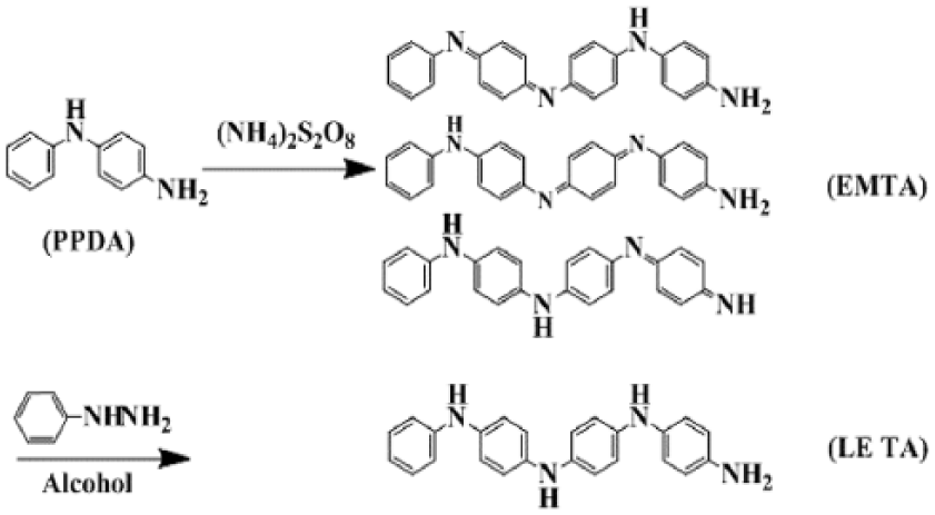
Synthetic route of TA.
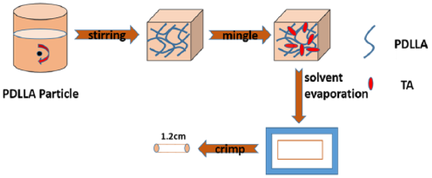
Sketch map of the PDLLA/TA conduit.
Characterization of TA, PDLLA, and PDLLA/TA
Figure 3 shows the FTIR spectra, mass spectra, ultraviolet absorption spectra, and thin-layer chromatography of TA. From Figure 3(a), for PPDA, the two bands at 3478 and 3389 cm−1 were the characteristic peaks of NH2 of the PPDA. The band at 3401 cm−1 was assigned to the N–H vibration absorption peak of EM TA. The peak at 3039 cm−1 was attributed to C–C of the benzene ring. The peaks at 1624 and 1528 cm−1 were the characteristic peaks of C–C of the benzene ring. The relative strength at around 1600 and 1500 cm−1 increased with the increasing number of quinone rings. The peak at 840 cm−1 was belong to 1, 4 replace of benzene ring. The peak at 1300 cm−1 was the characteristic peak of C–N. The peaks at 716 and 768 cm−1 were assigned to monosubstitution of the benzene ring. The successful synthesis of TA was also confirmed by MS (Figure 3(b)). From Figure 3(b), the highest peak at 365 nm in the MS was consistent with the molecular weight of TA. UV–Vis absorption spectrum showed peaks at 330 and 560 nm in Figure 3(c), which were assigned to EM TA, while the raw material PPDA and LE TA only had peak at 310 nm. Because EM TA contained quinone ring structure which was a sort of conjugated structure. The peak at 560 nm was attributed to the conjugated structure. In Figure 3(d), thin-layer chromatography proved the successful synthesis of EM TA and LE TA. The dot of them in thin-layer chromatography was labeled.
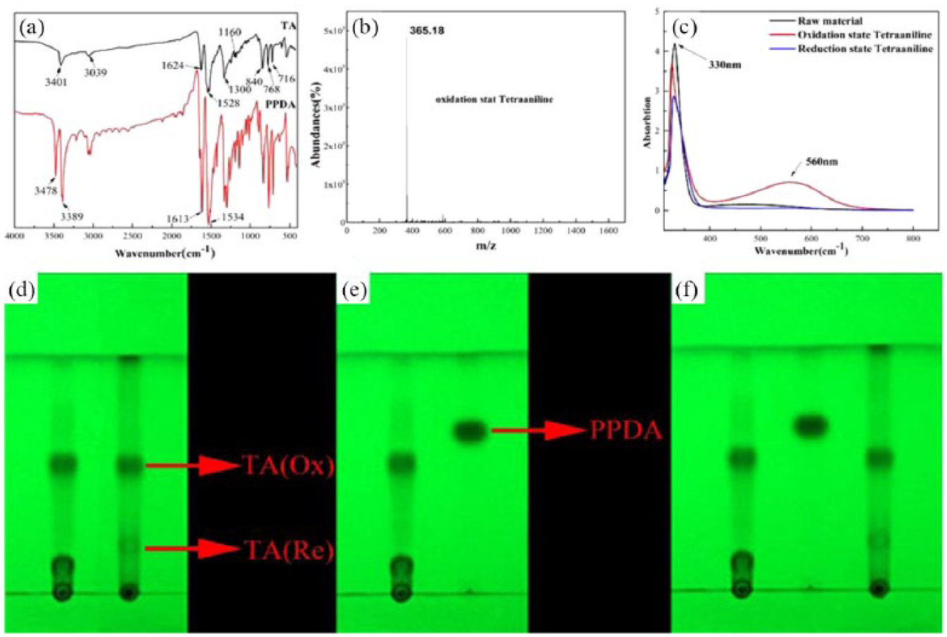
(a) FTIR spectra of N-phenyl-p-phenylenediamine (PPDA) and emeraldine salt of tetra-aniline (EM TA); (b) mass spectra of EM TA; (c) ultraviolet absorption spectra of PPDA, EM TA, and LE TA; (d) thin-layer chromatography of EM TA and LE TA, (e) EM TA and PPDA, and (f) EM TA, PPDA, and LE TA (from left to right); the eluting solvent is dichloromethane:ethyl acetate:ammonium hydroxide in the ratio of 100:15:1.
In Figure 4, the efficient preparation of the PDLLA/TA conduit was demonstrated by ATR-FTIR transmission spectra. From Figure 4, the band at 3401 cm−1 was the characteristic peak of TA which belongs to N–H. The bands at 3509 and 1774 cm−1 were the characteristic peaks of PDLLA. The peaks at 3509 and 1774 cm−1 were from the hydroxy and ester groups, respectively. All the above representative peaks were present in the PDLLA/TA spectrum, verifying that the PDLLA/TA was successfully synthesized.
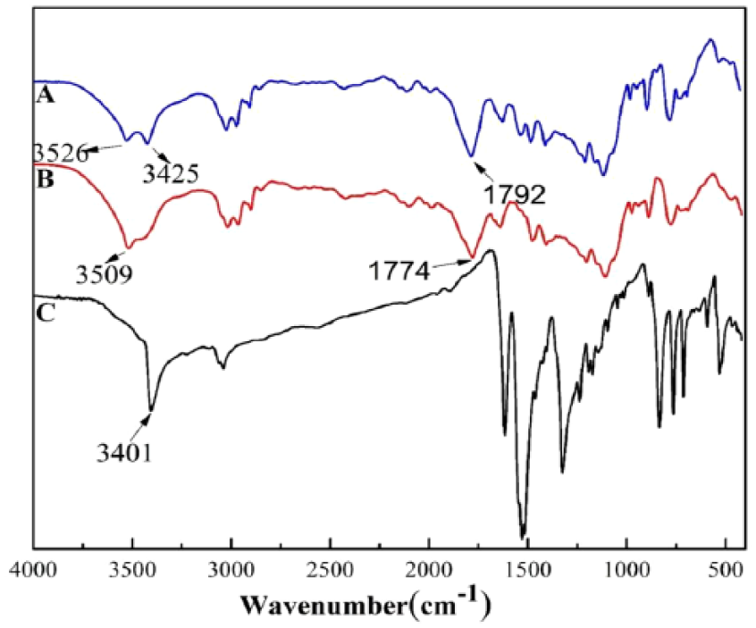
FTIR spectra of PDLLA and PDLLA/TA.
10% PDLLA/TA conduit with a length of 13 mm, an inner diameter of 4 mm, and an outer diameter of 3 mm was fabricated. The photographic images of 10% PDLLA/TA conduits are shown in Figure 5. Compared with the 5% and 15% PDLLA/TA films, 10% PDLLA/TA was equally distributed in PDLLA solutions in the preparation process. The surface of 10% PDLLA/TA was smoother than those of the other two. The SEM of the 10% PDLLA/TA films is shown in Figure 6.
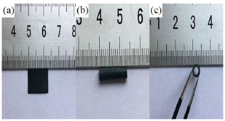
Images of (a) the PDLLA/TA film and (b, c) the PDLLA/TA nerve conduit.
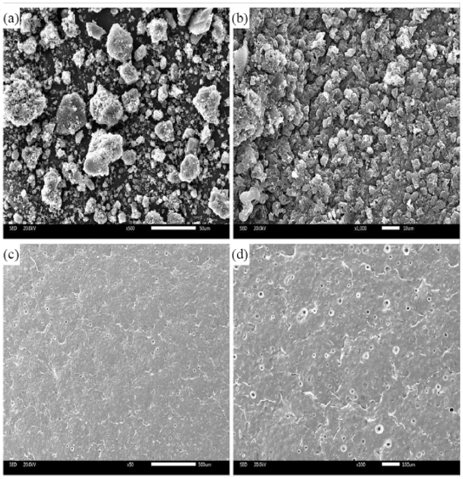
SEM of (a, b) TA and (c, d) 10% PDLLA/TA film; scale bars: 50 µm in (a), 10 µm in (b), 500 µm in (c), and 100 µm in (d).
The thermal property analysis
The thermal property analyses are shown in Figure 7(a) and (b). After adding TA, the heat stability of conduit was slightly improved. The phase transition temperature of TA was 160°C, that of PDLLA was 85.1°C, and that of PDLLA/TA was 164°C, and their temperatures were higher than the body temperature, so the polymer material was in the solid state under the endovascular environment. Materials in the glassy state were beneficial to maintain the size of the scaffold material stability and keep radial support of blood vessels. Compared with PDLLA, the glass transition temperature of the PDLLA/TA film was 82.5°C. Thermal stability of PDLLA/TA increased observably by adding TA. Meanwhile, the speed of weight loss of PDLLA/TA reduced slightly compared with pure PDLLA. The weight loss ratios of PDLLA and PDLLA/TA were 99.23% and 96.26%, respectively. There was no obvious difference between PDLLA and PDLLA/TA; it suggested good heat stability of PDLLA/TA. These data showed that TA was a heat-resisting polymer that could maintain high heat stability. The good thermal stability of PDLLA/TA made preparations for processing further.
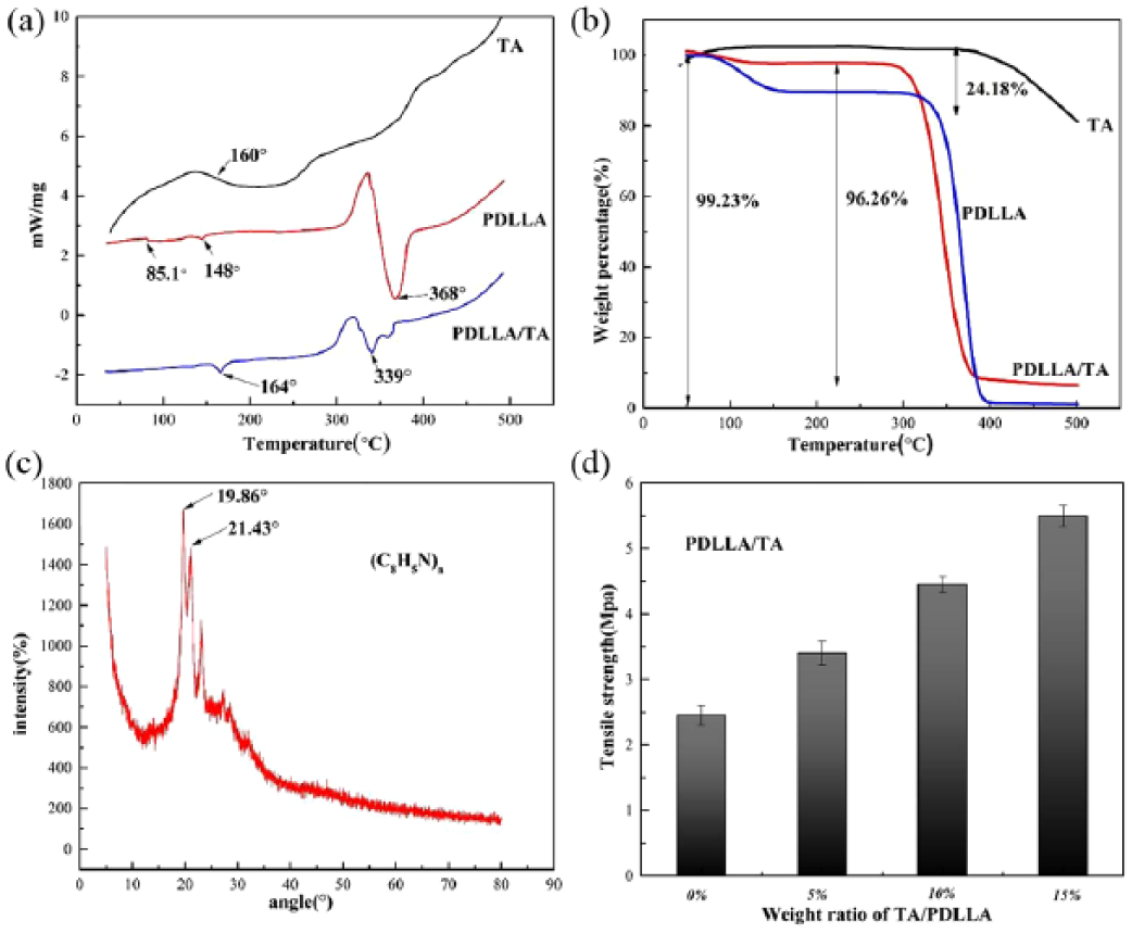
(a) DSC of TA powder, PDLLA, and PDLLA/TA films; (b) TG of TA powder, PDLLA, and PDLLA/TA films (the ratio of TA in PDLLA/TA is 10%); (c) XRD of TA; and (d) tensile strength of PDLLA/TA (5%, 10%, and 15%).
XRD of TA
Excellent crystallization was beneficial to improve the conductivity of TA. Therefore, a better crystallization of TA could enhance the bioelectric effect. Because of the high molecular structure regularity of TA, hydrogen bond interaction existed between molecules. The molecular chain number of TA was four. It had a repeat unit of PANi; therefore, it had crystallinity. The XRD peak was not tense enough, which signified that the crystallinity was not very well. As shown in Figure 7(c), according to the diffraction curve, the most intense peaks were centered around 19.6° and 21.04°. So it could be seen that the TA had a certain degree of crystallinity, suggesting that crystallinity was mainly ascribed to the periodicity parallel to the tetramer chain and corresponding to the periodicity perpendicular to the chain direction. These data also suggested that TA owned good conductivity in theory.
Conductivity
The conductivity of the PDLLA/TA films was tested by the four-point probe instrument; the result is shown in Table 1. PDLLA was state of insulation, while the PDLLA/TA had good electrical conductivity after doping with EM TA. The conductivity of different doping quantities was different and the conductivity of composite materials was enhanced with the increase of the content of TA. The conductivity of 5% PDLLA/TA was 0.00625 s/m, that of 10% PDLLA/TA was 0.01 s/m, and that of 15% PDLLA/TA was 0.025 s/m. These data were in the range of conductivity of the semiconductor (0.001–1 s/m). The conductivity of 10% PDLLA/TA could meet the need of nerve conduction. 16 However, the surface of 10% PDLLA/TA was smoother than the other two. A good tissue engineering material must have a smooth inner surface to avoid the influence of growth of regenerated nerve. Therefore, 10% PDLLA/TA was used to assess the interaction between the material and cells and in vivo animal study.
Conductivity of PDLLA/TA (the ratios are 5%, 10%, and 15%).
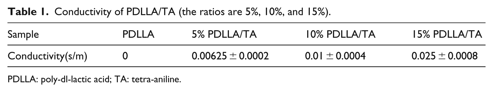
PDLLA: poly-dl-lactic acid; TA: tetra-aniline.
Mechanical property
The results of mechanical property are shown in Figure 7(d). The tensile strengths of PDLLA, 5% PDLLA/TA, 10% PDLLA/TA, and 15% PDLLA/TA were 2.45, 3.40, 4.45, and 5.50 MPa, respectively. The tensile strength of the conduit raised along with the increase of the TA content in PDLLA. The tensile strength of the PDLLA group fully met the requirement for tissue engineering. Due to the molecular structure of TA, the rigid structure of the benzene ring of TA could improve the mechanical properties of PDLLA/TA. This may be due to the intermolecular forces between TA and PDLLA, and in particular the carboxyl group in PDLLA formed hydrogen bonds with the amino group in TA to improve the mechanical property of PDLLA/TA, and the appropriate mechanical properties could ensure the growth of nerve fiber. The conduit would not be subjected to deformation according to the extrusion of the surrounding tissue; the conduit was pliable and tough. For suture of surgery, good compression modulus was beneficial and PDLLA/TA conduit had a certain resistance to pressure elasticity and support capacity.
Hydrophilicity
From Table 2, it can be seen that the CA of PDLLA (80°) was large due to the lack of hydrophilic groups. The PDLLA/TA group showed a lower CA (the CAs of 5%, 10%, and 15% PDLLA/TA were 78.5°, 62.5°, and 61.5°, respectively), because the amino group of TA was hydrophilic and could form a hydrogen bond with the hydroxyl group of the water molecule. The hydrophilicity group of PDLLA/TA was improved compared with PDLLA. With the amount of TA and the number of hydrogen bonding being increased, the hydrophilicity of the composite could be improved. Compared with 10% PDLLA/TA, the hydrophilicity of 15% PDLLA/TA did not change significantly. The phenomenon may explain that the number of hydrogen bonds formed had reached a certain amount, and the space structure of intrinsic molecule of the composite material was limited, so its hydrophilicity was not obviously improved. Good hydrophilicity of the material was advantageous to adhesion of cells and growth on the surface of the material. So PDLLA/TA could meet the need.
Contact angle of PDLLA/TA (the ratios are 5%, 10%, and 15%).
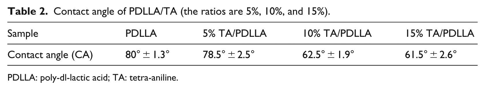
PDLLA: poly-dl-lactic acid; TA: tetra-aniline.
The degradation behavior
The PDLLA and PDLLA/TA conduits were placed in the PBS solution for a certain time; the pH value of the degradation solution was measured.
As shown in Figure 8(a), at the beginning of degradation (1 day), the pH of PDLLA/TA was higher than that of PDLLA, which was ascribed to the existence of TA. The hydrophilicity of PDLLA/TA was higher than that of PDLLA, so TA accelerated the degradation of PDLLA/TA and the pH of PDLLA/TA was lower than that of PDLLA at the beginning of degradation. Then, the pH values of PDLLA and PDLLA/TA continued to decrease because of their degradation. The phenomenon revealed the deficiency of PDLLA as a tissue engineering scaffold. Acidic environment would lead to severe inflammatory response. Compared with PDLLA, PDLLA/TA overcame the deficiency and the pH of PDLLA/TA began to rise at the seventh week. Maybe, TA contained the NH2 group which was alkaline and hydrophilic. So PDLLA/TA improved the problem of acidic environment of PDLLA in the process of degradation.
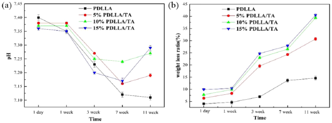
(a) pH change of the degradation liquid of PDLLA/TA flims from 1 day to 11 weeks; (b) weight loss ratio of PDLLA/TA films from 1 day to 11 weeks.
To assess their degradation property, the conduits were weighed. After the PDLLA and PDLLA/TA conduits were placed in PBS (pH = 7.4) for an extended time, as shown in Figure 8(b), there was a little change in their weights at the beginning (at 1 week) and it continued to decrease during the ensuing days (1–11 weeks). However, compared with PDLLA, the maximal weight loss ratio of PDLLA/TA could reach 40% across 11 weeks; this might be ascribed to the gradual release of the soluble components. When the poly-lactic-acid was degraded, the ester bonds in the main chain were hydrolyzed and disconnected. The molecular weight rapidly decreased, and when the molecular weight dropped to a certain extent, it could be dissolved in water. While the weight mass ratio of the PDLLA/TA group was higher than that of the PDLLA group all the time, the weight mass ratio increased with the content of increasing TA. That meant the addition of TA expedited the degradation of conduit, which maybe because the existence of TA accelerated the hydrolysis of ester bonds. The mechanism of degradation of TA would be further studied in the future.
It can be seen from Figure 9(a) and (b) that the surface of PDLLA conduit started to swell and curl after 6 weeks, then roughened gradually, and produced small cracks, while PDLLA/TA showed the appearance of a large hole after 6 weeks. As shown in Figure 9(c) and (d), after 12 weeks of degradation, PDLLA began to form hole; however, the number of holes of PDLLA/TA conduit increased and fused to form open pores together. Debris appeared and the samples became so brittle that even a slight touch would shatter the conduit, which indicated that the mechanical property of the sample became weaker after degradation. Compared with PDLLA, the degradation of PDLLA/TA was more serious at the same time point. For the PDLLA/TA conduit, low- and high-magnification SEM micrographs all showed prominent morphological changes during the degradation periods. The results suggested that the PDLLA/TA conduit had good biodegradability which the scaffold must possess.
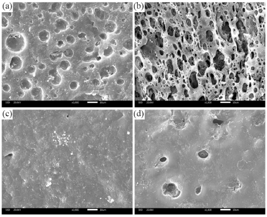
SEM of (a) 10% PDLLA/TA for 6 weeks; (b) PDLLA/TA for 12 weeks; (c) PDLLA for 6 weeks; and (d) PDLLA for 12 weeks. Scale bar: 10 µm.
Cell cytotoxicity
This study examined the cytotoxicity of PDLLA/TA conduit on RSC96 cells by CCK-8 assay. The result is shown in Figure 10. Cells were treated with PDLLA and 5%, 10%, and 15% PDLLA/TA. Compared with PDLLA, PDLLA/TA showed no significant difference in cell viability on the first day (p > 0.05). Cell growth of 10% PDLLA/TA significantly accelerated the viability of cells compared with 5% PDLLA/TA and 15% PDLLA/TA. 10% PDLLA/TA exhibited the strongest promoting effect on cell growth on the third and fifth days. Maybe 10% PDLLA/TA caused the electrostimulation effect. It could induce cell proliferation and growth along with damaging the cells. The ability to induce growth was higher than that to damage the cells. So the results proved the feasibility of 10% PDLLA/TA in vitro cell study.
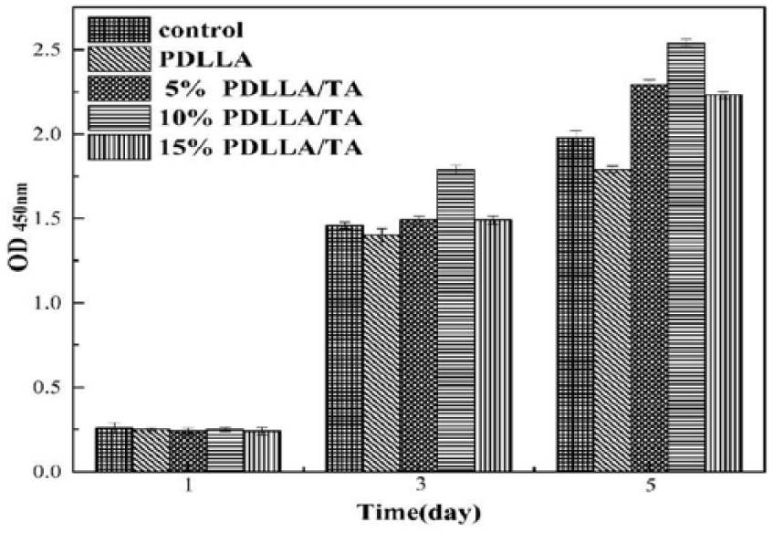
The cytotoxicity of PDLLA and PDLLA/TA which were cultured with RSC96 cells for 5 days.
Electrostimulation effect of PC12 cells
PC12 cells were selected to study the effect of 10% PDLLA/TA films combined with electrical stimulation on their behavior. From Figure 11, PC12 cells were able to adhere to the array of PDLLA and 10% PDLLA/TA films, which were viable throughout culture. There was no significant difference in the cell number throughout the experiments on any of the films (p > 0.05), indicating that the addition of TA did not have any significant effect on adhesion or proliferation. The PDLLA group which had no electrical stimulation had minimal levels of differentiation across the board in Figure 11(a). 200 mV and 1 h were the optimal parameters for electrical stimulation. The results are shown in Figure 12. The greater the OD value was, the stronger the viability of the cells was. Under the same stimulation time, the OD value was the maximum when the voltage was 200 mV, and the OD value was the maximum when the electrical stimulation time was 1 h and the voltage was constant. Cells with 10% PDLLA/TA stimulated with 200 mV voltage for 1 h exhibited more and longer neurites, as shown in Figure 11(c). On all the control films, approximately 11% of the cells formed neurites with a median length of around 6 µm. However, there was a clear increase in both the percentage of neurite-bearing cells and the median neurite length for the cells stimulated with 200 mV for 1 h after adding TA. The phenomenon suggested that electrostimulation could improve cell proliferation and growth preliminarily.
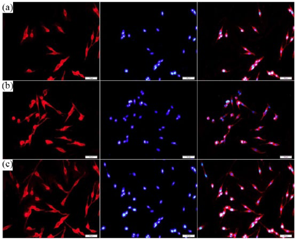
Fluorescent images of PC12 cells labeled by Dil (red) and DAPI (blue): (a) PDLLA, (b) 10% PDLLA/TA, and (c) 10% PDLLA/TA with cells stimulated with 200 mV for 1 h. Scale bar: 50 µm.
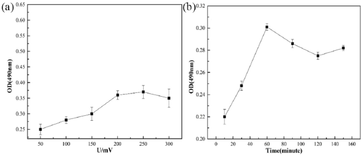
Proliferation of PC12 cells with different (a) voltage and (b) time.
Interaction of PC12 cells with the conduit
The interaction of PC12 cells with 10% PDLLA/TA was evaluated by SEM. From Figure 13, we could see PC12 cells that are cytoplasmically processed extending throughout the membrane. At a higher magnification, we could observe the feasible morphological aspect of these cells, namely, elongated cell body shape and bipolar projections. These data indicated that 10% PDLLA/TA membrane was a good substrate for survival, adhesion, and growth of these cells. The PC12 cells appeared to grow in large quantity on the 10% PDLLA/TA membrane. Following the cell adhesion, they could initiate scattering process which can be visualized in Figure 13. These results demonstrated the potential applicability of 10% PDLLA/TA in nerve tissue engineering.
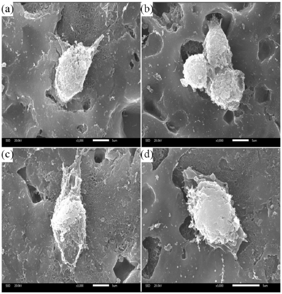
(a–d) SEM of PC12 cells and 10% PDLLA/TA film cultured together for 3 days. Scale bar: 5 µm.
Animal study analysis
The transplant images of the PDLLA and 10% PDLLA/TA films are shown in Figure 14. Representative images of histological sections of the implanted specimen at the retrieval time points of 1 and 4 weeks are shown in Figure 15. The analysis of inflammation was performed with ImageJ. There were many inflammatory cells after 1 week for the PDLLA and 10% PDLLA/TA films, but these two groups had no significant difference. It is ascribed to operative wound and acute immune response. So the two groups appeared with acute inflammatory reaction. After 4 weeks, a few neutrophils and inflammatory cell infiltration existed. These results proved that 10% PDLLA/TA film possessed good biocompatibility preliminarily. So, based on the inflammation analysis results, sciatic nerve defect experiments were projected to prove the feasibility of conduits in animal’s body. The image of nerve conduit sciatic nerve transplantation is shown in Figure 14(c). From Figure 14(d), 3 months later, while the sciatic nerve was undergoing tropistic growth, most of the nerve conduit was degraded, indicating that the rate of degradation of the nerve conduits matched the nerve growth rate. These results demonstrated that 10% PDLLA/TA had good biocompatibility and biodegradability further.
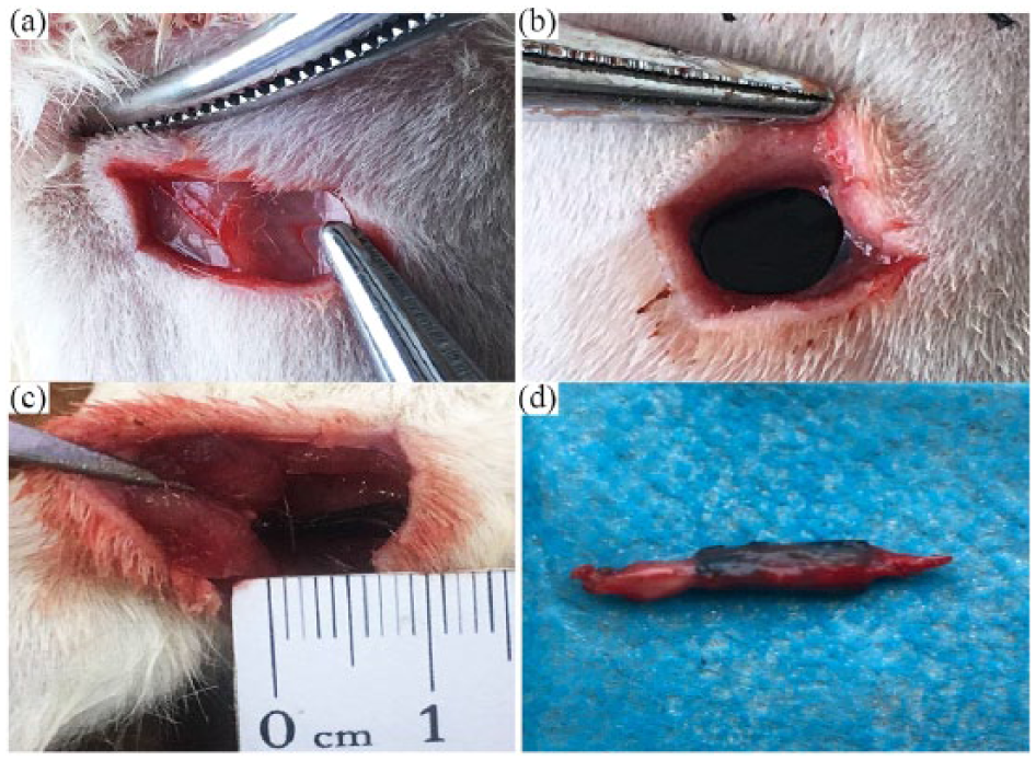
(a) Embedded PDLLA film, (b) embedded 10% PDLLA/TA film (size of films is 0.35 × 0.35 cm2 in the experiment), (c) sciatic nerve defect study of 10% PDLLA/TA nerve conduit, and (d) removed 10% PDLLA/TA nerve conduit.
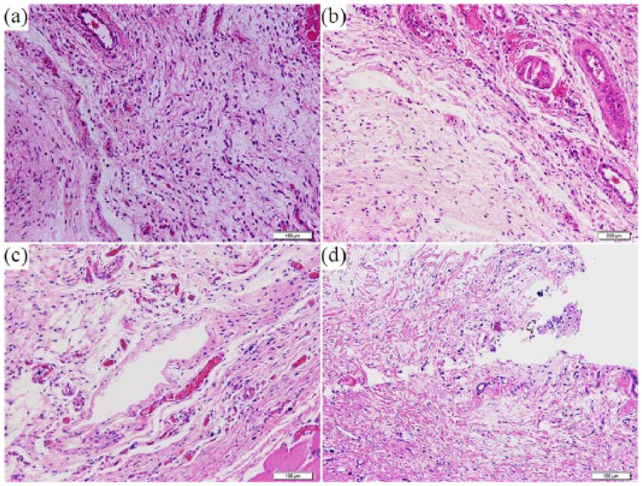
Electron microscopic images of HE tissue slice: (a) PDLLA group for a week, (b) PDLLA/TA group for a week, (c) PDLLA film for 4 weeks, and (d) PDLLA/TA film for 4 weeks.
Conclusion
In this study, the composite PDLLA/TA conduit was fabricated and the mechanical property, hydrophilicity, degradation behavior, electrical conductivity, and cytochemical properties of the conduit have been investigated by adding different amounts of TA for application in neural tissue engineering. In vitro cell culture studies exhibited higher proliferation, improved neurite outgrowth, and more cell differentiation on 10% PDLLA/TA than on PDLLA conduit after stimulating with 200 mV for 1 h. Results from 30 days of subcutaneous implantable experiments and 3 months of the sciatic nerve defect experiment indicated that the PDLLA/TA conduit had an ideal biocompatibility and biodegradability and was worth investigating deeply. The study may also provide an important experimental basis for the design and development of an electrostimulation effect nerve conduit.
Footnotes
Declaration of conflicting interests
The author(s) declared no potential conflicts of interest with respect to the research, authorship, and/or publication of this article.
Funding
The author(s) disclosed receipt of the following financial support for the research, authorship, and/or publication of this article: The financial support from the National Natural Science Foundation of China (Nos 51473130 and 51572206), Wuhan Huanghe Excellence Plan, Undergraduate Independent Innovation Project (2018-HS-A1-02), and Entrepreneurship Training Program of Wuhan University of Technology (Nos 20181049720018 and 20181049720009) is kindly acknowledged.