
Abstract
Today, an advanced wound dressing with the ability of blood clotting and antibacterial activity is the main subject of many studies to consider their necessity in modern society. In this study, it was aimed to present a novel scaffold with both abilities simultaneously. Poly(vinyl alcohol)/poly(lactic acid) nanofibrous scaffolds containing ceftriaxone antimicrobial agent (PVA-CTX/PLA) and tranexamic acid coagulant (PVA-CTX-TXA/PLA) were fabricated by electrospinning method. Morphology, antimicrobial activity, blood coagulation and bioavailability indexes, and swelling ability (gel formation) of produced samples were determined. Morphological results showed that the hybrid nanofibers were form successfully. The antibacterial efficiency of them against Gram-negative (Escherichia coli) and Gram-positive (Staphylococcus aureus) bacteria was more than 90% for PVA-CTX/PLA and it reached 100% for PVA-CTX-TXA/PLA. Both PVA-CTX-TXA/PLA and PVA-TXA/PLA scaffolds showed acceptable blood coagulation ability with an average absorption of 0.043 and 0.036 nm, respectively. PVA-CTX-TXA/PLA scaffolds had a gel formation ability of about 45 min. All scaffolds were successful in cell proliferation (L929 fibroblast cell) after 48 h.
Introduction
Wound dressing protects a wound from bacteria and prevents further infection, and also could enhance cell attachment, migration, and proliferation. Furthermore, a moist environment could support the wound fluids and growth factors around the wound, and thus affect cell migration and promote wound healing. 1 A non-toxic ideal wound dressing should maintain a moisture healing environment and antibacterial activity. 2
Various techniques have been reported for the fabrication of wound dressing materials. Recently, Muzzarelli et al. 3 formed a reinforced wound dressing material by using freeze-drying methods. Mi et al. 4 fabricated and characterized a sponge-like chitosan membrane as a wound dressing using the casting process. De Cicco et al. 5 reported nano-particulate powder by nanospray drying technology for wound dressing. In another study, Kobsa et al. 6 prepared electrospun nanofiber scaffolds for the treatment of cutaneous wounds.
Among these techniques, electrospinning is an easy and highly efficient method consisting of three major components: a high voltage power supply, a spinneret, and a grounded collector. 7 This method can produce nano- or microfibrous structures using natural and synthetic polymers that have unique characteristics such as high porosity, large surface area to volume ratio, and superior tensile strength. 8 Due to these outstanding properties, electrospun nanofibers have been widely used in biomedical applications for tissue engineering9–11 and wound dressing.12–14 Kyzioł et al. 15 presented a simple method of electrospinning of sodium alginate nanofibers loaded with ciprofloxacin hydrochloride, a fluoroquinolone antibiotic that is widely used for wound healing applications. The effect of an additional polymer, a surfactant, and alginate concentration on the formation of fibers was studied in detail. They found that the use of biopolymers, as well as non-toxic solvents and cross-linkers, makes the resultant scaffolds promising for various biomedical applications such as wound healing, regenerative medicine and drug delivery systems. In the study of Mohseni et al., 16 a highly porous wound dressing was developed from polycaprolactone (PCL) and poly(vinyl alcohol) (PVA) nanofibers with good handling, flexibility, and hydrophilicity, which provides requisite physical properties for wound dressings. Also, Mohseni et al. 17 developed antimicrobial wound dressings loaded with silver sulfadiazine (SSD) or silver nanoparticles (AgNPs) and make a direct comparison between these two groups of antimicrobial agents to evaluate their efficiency in the wound healing process. Jatoi et al. 18 presented a novel three-phase antibacterial wound dressing prepared from carbon nanotubes, AgNPs, and PVA nanofibers. The antibacterial test results confirmed excellent bactericidal and prolonged bacterial growth inhibition properties of the nanocomposites which suggest their suitability as a sustained antibacterial wound dressing biomaterial. Khan et al. 19 fabricated the electrospun cellulose acetate (CA)/SSD nanofibers for wound dressings of burn infections. In their study, the X-ray diffraction (XRD) spectra and water contact angle test indicated that CA/SSD nanofiber revealed the suitable water absorbency as required for scaffoldings.
Hemorrhage is uncontrolled bleeding from a wound. Various studies indicate that hemorrhage is a major cause of “potentially preventable” deaths in combat as well as civilian trauma. New methods and products for hemorrhage control are therefore a research priority to avoid potentially survivable deaths. A better understanding of the correlation between surface properties and their hemostatic potential (i.e. ability to form a blood clot) would result in an effective hemostatic wound dressing.20–22 Wiegand et al. 23 implemented several tests to evaluate the efficacy of cotton gauze (Gazin®, Lohmann & Rauscher, Germany), collagen The Suprasorb C Collagen Wound Dressing (SC) (Suprasorb® C, Lohmann & Rauscher) and collagen The Puracol™ (collagen PC) Collagen Wound Dressing (PC) (Puracol™, Medline Industries, Inc., USA), and oxidized regenerated cellulose (Tabotamp®, Johnson & Johnson, USA) for enhancing blood clotting, coagulation, and platelet activation. They used four wound dressing with acknowledged hemostyptic properties. They concluded that collagen-based dressings are ideal hemostyptic wound dressings with a very good benefit/safety ratio. Liao et al. [24] Electrospun nanofibrous mat for wound dressing application was successfully prepared from PCL, cellulose acetate, and dextran blend solution. The results showed that PCL-CA-dextran- tetracycline hydrochloride (TCH) mat had good bioactivity, blood clotting ability, and effective antibiotic activity against bacteria.
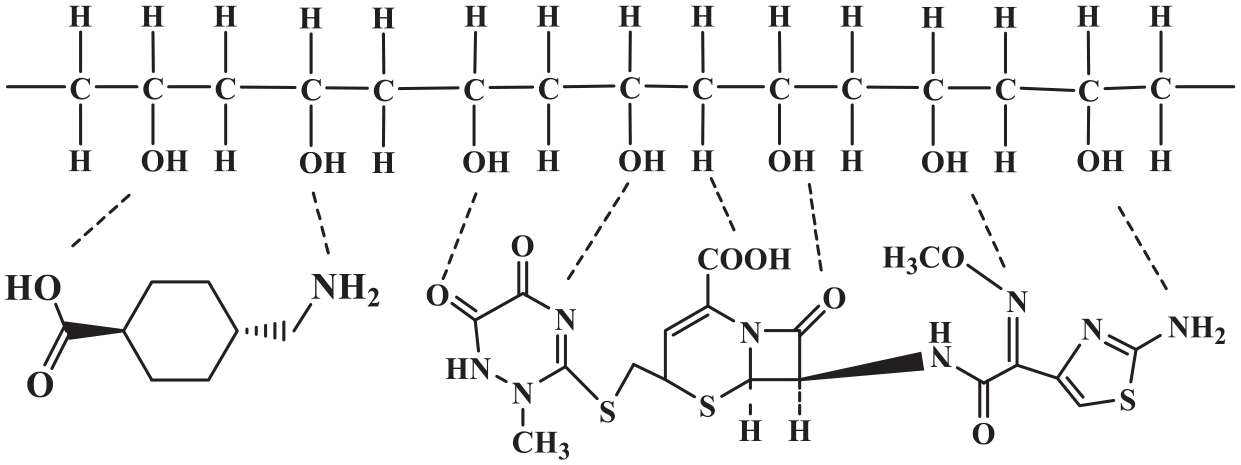
Considering the promotion of community standards, there is a need for products that can simultaneously perform several operations together. This is also evident in wound dressing. In this regard, little work has been done on wound dressing which can act both as a wound dressing and as a blood coagulant in that unit. In the present study, the purpose was wound healing production in order to apply blood coagulation, gel formation, antimicrobial activity, and biocompatibility. Using two polymers of PVA and PLA (poly(lactic acid)) with ceftriaxone and tranexamic acid, nanofibers were produced by electrospinning. To observe the morphology of the samples, a scanning electron microscopy (SEM) was used and antimicrobial activity of these scaffolds was performed using standard American Association of Textile Chemists and Colorists (AATCC) 100-2004. MTT test (ISO 10993-5) was used to examine the proliferation of cells cultured on samples to obtain the biocompatibility of the samples. Also, by measuring the amount of blood hemoglobin absorption at 540 nm and investigating the activation of blood platelets, the ability to coagulate the scaffolds was studied. By measuring the scaffold swelling percentage of PVA-CTX-TXA/PLA, the ability of gel formation was considered.
Experimental section
PVA/PLA nanofibers loaded with the drug
PVA (Mw = 89,000, Sigma-Aldrich, USA) solution was prepared by dissolving 5 wt% PVA in distilled water for 4 h at 85°C. PLA (Mw = 60,000, Sigma-Aldrich, USA) solution was prepared by dissolving 7% wt/wt PLA in N,N-Dimethylformamide (DMF, Sigma-Aldrich)/Acetone (Sigma-Aldrich) (8:2) mixture solvent for 2 h at 45°C. To load the drugs in PVA, Ceftriaxone (Sigma-Aldrich) (8 µg/mL) and Tranexamic acid (Caspian Tamin, Iran) (20 mg/mL) added to the prepared polymer matrix directly and stirred for 2 h.
Prepared solutions were placed in a syringe for electrospinning in a two-nozzle system. PLA solution was inserted into the first syringe and PVA solutions were placed in the second syringe in the form of PVA-CTX, PVA-TXA, and PVA-CTX-TXA solutions . Electrospinning carried out in a voltage of 20 kV, 12 cm from tip to collector and feed rate of 1.2 mL/h, and fabrication of nanofibers were collected on an aluminum foil.
Characterization
The Fourier-transform infrared spectroscopy (FTIR) spectra of nanofibers were obtained by a spectrophotometer (Thermo Nicolet Nexus 870 FTIR from Nicolet Instrument Corp., USA). The morphology of electrospun nanofibers was observed using SEM (LEO1455VP, England). The fiber diameter was determined using ImageJ (National Institutes of Health (NIH), USA) software. The viscosity of polymer solutions was measured using Brookfield’s viscometer DV-II + PRO at a temperature of 24°C at 100 r/min. The conductivity was measured using a Multimeter (Fluke-289/FVF True-rms Logging Multimeter Combo Kit with TrendCapture).
Study of platelet activation and blood coagulation tests
The blood clotting studies were done based on reported literature. 25 For this purpose, the dressings were cut to 1 × 1 cm2 dimensions and placed in glass bottles; 0.1 mL of human blood was mixed with anticoagulant agent acid–citrate–dextrose at a ratio of 9:1 added to each composite nanofiber mat and placed in a 25-mL plastic Petri dish, which was followed by the addition of 10 µL of 0.2 M CaCl2 solutions to initiate blood clotting and PVA/PLA mat was used as negative control. These mats were then incubated at 37°C for 10 min; 15 mL of distilled water was then added dropwise without disturbing the clot. Subsequently, 10 mL of solution was taken from the dishes and was centrifuged at 1000 r/min for 1 min. The supernatant was collected for each sample and kept at 37°C for 1 h; 200 µL of this solution was transferred to a 96-well plate. The optical density was measured at 540 nm using a plate reader (Dynex Technologies, USA).
The dressings of PVA/PLA, PVA-CTX/PLA, PVA-TXA/PLA, and PVA-CTX-TXA/PLA were cut to 1 × 1 cm2 dimensions to evaluate platelet activation . Platelet-rich plasma (PRP) was isolated from the blood by centrifugation of blood at 2500 r/min for 5 min; 100 µL of PRP was poured onto the composite nanofiber mats and incubated at 37°C for 20 min. The composite nanofiber mats were then washed three times with phosphate-buffered saline (PBS) solution and fixed using 0.1% glutaraldehyde solution. The mats were dried and SEM images were taken.
The swelling ability of the electrospun mat
All the PVA/PLA, PVA-CTX/PLA, PVA-TXA/PLA, PVA-CTX-TXA/PLA dressings were cut into 2×2 cm2 and were dried and weighed in a vacuum oven at 37 °C for 24 hr. Then, the dressings were immersed in phosphate buffer solution (PBS) at pH: 7.4 at room temperature for 12 hr and distributed over a period of time after the excess water were omitted carefully with filter paper.
The percentage of dressing swelling was calculated using equation (1)
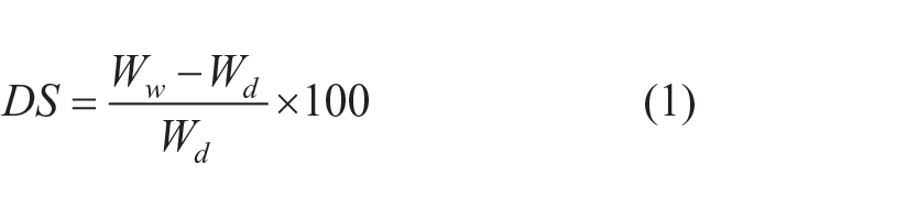
where Ww and Wd are wet and dry dressings, respectively.26
Antibacterial activity of the electrospun mats
In order to consider the examination, first, the agar culture medium for the growth of bacteria was prepared. Thus, 40 g of Nutrient agar powder were mixed with 1000 cc distilled water. The spout was sealed with sterilized gas in order to prevent bacteria from entering into the culture medium and then heat it . The solution of the culture medium was boiled, and the color of the lamina color turned into a clear color. Then, the culture medium transported in an autoclave at a temperature of 120°C for 30 min to sterilize.
After sterilizing all the instruments, the 0.5 McFarland )MC( solution was prepared by mixing 9.95 mL of 1% sulfuric acid solution and 0.05 mL of barium chloride (1.175%). Escherichia coli (AATCC 11303) and Staphylococcus aureus (AATCC 25938) were used to determine the antibacterial properties. Accordingly, bacterial suspensions with a concentration of 1.5 × 10⁸ CFU mL−1 were prepared and added directly to the samples with a dimension of 1 × 1 cm2 for 1 h . They were then incubated at 37°C for 72 h. Finally, the number of bacterial colonies in culture media was counted, and by using the formula below, the percentage of bacteria was reduced [27] (equation (2))
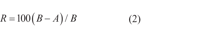
where R is percent reduction, A is the number of bacteria recovered from the inoculated treated test specimen swatches in the jar incubated over desired contact period, and B is the number of bacteria recovered from the inoculated treated test specimen swatches in the jar immediately after inoculation (at 0 contact time). 28
MTT test
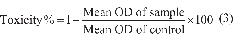
The toxicity of cellular PVA/PLA, PVA-CTX/PLA, PVA-TXA/PLA, and PVA-CTX-TXA/PLA dressings to L929 fibroblast cells was evaluated. All dressings were cut in sterile conditions. This test was performed directly according to standard ISO-10993-5. The culture conditions were incubated with 5% carbon dioxide gas, 90% humidity, and 37°C culture temperature. The cell culture plate was also selected as a negative control (cytotoxic non-response). The diphenyl tetrazolium bromide test was used to evaluate cell proliferation. First, the specified number of cells was cultured on the surface of each sample. Dimethylthiazol diphenyl tetrazolium bromide test was used to evaluate cell proliferation. First, the specified number of cells was cultured on the surface of each sample. In this test, 1 × 10⁴ cells per well were poured into 96 wells per cell. After 3 h, the cells are attached to the specimens, and the medium can be added to cover the whole surface of the sample. The same trend was taken for the control sample in a similar way. After the specified period of time, the culture medium was removed, and the 200 µL MTT solution was injected at 0.5 mg/mL to each well. The same trend was taken for the control sample in a similar way. After the specified time, the culture medium was removed from the cell culture, and 200 µL of MTT solution was injected at 0.5 mg/mL to each well. After 4 h, the solution was removed to the cells, and the isopropanol was added to dissolve the purple crystals. The absorbance was then measured at 570 nm.
The percentage of biocompatibility was calculated as follows (equation (3))

Cell images were also evaluated by reverse optical microscopy. For this purpose, after 24 and 48 h, samples of cell growth were placed under a microscope, and their cell growth was studied.
Results and discussion
Viscosity and morphology of electrospun mats
The viscosity of PVA, PLA, PVA-CTX, PVA-TXA, and PVA-CTX-TXA solutions was presented in Table 1.
Viscosity and conductivity of polymeric solution components.
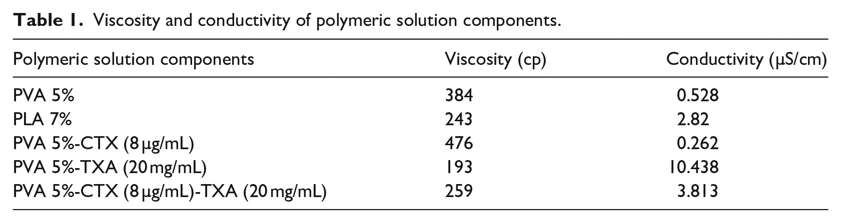
As shown in Table 1 and Figure 1, the PLA polymer solution has a viscosity of 243 cp and PVA polymer solution has a viscosity of 384 cp, in which, by adding ceftriaxone to PVA, the viscosity of the polymer solution has increased due to the size and structure of ceftriaxone molecules that cause to have more interaction with the surrounding molecules and increase the viscosity. And the other reason for the increase in viscosity is the use of ceftriaxone in powder form. Also, with an increase of the tranexamic acid drug, the viscosity of the polymer solution decreased due to disturbing chain entanglements of the polymer chains and using the form of the tranexamic acid solution. 29
Interaction of poly(vinyl alcohol) with tranexamic acid and ceftriaxone.
SEM images of electrospinning nanofibrous scaffolds are shown in Figure 2. By considering Figure 2(a) and (b), PVA nanofiber with an average diameter of 250 ± 84 nm and PLA nanofiber with an average diameter of 114 ± 44 nm are produced uniformly and without the bead. Also, it is observed in Figure 2(c) that hybrid PVA/PLA nanofiber is successfully formed. 30 As it can be seen, nanofibers with a larger diameter belong to the PVA polymer, whereas thinner nanofibers belong to the PLA polymer. By adding TXA to the PVA polymer solution, due to the low viscosity of the PVA-TXA solution, beads appeared in PVA-TXA/PLA scaffolds (Figure 2(d)). Also, the addition of TXA to PVA resulted in an increase in conductivity in the polymer solution. Subsequently, the solution was subjected to a larger tensile force using an electric field that produced thinner fibers in the PVA-TXA component.31,32 Because ceftriaxone was powdered and added to PVA, the high viscosity of the PVA-CTX polymer solution resulted in the formation of uniform and low-bead nanofibers (Figure 2(e)). 33 As observed in Figure 2(f), by adding both drugs to the PVA polymer solution, due to the viscosity and electrical conductivity in the PVA-CTX-TXA and PLA have been approximately close together, so it can be seen that all of the nanofibers are produced approximately with equal diameters.
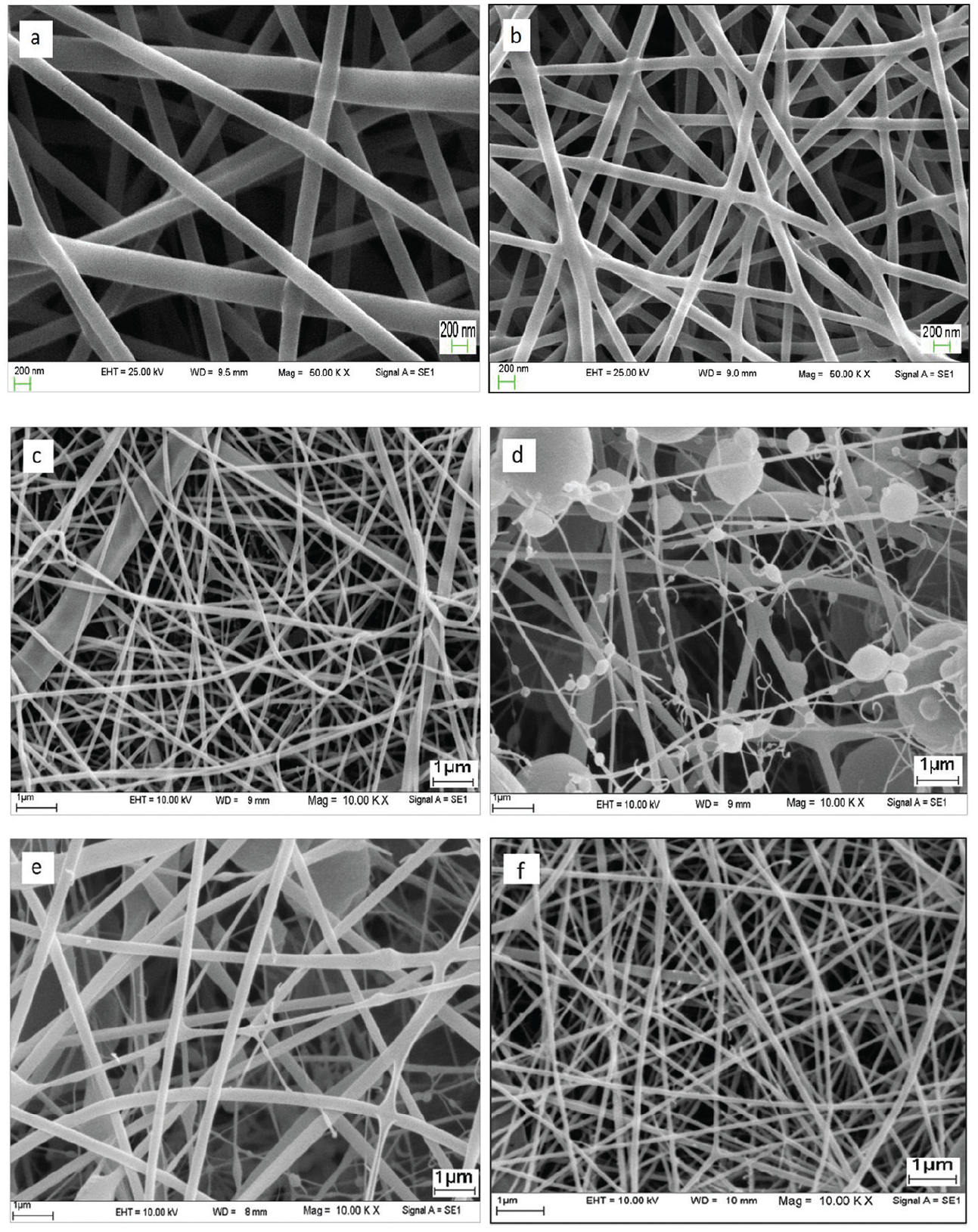
Images of electron microscopy of nanofibers loaded with the drug: (a) PVA 5%, (b) PLA 7%, (c) PVA 5%/PLA 7%, (d) PVA 5%-TXA (20 mg/mL)/PLA, (e) PVA 5%-CTX (8 µg/mL)/PLA, and (f) PVA 5%-CTX (8 µg/mL)-TXA (20 mg/mL)/PLA.
In general, all hybrid scaffolds have a low average diameter, in which this low diameter of nanofiber has a good advantage such as more surface area to volume ratio, the better interaction of nanofibers with cells, more control of the humidity wound and more ability for loading the drugs in pores, and, finally, increasing the antibacterial properties. 34
FTIR spectrum
The FTIR spectra of PVA/PLA, PVA-TXA/PLA, PVA-CTX/PLA, and PVA-CTX-TXA/PLA scaffolds are shown in Figure 3. In the FTIR spectrum of PVA/PLA, the characteristic peaks appeared at 2960, 1729, 1458, 1362, 1162, and 870 cm−1, which represent the symmetrical stretching mode of the C–H bond, C=O stretching of ester group, –CH bending in –CH3, −CH3 symmetric deformation, C–O stretching, and –C–C stretching, respectively.35,36 The band around 3448 cm−1 is attributed to the presence of water that is absorbed by PVA molecular chains. 37 In the PVA-TXA/PLA scaffold spectrum (Figure 3(b)), the bands appearing in 3431 cm−1 are related to the stretching vibration peak of its side hydroxyl groups, 38 and a band at 1539 cm−1 can be ascribed to NH3+ deformational mode in tranexamic acid. Signals at 1637 and 1382 cm−1 are assigned to the asymmetric and symmetric stretching mode of –COO–, respectively. 39 Also the absorption peak at 1093 cm−1 is related to C‒O=C stretching.40 Also, as shown in Figure 3(c), the band at 2949 cm−1 is assigned to stretching vibrations of C–H group, and stretching vibration of carbonyl groups (C=O) appeared at 1734 and 1627 cm−1. The bands at 1368 and 1044 cm−1 could be assigned to stretching vibrations of C–N and C–O, respectively.41 Another moderate intensity peak is at 1469 cm−1, which might be related to the bending vibration of CH3. The bands appearing in the 3400 cm−1 range belong to all types of hydrogen-bonded OH groups. The peak appearing in 3440 cm−1 is related to this group. Also, the courier appearing in area 1170 cm−1 was of ester specification.38,40
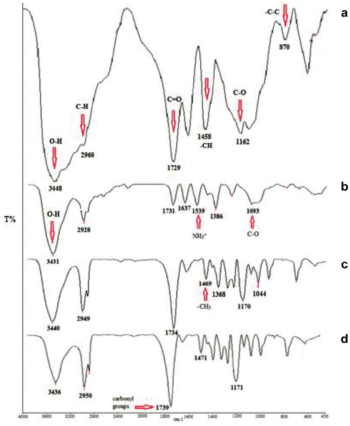
The FTIR spectra of electrospun: (a) PVA/PLA, (b) PVA-TXA/PLA, (c) PVA-CTX/PLA, and (d) PVA-CTX-TXA/PLA.
It can be seen that all the characteristic peaks of PVA/PLA, PVA-TXA/PLA, and PVA-CTX/PLA were visible in PVA-CTX-TXA/PLA scaffold, and some peaks were being overlapped (Figure 3(d)). When compared with pristine PVA/PLA, the peak intensities were observed to be decreased with CTX and TXA content. This may be due to the formation of hydrogen bonds among the components, and the interhydrogen bonds formed between two different macromolecules were stronger than those formed between the molecules of the same polymer. 2
Platelet activation and the ability to coagulate blood
In order to evaluate the potential of nanofibrous bone grafting, a general review of blood coagulation was performed. After adding blood, the nanofibrous coating was completely covered with blood. After 10 min of incubation, the dressings showed complete blood coagulation. The red blood cells that were trapped in the clot were hemolyzed with water. The absorbance of the hemoglobin solution was recorded at 540 nm. The amount of absorption above the hemoglobin solution indicates a slower rate of coagulation of the blood. 42 According to Figure 4, three samples including PVA/PLA, PVA-TXA/PLA and PVA-CTX-TXA/PLA, were evaluated and the absorption rate of PVA-CTX-TXA/PLA dressing was upper than PVA-TXA/PLA dressing which represents a slower rate of blood coagulation which is due to the effect of ceftriaxone drug . But compared to the PVA/PLA sample, without a drug, it has a much lower absorbance and, given the clot formed in Figure 5, it can be concluded that the PVA-TXA/PLA and PVA-CTX-TXA/PLA have good coagulation.
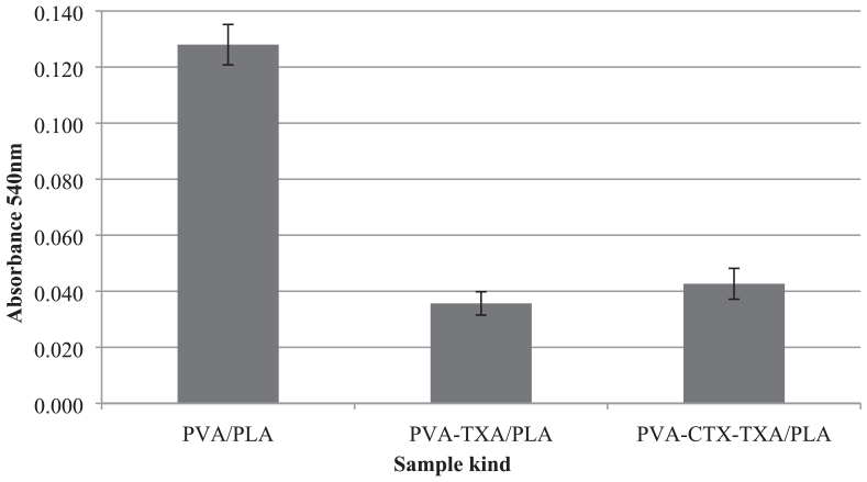
Absorption value of hemoglobin solution at 540 nm.
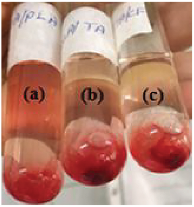
Blood clotting coagulation process: (a) PVA/PLA, (b) PVA-TXA/PLA, and (c) PVA-CTX-TXA/PLA.
Also, SEM images from Figure 6 show that active platelets spread throughout PVA-TXA/PLA and PVA-CTX-TXA/PLA nanofibers. Uniform distribution of platelets along the nanofibers indicates complete adhesion of the platelets and therefore the ability to coagulate blood. 43
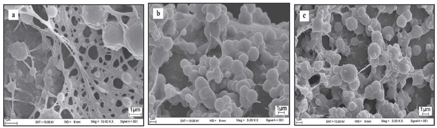
SEM images of platelet activation: (a) PVA/PLA, (b) PVA-CTX-TXA/PLA, and (c) PVA-TXA/PLA.
Swelling
Besides all other properties, a dressing has to absorb the exudates on the wound surface and provide a moist wound environment toward the wound bed. Water uptake ability of a biomaterial is an important factor for cell seeding which affects distribution of cell suspension throughout the material and a transfer efficiency of oxygen and nutriment. 44 In this study, the dressings degree of swelling was calculated using the common formulas of Swelling called Flory-Huggins. Investigation of swelling by immersed dressing in a PBS solution at pH: 7.4 is shown in Figure 7, after a specified period of time. According to this figure, the dressing loaded with TXA showed a slightly lower degree of swelling than the dressing loaded with CTX, and dressings containing both drugs have the highest degree of swelling. It was also found that an increase in the drug caused an increase in the degree of swelling in the dressings. As it can be seen, with increasing duration, the degree of swelling increased until it reached a constant rate. It was also found that during 45 min, it reached the highest swelling value. This ability to high swelling in the short term reflects the nature of the hydrophilic nature of the dressing, and it is possible to absorb wound secretions in a short time.
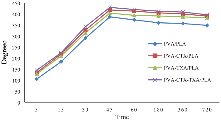
Degree of swelling (%) of PVA/PLA, PVA-CTX/PLA, PVA-TXA/PLA, and PVA-CTX-TXA/PLA.
Antimicrobial test
Prevention of bacterial infection has great importance in treating wounds, and antibacterial activity is one of the main requirements for dressing the wound because the wound is highly exposed to contagious bacteria. 24 To evaluate the antimicrobial properties of PVA/PLA, PVA-CTX/PLA, and PVA-CTX-TXA/PLA dressings, the number of colonies formed on the culture plates was counted. As shown in Figure 8, electrospinning nanofibrous of PVA/PLA without a drug showed no activity against S. aureus and E. coli bacteria. PVA-CTX-TXA/PLA dressing also showed strong antibacterial activity against both Gram-positive and Gram-negative bacteria. In the PVA-CTX /PLA dressing, the number of colonies formed on the culture plates significantly decreased. In general, the use of antibiotic ceftriaxone results in antimicrobial activity against Gram-negative bacteria E. coli and Gram-positive bacteria S. aureus. 45 However, the inhibitory effect on E. coli (Gram-negative bacteria) is higher than the S. aureus (Gram-positive bacteria), due to the greater thickness of the S. aureus cell wall than E. coli. 46
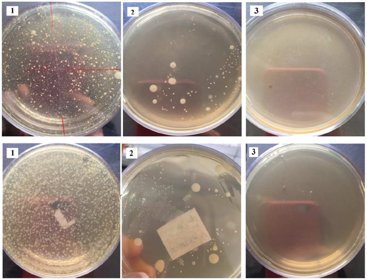
Evaluation of antimicrobial properties of (a) Gram-negative bacteria E. coli and (b) Gram-positive bacteria S. aureus: (1) PVA/PLA, (2) PVA-CTX /PLA, and (3) PVA-CTX-TXA/PLA.
Cell biology and cell morphology
In this study, the viability assay was measured for 24 and 48 h. The results of the cytotoxicity analysis of scaffolds PVA/PLA, PVA-CTX/PLA, PVA-TXA/PLA, and PVA-CTX-TXA/PLA are depicted in Figure 9. According to ISO 10993-5, samples with cell viability larger than 75% can be considered as non-cytotoxic 44 so that revealing in all scaffolds no toxicities and good biocompatibilities of composite nanofibers toward cells. The results showed that PVA/PLA scaffolds have higher cell numbers and higher cellular compatibility than those with the drug. 47 Also, it was observed that PVA-CTX/PLA scaffolds showed 80% and 88% viability after 24 and 48 h and PVA-TXA/PLA scaffolds showed 78% and 85% viability after 24 and 48 h, respectively. The reason is the potential of the low toxicity of ceftriaxone and increased cell death by the tranexamic acid drug.48,49 Consequently, the simultaneous use of two drugs in the production of scaffolds reduced the proliferation of cells and subsequently reduced the degree of cell compatibility. Based on the results, in comparison to the increase in compatibility after 24 and 48 h, it was observed that the viability of PVA/PLA scaffold was increased about 13% and the viability of PVA-CTX/PLA, PVA-TXA/PLA, and PVA-CTX-TXA/PLA scaffolds was increased about 10%.
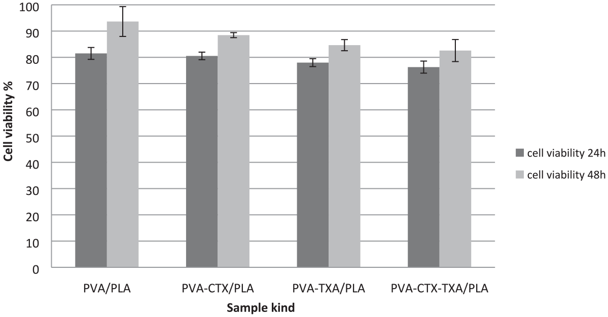
Cytotoxicity test on different mats after 24 and 48 h in L929 cells.
In examining the morphology of cell growth from microscopic images in the form, it is concluded that the growth rate of the L929 fibroblast cell increased after 24 to 48 h and subsequently increased cell compatibility (Figure 10). 50
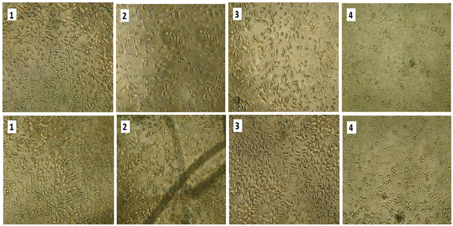
L929 fibroblast cell growth after (a) 24 h and (b) 48 h: (1) PVA/PLA, (2) PVA-TXA/PLA, (3) PVA-CTX/PLA, and (4) PVA-CTX-TXA/PLA.
Conclusion
In the present study, we successfully obtained drug-loaded PVA/PLA-blended composite nanofibers by using the electrospinning process. Obtained scaffolds were characterized by various methods. PVA/PLA loaded by tranexamic acid composite nanofibrous scaffolds showed enhanced blood clotting and excellent platelet activation ability. It was also concluded that the use of ceftriaxone antibiotics induces antimicrobial activity against E. coli (Gram-positive bacteria) and S. aureus (Gram-negative bacteria). In examining the swelling test on PVA-CTX-TXA/PLA nanofibers, it was found that this dressing has a high hydrophilic nature and can absorb wound secretions in a short time. In vitro cytotoxicity studies revealed that the scaffolds showed enhanced cell viability. All these studies indicated that these advanced PVA/PLA-drug composite nanofibrous scaffolds can be used for burn, chronic, and diabetic wound infections.
Footnotes
Declaration of conflicting interests
The author(s) declared no potential conflicts of interest with respect to the research, authorship, and/or publication of this article.
Funding
The author(s) received no financial support for the research, authorship, and/or publication of this article.