
Abstract
Zingerone loaded chitosan nanoparticles (Z-NPs) were developed to deliver the Zingerone across cell membrane and to further enhance its anti-virulence property. The Z-NPs were characterized with respect to size, percentage entrapment efficiency (% EE), zeta potential and percentage drug release. Further the Z-NPs were evaluated for antioxidant DPPH assay, antibiofilm, anti-virulence activities, and gene expression profiles. The developed Z-NPs showed an average size of 390 nm, zeta potential of +56.6 mV, 67% drug entrapment efficiency, and exhibited pH dependent controlled release of Zingerone over a period of 5 days (up to 80%). The Z-NPs retained the antioxidant effect of Zingerone as assessed by DPPH scavenging assay. Evaluation of nanoformulation for anti-virulence potential against Pseudomonas aeruginosa depicted significant reduction in swimming, swarming, and twitching motilities along with quorum sensing inhibition and eradication of biofilms. Decrease in expression of quorum sensing (QS) genes was also observed in the presence of Z-NPs. The results of the present study revealed that Z-NPs could be exploited as a promising anti-virulence candidate against P. aeruginosa infections.
Introduction
Pseudomonas aeruginosa causes opportunistic infections and is a major threat to mankind due to increasing incidences of multi-drug resistance and biofilm formation. Biofilm formation is linked to a form of inter-bacterial communication known as quorum sensing (QS) in which quorum sensing signal molecules (QSSM) are known to regulate virulence and gene expression of P. aeruginosa. Given the advent of multi-drug resistance in bacteria, over the past decade, bioactive plant based natural products have caught the attention of researchers as novel therapeutic agents due to their antibacterial activity especially against multi-drug resistant Gram positive and Gram negative bacteria. Some of the plant based bioactives (like ajoene, gingerol, thymol, etc.) have significant antioxidant, 1 anti-inflammatory, 2 antimicrobial, 3 and anti-cancer properties. 4 Zingerone [4-(4-hydroxy-3-methoxyphenyl) butan-2-one], also called “vanillyl acetone,” has gained significant attention for its potential therapeutic use against variety of human diseases. It has been shown to reduce endotoxin induced acute lung injury in mice and also suppressed liver inflammation induced by antibiotic mediated endotoxemia. 5 However, low aqueous solubility, and poor bioavailability limits the full therapeutic use of this molecule. 6 Hence, to utilize the full therapeutic benefits of this molecule, drug delivery strategy needs to be developed.
In recent times, there is an increasing interest in nanotechnology based formulations for effective drug delivery. 7 Some beneficial properties of nanocarriers including small size and high surface-to-volume ratio allow them to gain access into the microorganisms. Direct interaction of drugs with microbial cell membranes/walls or key proteins/enzymes may inhibit pathogen growth and result in cell death. 8 More importantly, the positively charged nanoparticles, improve the interaction with negatively charged membranes of microorganisms which results in decreased osmotic stability, membrane disruption, and eventual leakage of intracellular elements. 9 Many polycationic biopolymer, such as chitosan are being used to take advantage of positively charged colloidal particles in the range 10 to 1000 nm. 10 Chitosan is deacetylated chitin derivative biopolymer in which reactive amino groups provide condition for electrostatic interactions. 11 In addition to antimicrobial property,12,13 it is also beneficial due to its biodegradable and biocompatible nature. Dextran sulfate, which is negatively charged, is often used with chitosan to reduce cell toxicity caused by the high positive charge of chitosan. 14 The chitosan-dextran sulfate combination was used to synthesize Zingerone nanoparticles (Z-NPs), which eventually were formed by electrostatic interaction between sulfate groups of dextran sulfate and amino groups of chitosan. This is one of the simplest methods for synthesizing nanoparticles without using external cross-linking agents. 15 The stable nanoparticles are highly adjustable, in terms of zeta potential, which can be achieved by modification of polymers’ ratio and pH. 16
The present investigation is focused at the development of nanocarriers employing the macromolecules like chitosan and dextran sulfate, for the delivery of Zingerone across the pathogen and evaluating the anti-virulence potential of such nanoformulation against P. aeruginosa.
Material and methods
Bacterial strains
Standard strains of P. aeruginosa PAO1, and E. coli MG4 (pkDT17), were obtained from Dr Barbara H Iglewski, Department of Microbiology and Immunology, University of Rochester, New York, USA and maintained in nutrient agar stabs at 4°C. Agrobacterium tumefaciens A136, was obtained from Dr J Handlesman, Wincoinsin University USA. Both these strains were employed as biosensor strains. Pure Zingerone was procured from Gogia Chemicals Ltd. India.
Standardization of chitosan-dextran sulfate for nanoparticle formulation
Blank nanoparticles were synthesized using chitosan and dextran to encapsulate zingerone following the method of Anitha et al. 16 Different concentration of chitosan solution 0.075% to 0.1% (w/v) were prepared in 1% acetic acid and dextran sulfate 0.075% to 0.1% (w/v) solutions were made in distilled water. Dextran sulfate solution was added dropwise to chitosan solution under vigorous stirring in different volume ratios 3:1, 3:2, and 3:3. At volume ratio 3:1, nanoparticles were formed almost instantaneously at room temperature. These preparations were checked for the presence of opalescence. The concentrations giving opalescence were selected for nanoparticle preparation. Suspension was stirred for 15 min, and then nanoparticles were separated from the suspension by centrifugation at 20,000 rpm for 20 min (REMI, PR-24, Mumbai, India), followed by washing twice in distilled water and re-suspended in 10 mM phosphate buffer saline.
Preparation of Zingerone nanoparticles
Z-NPs were prepared by using ion-gelation method. 16 Different concentrations of Zingerone (1 mg, 2 mg, 3 mg, 4 mg, and 5 mg/mL), dissolved in methanol, were added to chitosan solution and mixed thoroughly by stirring. Dextran sulfate solution was added drop wise to chitosan solution under vigorous and continuous stirring in a volume ratio of 3:1 (chitosan:dextran sulfate) to ensure proper mixing. The final mixture was warmed (40°C) in a water bath and allowed to stir for 30 min. To obtain the opalescent suspension of nanoparticles spontaneously, methanol was evaporated. The drug-loaded nanoparticle suspension was centrifuged at 20,000 rpm for 30 min followed by washed twice in water to remove unencapsulated compound. The pellet was then re-dispersed in 10 mM phosphate buffered saline (PBS).
Characterization of Zingerone nanoparticles
Mean particle size, polydispersity index (PDI) and zeta potential of the prepared Z-NPs were determined by using Zetasizer (Zetasizer 2000HS, Malvern Instruments Limited, UK) at a temperature of 25°C. For this purpose, final nanoparticle suspension was diluted 10 to 20 times with deionized water and kept in polystyrene cuvettes for measurement.
Energy dispersive X-ray spectroscopic analysis
The energy dispersive X-ray spectroscopic (EDS) analysis of Z-NPs was also done to confirm the entrapment of zingerone inside the nanoparticles and to confirm the purity of the nanoformulation. The EDS spectra of all the formulation ingredients including Zingerone, dextran sulfate, chitosan blank NPs, and Z-NPs were recorded on FESEM (SU8010 HITACHI, Japan) with X-ray source of Bruker X FLASH 6130, USA.
Determination of percent entrapment efficiency
The percent entrapment efficiency (% EE) of Zingerone within the Z-NPs was determined by indirect method. Nanoparticles suspension was centrifuged at 20,000 rpm for 20 min. Supernatant was collected and absorbance of free Zingerone at 254 nm using UV spectrophotometer was taken. Amount of un-entrapped Zingerone (free zingerone) was calculated from the standard calibration curve of zingerone. Entrapment efficiency [%EE] was calculated based on the ratio of amount of drug present in the Z-NPs to the concentration of drug used during the loading process.
In vitro drug release studies
The in vitro drug release profile of Zingerone from Z-NPs was carried out at two different pH (5 and 7.4) using acetate buffer and phosphate buffer saline (PBS) respectively as release medium. Biological medium [in the form of artificial urine pH 6.5] was also used as one of the release medium to study the stability of nanoformulation. Briefly, a known quantity of Z-NPs was taken in dialysis bags (cellulose membrane, MW cut-off 10,000 Da, Sigma-Aldrich chemical Co. Ltd., St Louis, USA). After tying both the ends, each dialysis bag was immersed in different beakers containing 100 mL of PBS (10 mM), 100 mL of acetate buffer and 100 mL of artificial urine media [comprising of potassium chloride (0.2 g/L), sodium chloride (8 g/L), di-sodium hydrogen phosphate (1.14 g/L), potassium dihydrogen phosphate (0.2 g/L), milli-Q water up to 1 L]. 17 These were incubated in a water bath shaker at 37°C. At defined time intervals, 3 mL of sample was withdrawn from each dialysis chamber. Samples were then centrifuged and the supernatant was analyzed for Zingerone content by UV spectrophotometer. Simultaneously, fresh medium was replaced back in the same amount. 18 The percentage Zingerone leakage from Z-NPs in urine medium after 1 h was taken as a means of predicting stability.
Stability of Z-NPs at different temperatures
Physical stability was checked using wavelength scan of Z-NPs using UV spectrophotometer to determine λmax and changes were observed in terms of opalescence versus aggregation. Z-NPs formulation was stored at different temperatures (−20°C, 0°C, 4°C, and 30°C) from 0 h to 6 months and the nanoparticles were assessed after specified time period for any change in their physical appearance including particle aggregation, phase separation, etc.
Quorum sensing inhibition activity of Z-NPs
The quorum sensing inhibition (QSI) activity of Z-NPs was assessed by qualitative screening using the reporter strain E. coli MG4 and Agrobacterium tumefaciens A136 by the method of Vattem et al. 19 The QSI was checked at different temperatures (−20°C, 0°C, 4°C, and 30°C). Diameters of turbid colorless halos around the wells containing test agents were determined which depicted the QSI activity of Z-NPs. Zingerone free nanoparticles and Zingerone alone were kept as controls.
Effect of Z-NPs on quorum sensing signal molecules
To evaluate the effect of Z-NPs on the inactivation of QSSMs, P. aeruginosa PAO1 was allowed to grow in the presence and absence of Z-NPs and Zingerone alone for 24 h at 37°C. Ethyl acetate extract of all untreated and treated supernatant of P. aeruginosa were extracted and TLC overlay method was carried out using A. tumefaciens A136 to identify QSSMs. 20 Synthetic QSSM, 3O-C12-HSL was also loaded on TLC plates as positive control.
Determination of Minimum Inhibitory Concentration of Z-NPs
The minimum inhibitory concentration (MIC) of Zingerone and Z-NPs against P. aeruginosa PAO1 were determined by microdilution method. About 100 µL of Z-NPs (5 mg/mL of Zingerone with entrapment efficiency of 67%) and 100 µL of Zingerone (30 mg/mL) was incorporated into the plate and the lowest dilution was achieved by double serial dilution. For blank NPs, the highest concentration of chitosan incorporated into the plate (1000 µg/mL) and lowest (10 µg/mL) was achieved by double serial dilution and incubated at 37°C for 24 h. Next day visible growth was observed. Luria broth (LB) without P. aeruginosa PAO1 was taken as negative control, while P. aeruginosa grown in LB was taken as positive control. The lowest concentration at which no visible growth was observed was taken as MIC. 21
Anti-virulent activity of Z-NPs
To estimate the production of virulence factors, P. aeruginosa, PAO1 was inoculated overnight into LB with Zingerone and Z-NPs separately at 37°C. The suspension was centrifuged at 10,000 rpm for 15 min. Cell free supernatant was separated for the analysis of virulence factors. Sterile medium served as control.
Pyocyanin
Pyocyanin was measured using the method of Huerta et al. 22 Briefly, 3 mL of culture supernatant was mixed in 1.2 mL of chloroform and incubated for 30 min at room temperature. The absorbance of chloroform layer having pyocyanin was measured at 690 nm. The concentration of pyocyanin was expressed in μg/mL.
Hemolysin
Hemolysin was estimated quantitatively in cell free supernatant by the method of Lankisch and Vogt. 23 In brief, 2% human RBCs suspension was added in 1.5 mL of cell free supernatant. After incubating the mixture at 37°C for 2 h, the absorbance was taken at 545 nm. Amount of hemolysin was expressed in terms of hemoglobin released (mg/mL).
Protease
To quantify protease production, the method of Visca et al. 24 was followed. Briefly, 1.5 mL cell culture supernatant was diluted in 10 mM Tris-HCl buffer (pH 7.5). This supernatant was incubated with 15 mg hide azure powder for 1 h under shaking conditions at 37°C. Centrifugation was done at 3500 rpm and absorbance was taken at 595 nm.
Elastase
Elastin congo red was used as a substrate for determination of electrolytic activity by the method of Visca et al. 24 Briefly, 5 mg of elastin congo red was added in 1 mL of cell free supernatant with 100 mM Tris-HCl buffer (pH 7.0). Mixture was then incubated at 37°C for 2 h under shaking conditions. To stop reaction, 1 mL of 0.7 M sodium phosphate buffer (pH-6.0) was added and absorbance was taken at 495 nm after centrifugation. Elastase activity was expressed in units per litre (U/L).
Alginate
The method of Mathee et al. 25 was followed to estimate the amount of alginate in the culture supernatant of P. aeruginosa. In brief, alginate was precipitated in cell free supernatant with equal volume of 2% (w/v) cetylpyridium chloride (prepared in distilled water). The mixture was centrifuged at 10,000 rpm for 10 min at room temperature. Alginate pellet was suspended in 5 mL of 1 M NaCl. The mixture was precipitated again with 5 mL of 2-propanol followed by centrifugation at 10,000 rpm for 10 min. Depending on alginate quantity, pellet was resuspended in saline. Alginate concentration was estimated using carbazole/borate method after taking absorbance at 500 nm.
Motility assay
Swimming motility
Media plates containing 1.0% tryptone, 0.5% NaCl, and 0.3% agarose were point inoculated with overnight culture of P. aeruginosa PAO1 grown in presence of Zingerone and Z-NPs. After incubation at 30°C for 24 h, swimming motility was determined by measuring the radius of circular expansion of bacterial migration from the point of inoculation. 26
Swarming motility
Nutrient agar (8 g/L) supplemented with glucose (5.0 g/L) was prepared and plates were point inoculated with overnight culture of P. aeruginosa PAO1 grown in presence of Zingerone and Z-NPs. After incubation at 37°C for 24 h, swarming motility was determined by measuring circular turbid zones.
Twitching motility
Media plates containing agar layer of Luria broth (1.0% agar) were prepared and stabbed with toothpick up to bottom of the petri dish with overnight culture of P. aeruginosa PAO1 grown in presence of Zingerone and Z-NPs. After incubation at 37°C for 48 h, a hazy zone of growth at the interface between the agar and polystyrene surface was observed.
Effect of Z-NPs on expression profiles of key QS genes
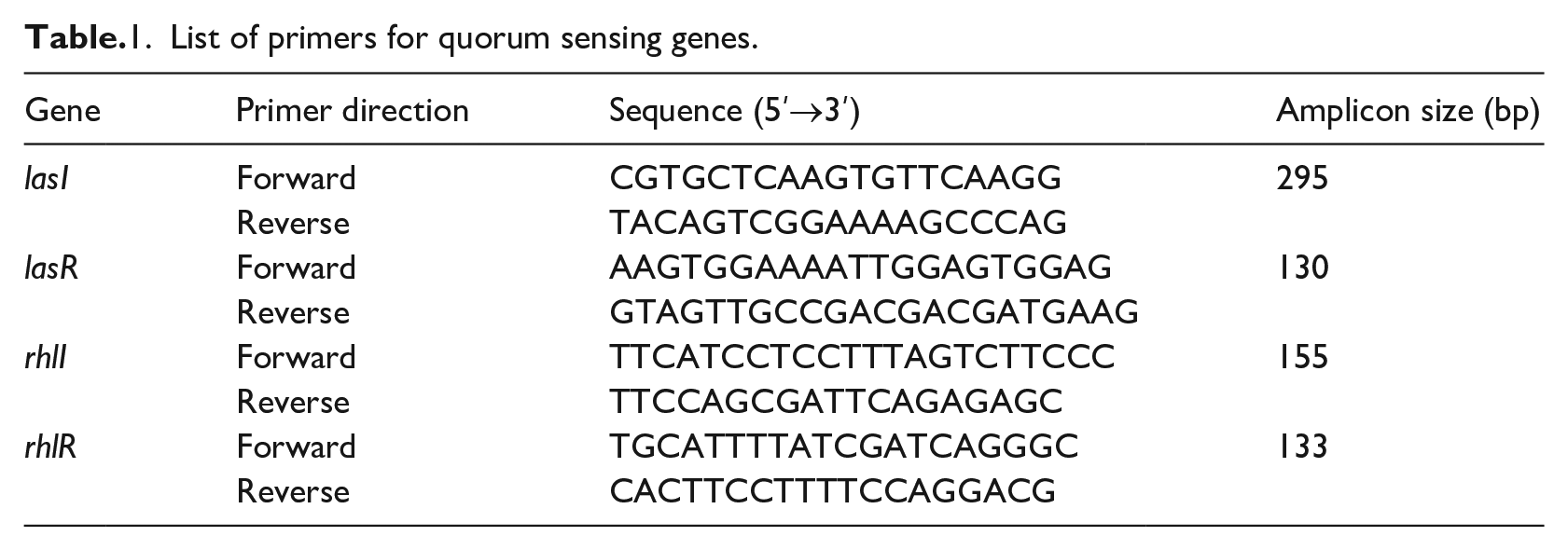
The quantitative real time polymerase chain reaction (qRT-PCR) was performed to assess the effect of Z-NPs on the expression of QS regulated genes. P. aeruginosa PAO1 was grown in Luria broth with shaking conditions at 37°C to obtain log phase cells. The grown cells were adjusted to optical density (OD) of 0.5 for RNA isolation. Similarly test sample was grown in presence of Z-NPs (5 mg/mL) and log phase cells were separated to isolate RNA. RNA was extracted by TRIzol reagent method. DNase was used to remove residual DNA. Agarose gel electrophoresis was used to check quality of extracted RNA and later quantified by measuring the absorbance at 260/280 nm using Nanodrop spectrophotometer (2000-C Thermo Scientific, USA). The cDNA synthesis was carried out using cDNA kit (ThermoFisher Scientic, USA) according to the manufacturer protocol. To study the expression of target genes, qRT-PCR was performed with SYBR green qPCR master mix. 16S RNA gene was used as an internal control to calculate the relative expression of target genes. List of primers used is given in Table 1. The fold change in gene expression was determined in terms of 2−∆∆CT. Data was compiled after performing three independent experiments.
List of primers for quorum sensing genes.
Anti-biofilm effect of Z-NPs
Biofilm inhibition assay
Biofilm formation was carried out using 96 well microtiter plates (Corning, NY, USA) for 7 days at static conditions. P. aeruginosa cells (1 × 10⁷ CFU/mL) were grown in absence and presence of Zingerone and Z-NPs at 37°C for 24 h. Loosely adhered planktonic cells were removed by washing three times with PBS. Crystal violet (0.1% w/v) (prepared in distilled water) was used to stain the adhered cells for 15 min. Excess dye was rinsed with PBS washing followed by elution with 95% ethanol. Absorbance of the sample was taken at 600 nm. 27
Biofilm eradication assay
Biofilms were generated in 96 well microtiter plates under static conditions for 7 days. Next day, the established biofilms were treated with Zingerone and Z-NPs. Media was drained from wells and adhered cells were stained with crystal violet (0.1% w/v) prepared in distilled water for 15 min at room temperature followed by washing with sterile PBS to remove excess dye. The stained biofilms were eluted with 95% ethanol and absorbance of the sample was taken at 600 nm.
Anti-oxidant activity of Z-NPs
The DPPH (2, 2-diphenyl-1-picrylhydrazyl) radical scavenging activity of the Z-NPs and Zingerone was determined quantitatively. The DPPH free radical based electron-transfer produces a violet solution in ethanol. This free radical, which is stable at room temperature, gets reduced in presence of an antioxidant molecule, giving rise to colorless ethanol solution. The antioxidant effect is proportional to the disappearance of DPPH free radical in the test samples. The reaction mixture consisted of 0.5 mL Z-NPs, 3 mL absolute ethanol, and 0.3 mL of 0.5 mM DPPH. The contents were mixed vigorously and incubated for 100 min at room temperature in dark. The mixture was observed for color change and absorbance was measured at 517 nm. The DPPH scavenging activity was calculated according to given formula. 28
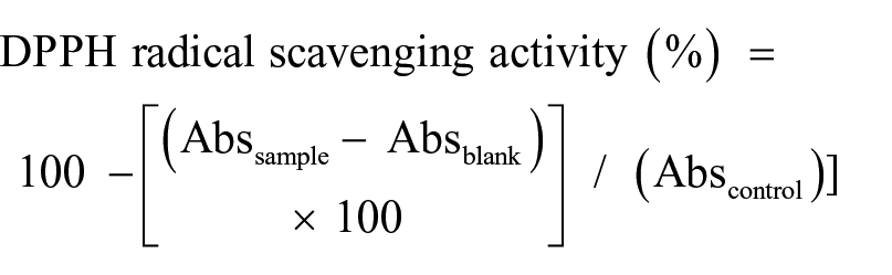
Where:-
Abssample is the absorbance of DPPH radical + sample;
Absblank is the absorbance of sample + methanol;
Abscontrol is the absorbance of DPPH radical + methanol
Statistical analysis
All the experiments were performed in triplicates and repeated on different days. Data of treated and untreated groups were compared by using student-t test. The p values were calculated and p < 0.05 was considered as significant.
Results
Synthesis of Z-NPs
Zingerone nanoparticles were prepared in order to increase stability, enhance delivery of the Zingerone across cell membrane and thus improve its anti-virulence property. Z-NPs were prepared by using the ion-gelation method. Concentration of zingerone to be entrapped into chitosan-dextran sulfate nanoparticles was selected based on the entrapment efficiency of the formulations using different concentration of Zingerone. As the concentration of Zingerone increased from 1 mg/mL to 4 mg/mL, the entrapment efficiency decreased (data not shown). At 5 mg/mL concentration of Zingerone, the Z-NPs showed best entrapment efficiency. Opalescent formulation of nanoparticles was observed with chitosan and dextran sulfate (0.1%) at 3:1 (v/v).
Particle size, zeta potential, polydispersity index, entrapment efficiency, and morphology of Z-NPs
Developed nanoformulation was found to be nanometric in size (392 nm), with PDI < 0.4, indicating a uniform size distribution of the nanoparticles. Z-NPs had zeta potential of +56.6 mV, which indicates the positive surface charge on the particles, which may help to adhere the Z-NPs to the biological membrane. The entrapment efficiency was found to be around 67%. The characteristics of different Z-NPs formulations are depicted in Table 2.
Characterization of zingerone nanoparticles (Z-NPs).
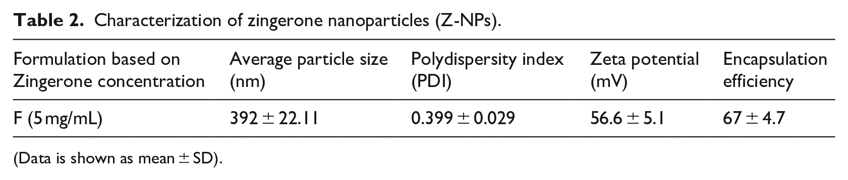
(Data is shown as mean ± SD).
Energy-dispersive X-ray spectroscopy analysis
Zingerone, being a simple molecule, comprises of only “C” and “O” atoms, while chitosan is made up of “C,” “O,” and “N”. The presence of Na in the blank NPs was attributed to the presence of dextran sulfate sodium. It was further observed that % of “C” was reduced, while % of “O” was increased in Z-NPs, as compared to free Zingerone, due to the complex nano-assembly formation (Table 3). Similarly the presence of elements like “N,” “S,” and “Na” in Z-NPs confirmed the encapsulation of Zingerone in chitosan and dextran sodium sulfate. The absence of any other element confirmed the purity of the developed system (Figure 1(a)–(e)).
Percentage density of the elements present in the tested compounds.
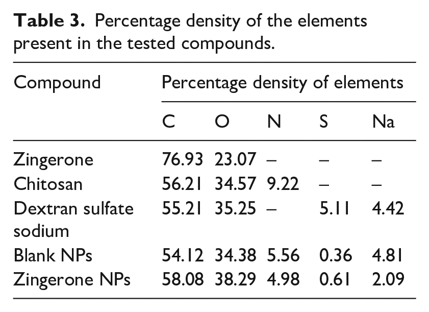
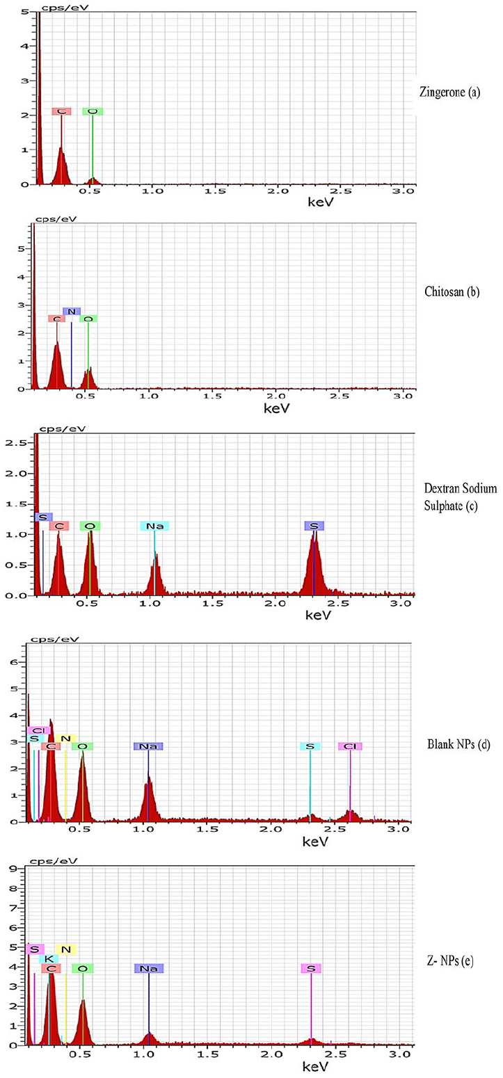
EDS analysis spectra of (a) Zingerone, (b) chitosan, (c) dextran sulfate sodium, (d) blank NPs, and (e) Z-NPs.
In vitro release profile of Z-NPs
In vitro release study of Zingerone from Z-NPs was carried out at different pH to (1) To confirm the encapsulation of Zingerone; (2) To understand the release behavior of Zingerone from chitosan nanoparticles; and (3) To determine the optimum condition (pH of medium) for release of Zingerone from the nanoparticles. The initial fast release was observed till the first 12 h. Zingerone was released up to 53% and 44% in buffer solutions with pH-5 (sodium acetate buffer) and pH-7.4 (PBS), respectively. After 24 h, the release rate was slow, but sustained release was observed which lasted for about 120 h (5 days). Drug release was up to 80% and 78% at pH 5 and pH 7.4, respectively (Figure 2). In vitro release of Zingerone from Z-NPs in urine medium at 37°C for 60 min was observed to be less than 10% (actual value is 9.87%). These results indicated that the formulation was stable in this medium as well.
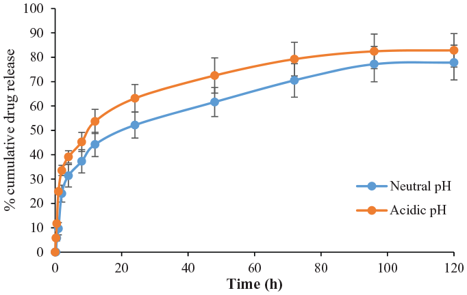
Drug release profile of Z-NPs at neutral pH (7.4) and acidic pH (5.0).
Stability studies of Z-NPs
Physical stability of Z-NPs was assessed by observing the change in opalescence versus aggregation and by measuring the absorbance of nanoparticle formulation kept at different temperatures (−20°C, 0°C, 4°C, and 30°C). No change in absorbance as well as physical appearance of the formulation was observed at different temperatures.
Anti-quorum sensing activity of Z-NPs
The quorum sensing inhibition (QSI) of Z-NPs was assessed by qualitative screening using the reporter strain E. coli MG4 and A. tumefaciens (A136) (Figure 3(a) and (b)). Bigger zones of inhibition was observed around the wells containing Z-NPs, indicating either inhibition in production or inactivation of QSSMs. The diameter of colorless zone of inhibition, which is an indicator of QSI activity, remained almost the same irrespective of temperature settings which confirmed that different temperature conditions do not affect the quorum sensing inhibition property of Z-NPs. The role of Z-NPs and free Zingerone in the inactivation of QSSMs was determined using TLC overlay method. TLC profile of treated QSSMs with Z-NPs showed the complete absence of QSSMs spots in chromatogram overlaid with A. tumefaciencs A136 as compared to untreated control. However, a single spot of QSSMs was observed in extract obtained after treatment with Zingerone (Figure 4).
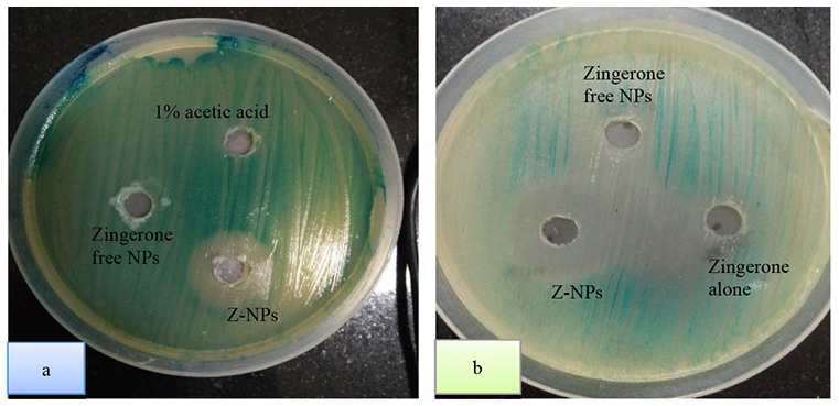
Quorum sensing inhibition activity of Zingerone, blank nanoparticles, and Zingerone nanoparticles (Z-NPs) formulation using reporter strain (a) E. coli (MG4) and (b) Agrobacterium tumefaciens (A136).
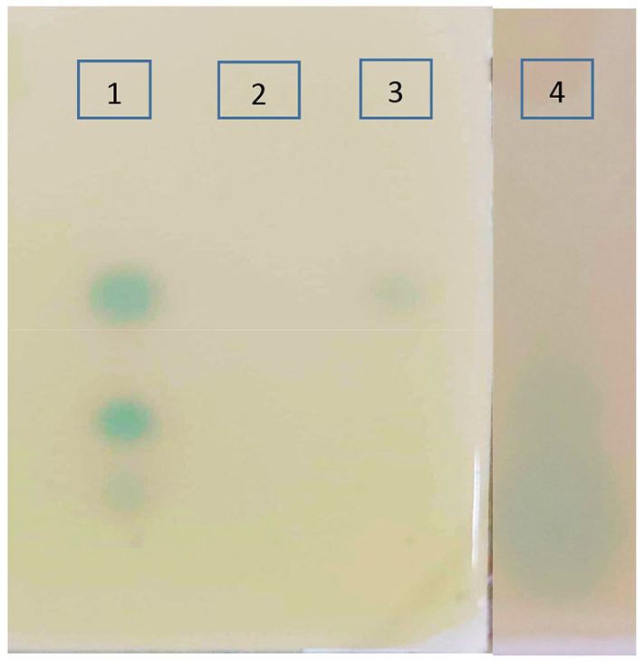
TLC overlay profile indicating the effect of Zingerone nanoparticles (Z-NPs) on quorum sensing signal molecules QSSMs. Lane 1-Ethylacetate extract of untreated PAO1, Lane 2-Ethyl acetate extract of PAO1 which was treated with Z-NPs, Lane 3-Ethyl acetate extract of PAO1 treated with free zingerone, and Lane 4-Synthetic 3-oxo-C12-HSL.
Antibacterial activity of Z-NPs
MIC of Zingerone and Z-NPs and was determined against P. aeruginosa PAO1 by using micro-dilution method. MIC of 30 mg/mL, 500 µg/mL, and 60 µg/mL was observed for Zingerone alone, blank NPs, and Z-NPs, respectively indicating that Z-NPs had significantly lower MIC value as compared to Zingerone (Figure 5). Further Sub-MIC of Zingerone and Z-NPs was selected on the basis of growth profile analysis of P. aeruginosa. It was observed that 10 mg/mL of Zingerone and 30 µg/mL of Z-NPs did not have any negative effect on growth as compared to untreated control and hence these were selected as Sub-MIC values. All further experiments were carried out at Sub-MIC value of 10 mg/mL of Zingerone and 30 µg/mL of Z-NPs.
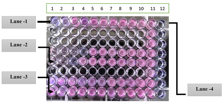
Growth of P. aeruginosa PAO1 in presence of Zingerone alone (Lane-1), Zingerone nanoparticles (Z-NPs) (Lane-2), and Blank nanoparticles (Lane-3), column 11 served as positive control and column 12 served as negative control.
Anti-virulent activity of Z-NPs
Reduction in levels of all the virulence factors produced by P. aeruginosa was observed in the presence of Zingerone alone. However, further significant reduction in (p < 0.01) production of all the virulence factors (alginate, pyocyanin, cell bound hemolysin, cell free hemolysin, elastase, and protease was observed in presence of Z-NPs (Figure 6(a)–(f)). Alginate was also significantly reduced (71%) in Zingerone alone and (80.3%) in Z-NPs respectively (Figure 6(a)). Maximum cell free hemolysin (0.39 mg/mL) was produced by P. aeruginosa PAO1 and Z-NPs was able to reduce the level up to (0.094 mg/mL) as compared to Zingerone alone (Figure 6(d)). Similarly, all the extracellular enzymes; (elastase and protease) (Figure 6(e) and (f)) were found to be significantly reduced in the presence of Zingerone nanoparticles.
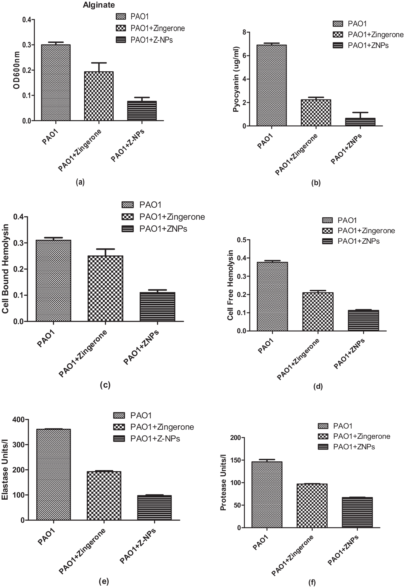
Effect of Zingerone and Z-NPs formulation on (a) alginate, (b) pyocyanin, (c) cell bound hemolysin, (d) cell free hemolysin, (e) elastase, and (f) protease production by P. aeruginosa.
Reduction of motility phenotypes by Z-NPs
P. aeruginosa showed characteristic motility phenotypes like swimming, swarming, and twitching in the form of smooth spreading, dendritic appearance, and circular zones, respectively. After the treatment with Z-NPs, a significant reduction in swimming, swarming, and twitching motilities was observed in the presence of Z-NPs as compared to untreated control (Table 4).
Estimation of motility (Zone diameter in mm) exhibited by P. aeruginosa grown in absence and presence of Zingerone and ZNPs.
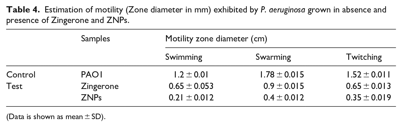
(Data is shown as mean ± SD).
Down-regulation of quorum sensing genes by Zingerone nanoparticles
Expression of QS-regulated genes including lasI, lasR, rhlI, and rhlR was estimated by qRT-PCR. It was observed that maximum downregulation took place in lasI gene (4.16 fold), as compared to lasR, rhlI, and rhlR (<2 fold) in comparison to the untreated control and internal control (Figure 7).

Inhibition of QS genes in P. aeruginosa PAO1 by Zingerone nanoparticles (Z-NPs). Expression profiles are shown for lasI, lasR, rhlI, and rhlR. The assays were independently repeated at least three times, and the data shown are representative of fold change.
Anti-biofilm efficacy of Zingerone nanoparticles
Biofilm inhibition
Biofilm inhibition ability of Zingerone and Z-NPs were evaluated. P. aeruginosa biofilms were allowed to form in absence (control) and in presence of Zingerone and Z-NPs. The blank NPs did not show any anti-biofilm effect (Data not shown). Weak effects on biofilm formation were observed up to fourth day however, there was pronounced decrease in biofilm mass after fourth day. Free and encapsulated Zingerone inhibited biofilm formation. However, maximum reduction in biofilm formation was observed with Z-NPs treated biofilms as compared to untreated control.
Biofilm eradication
The preformed biofilms, young (1 day), old (4 days), and mature (7 days) biofilms were treated with Z-NPs and Zingerone alone (Figure 8(b)). Z-NPs were able to significantly eradicate (p < 0.001) 1 day old, 4 days old, and 7 days old established biofilms as compared to control group, where blank NPs had no effect on pre-formed biofilms (Data not shown).
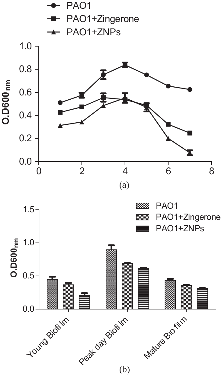
(a) Inhibition of biofilm formation by standard strain PAO1 of P. aeruginosa in absence and presence of Zingerone and Zingerone nanoparticles. (b) Biofilm eradication of preformed biofilms of P. aeruginosa PAO1 in absence and presence of Zingerone and Zingerone nanoparticles (Z-NPs).
Antioxidant effect of Z-NPs
The antioxidant effect of Z-NPs was assessed by the DPPH scavenging assay. It was observed that, color of DPPH solution changed from deep violet to pale yellow upon addition of Z-NPs, indicating its free radical scavenging capability. Blank NPs were taken as negative control. Antioxidant activities of Zingerone and Z-NPs were also calculated quantitatively by taking absorbance at 517 nm and the values were found to be 86.5% and 85.18%, respectively. Thus, it was concluded that the antioxidant activity of Zingerone was retained inside the Z-NPs.
Discussion
In the present study, chitosan nanocarriers of Zingerone were prepared in order to increase it’s the bioavailability of Zingerone, since its therapeutic potential and clinical applicability may be hindered by low availability and rapid degradation at the target site. Nanoparticles may help in delivering the encapsulated Zingerone across the cell membrane of P. aeruginosa, and thus eventually increase its anti-virulence properties as well. Zingerone, besides being a powerful antioxidant, possess antibacterial and anti-virulence properties. 29 Considering the vast benefits and the applications of nanocarriers in the drug delivery at various target sites, it was hypothesized that the nanoformulation of Zingerone will help to overcome all these challenges. Nanoparticles of Zingerone (Z-NPs) were prepared by ionic gelation method, in which electrostatic interaction between protonated amino groups of chitosan and sulfate groups of dextran sulfate occurred. Opalescence was taken as an indicator of nanoparticle formation, as it occurs due to the interaction between chitosan and polyelectrolytes. Low concentration of polyelectrolytes resulted in formation of transparent solution, whereas high concentration of polyelectrolytes resulted into aggregate formation, due to excessive crosslinking leading to opalescence. 30 Although, different concentrations of chitosan and dextran were formulated, but the concentration of (0.1%) of Chitosan and dextran sulfate each, showed better entrapment efficiency (67%) of Zingerone inside nanoparticles. Hence, this particular concentration was selected for development of formulation.
The prepared nanoformulation was further characterized with respect to size, PDI, zeta potential, and % entrapment efficiency. Size of nanoparticles has been shown to affect the rate of phagocytosis. Moreover, enhanced permeability and retention effect are attributed to the optimum size of the nanoparticles, which may vary depending upon its application. 31 In the present study the particle size of Z-NPs was found to be 392 nm. Sadat et al., 32 reported that NPs with the size range from 200 to 500 nm were found unaffected by splenic filtration mechanism. PDI index of nanoformulation was found to be 0.399 indicating, homogenous particle size distribution. The EDS data also confirmed that NPs were formed due to the interaction between the sulfate groups of dextran sulfate and amino groups of chitosan. Z-NPs were found to have zeta value of +56.6 mV, indicating its cationic charge. Positive charge on the surface of Z-NPs may endorse more cellular attachment of nanoparticles leading to higher uptake either by endocytosis or by direct penetration, since cationic surface of Z-NPs interact with anionic terminal of phospholipid, proteins and glycans on bacterial cell surface, due to the electrostatic interactions.33,34 Zeta potential also indicates the stability of colloidal dispersions. Colloidal carriers with high zeta potential (more than ±30 mV) are electrically stabilized as compared to NPs with low zeta potential which tend to aggregate.
The ability of nanocarriers to release its content, is considered as a significant factor. In the present study, in vitro release profile of Zingerone was found to be biphasic in nature, at both the tested pH 5 and 7.4. Initially, the fast release was observed up to 12 h followed by slow and sustained release up to 120 h. The release mechanism of drug may involve either diffusion of drug molecules through polymer matrix or through polymer degradation. 35 The initial drug release might be due to the release of surface bound drug to the nanocarriers, while the later sustained release can be attributed to diffusion of Zingerone dispersed into polymer matrix. The drug release was found to be slightly more in acidic pH than neutral pH. Initial drug release can be advantageous to show early effect of drug, followed by slow and sustained drug release which help to maintain the drug in therapeutic index. Due to hydrophilic nature of chitosan, the release medium can easily penetrate into the particles and dissolve the entrapped Zingerone. Similar trends of drug release from chitosan based preparations were observed, indicating the efficacy of such preparations in targeted delivery of drug.35,36 Nanoparticle formulation in terms of physical integrity, was found to be stable till 6 months at all incubation temperatures and it was also found to be stable at pH 6.5 for 24 h in biological media. Z-NPs were further tested in vitro for their antibacterial, anti-quorum sensing, and anti-biofilm potential. The MIC values of chitosan and Zingerone alone as well as Z-NPs indicated their antibacterial potential. Further, significant reduction in the MIC value of Z-NPs as compared to chitosan and Zingerone alone indicated that entrapped Zingerone was more effective against P. aeruginosa depicting the improved antibacterial effect. Increase in antibacterial efficiency of Z-NPs might be attributed to several factors such as increased permeability of bacterial cell membrane coupled to a significant membrane depolarization.
Quorum sensing plays a vital role in the pathogenesis of P. aeruginosa, as various virulence phenotypes are regulated by this system. Z-NPs showed inhibition of quorum sensing at all temperatures tested in the present study. The QS inhibition observed in the present study might be attributed to the presence of isothiocynate linkage present in the nanoparticles, which interacted with entrapped Zingerone. The latter has been known to possess QSI property of its own. 37 Quorum sensing systems (Rhl/R, LasI/R, and PqsR/mvfR) of P. aeruginosa are regulated by BHL (Rhl), OdDHL (Las), and PQS (Pqs) quorum sensing signal molecules. The Effect of Z-NPs on the production of these signal molecules was also evaluated. It was observed that Z-NPs showed inhibition in the production of signal molecules, which was indicated by less/no blue color spot in TLC chromatogram in comparison to Zingerone alone. Various workers have proposed that anti-virulence activity of plant extract is attributed to their quorum sensing inhibitory potential. 38 Inhibition of signal molecules and receptor binding activity are the most accepted theories of anti-quorum sensing activity of phytochemicals. 19
Interestingly, in the present study, Z-NPs could significantly suppress all the virulence factors including protease, elastase, pyocyanin, and hemolysin production which are directly related to virulence of P. aeruginosa. All these virulence factors are regulated by QS system. 39 Results of the present study, as well as earlier reports 37 of Zingerone alone has also shown its inhibitory potential on the virulence factors. Since, Z-NPs inhibited the production of many virulence factors, decrease in the expression of quorum sensing regulated genes, such lasI, lasR, rhlI, and rhlR was also expected. Downregulation of all the genes was observed in the presence of Z-NPs by qRT-PCR. Results of the present study confirmed that Z-NPs affected both Las and Rhl system of P. aeruginosa, however, a pronounced effect was observed with Las system. It is well documented that Las and Rhl systems control the biofilm formation and various virulence factors. Elastase is mainly responsible for pathogenesis of P. aeruginosa, as it is involved in host tissue damage, while, rhamnolipids play crucial role in biofilm formation. Biofilm formation is a result of adherence of microorganisms which is mediated by flagellar motilities like (swimming and swarming) and twitching motility in later stages. Physiological properties of bacteria such as motility phenotype and biofilm formation are known to get altered by many phytochemicals. 37 Mycofabricated silver nanoparticles possessing QSI properties were also shown to reduce motilities and other virulence factors produced by P. aeruginosa. 40 Along with anti-virulence effect, the anti-biofilm efficacy of Z-NPs in the present study indicated that nanoformulation of Zingerone has good anti-biofilm effects. Chitosan based nanoparticles have the capacity to depolarize cell membrane of biofilm cells which in turn increase permeability to nanoparticles. 39 In the present study, reduction in motility phenotypes in presence of Z-NPs could have resulted in ineffective movement of organism, leading to failure to obtain nutrients from the surrounding environment and finally resulting in reduced biofilm formation by P. aeruginosa. It is hypothesized that the anti-quorum sensing effects of Z-NPs might be responsible for anti-motility as well as anti-biofilm effects. Since, Z-NPs can influence both Las and Rhl systems, these could be exploited efficiently in controlling P. aeruginosa infections. Apart from antibiofilm activity, Z-NPs also showed strong antioxidant activity as compared to Zingerone alone. In a previous report, it was confirmed that Zingerone has a potent free radical scavenging effect. 28 This activity of Zingerone may help in removing all the free radicals generated due to tissue damage during infection.2,28 In conclusion, the developed Z-NPs successfully reduced the production of various virulence phenotypes and biofilm formation, which was also confirmed by gene expression profiles of key QS genes circumventing the difficulties inherent to free Zingerone. We hypothesize that, Z-NPs have the potential to serve as a novel agent with anti-virulence potential, hence making it a promising therapeutic agent.
Conclusion
Zingerone is a powerful antibacterial, anti-virulence, and anti-inflammatory agent. Its therapeutic benefits have not been utilized properly, due to limitations such as low solubility and potential rapid degradation in biological fluids. To overcome these drawbacks, Z-NPs were developed, using ion gelation method. The results of the present study indicated that Z-NPs besides being stable, also possessed quorum sensing inhibition activity shown by inactivating QSSMs and downregulating gene expression of key QS genes. In addition Z-NPs inhibited biofilm formation and showed anti-oxidant activity suggesting that Z-NPs nanoformulation could have potential applications and can be employed as therapeutic agent against the infections caused by P. aeruginosa.
Footnotes
Declaration of conflicting interests
The author(s) declared no potential conflicts of interest with respect to the research, authorship, and/or publication of this article.
Funding
The author(s) disclosed receipt of the following financial support for the research, authorship, and/or publication of this article: We acknowledge the financial support provided under INSPIRE programme of Department of Science and Technology (DST), Government of India (DST-INSPIRE # IF140915) fellowship to the first author.