
Abstract
Betulinic acid has demonstrated colitis-suppressing properties; however, its poor water solubility limits clinical application. This study investigates the effects of a nanoscale drug delivery system for betulinic acid on dextran sodium sulfate (DSS)-induced colitis and associated anxiety-like behaviors. Betulinic acid-loaded carboxymethyl chitosan/sodium alginate (BA-CS-SA) microspheres were characterized using scanning electron microscopy and dynamic light scattering. A DSS-induced ulcerative colitis mouse model was established, and the therapeutic effects of BA-CS-SA were evaluated based on colon length, weight, histopathological analysis, qPCR, and Western blot. Anxiety-like behaviors were assessed using the open field test and elevated plus maze test. Inflammatory factor levels were measured using enzyme-linked immunosorbent assays. In vitro drug release assays confirmed the effective release of betulinic acid in artificial colonic fluid. BA-CS-SA treatment significantly improved colon length and weight while reducing spleen length compared to free betulinic acid. BA-CS-SA also led to a greater reduction in histological scores and a more pronounced increase in Claudin-1 levels. Furthermore, BA-CS-SA microspheres improved anxiety-like behaviors, as evidenced by increased time spent in the center and open arms. Mechanistically, BA-CS-SA microspheres more effectively suppressed TNF-α and IL-1β expression in colon and cortex tissues compared to free betulinic acid. BA-CS-SA microspheres provide superior protection against DSS-induced colitis and anxiety-like behaviors compared to free betulinic acid, primarily through enhanced anti-inflammatory effects.
Introduction
Inflammatory bowel disease (IBD) is a chronic condition characterized by intestinal inflammation and nonspecific symptoms, such as weight loss, diarrhea, and blood in the stool, and accompanied by mood disorders, such as anxiety and depression. 1 IBD development involves dysregulated intestinal microbiota homeostasis, impaired intestinal barrier function, and colonic tissue damage triggered by excessive inflammation. 2 The most common forms of IBD are crohn’s disease and ulcerative colitis (UC), affecting approximately 3.9 million females and 3.0 million males globally.3,4 Dextran sodium sulfate (DSS) is known to induce intestinal inflammation, resulting in the excessive release of proinflammatory cytokines, such as tumor necrosis factor (TNF)-α, interleukin (IL)-1β, and IL-6. The predominant experimental animal model for IBD is DSS-induced colitis due to its close resemblance to the pathophysiology of human IBD. 5 Recent studies definitively established that DSS-induced anxiety and depression were directly correlated with neuroinflammation, oxidative stress, and synaptic impairments, in addition to the levels of brain-derived neurotrophic factor (BDNF).6 –8 It was well-established that IBD significantly impacted quality of life and increases the risk of colorectal cancer if not effectively managed. 9 Thus, strategies to inhibit inflammation, enhance intestinal barrier function, and regulate intestinal flora were promising for the clinical prevention and treatment of IBD.
Betulinic acid, a natural plant-derived pentacyclic triterpenoid acid, was demonstrated to have anti-inflammatory and antioxidant properties. 10 It exhibits colitis-suppressing activities 11 and a profile complementary to the current therapies for IBD, with established efficacy and safety at doses of 20 and 50 mg/kg/day in murine colitis models. 12 Typically, betulinic acid is harmless to normal tissues within a specific dosage range. 13 Betulinic acid possesses significant biological activity; however, its poor water solubility and short in vivo half-life significantly hinder its clinical efficacy.14,15 Researchers have successfully developed biodegradable polymer nanoparticles of betulinic acid for cancer treatment. 16 However, there is currently no reported nanoscale drug delivery carrier for betulinic acid specifically designed for anti-colitis therapy.
Over the past two decades, drug delivery systems developed from natural biopolymer-based polyelectrolyte complexes have garnered significant attention. 17 The chitosan–sodium alginate complex (PEC) is primarily formed by mixing oppositely charged polyelectrolytes in solution through electrostatic interactions. To improve the structural integrity and stability of the matrix, additional ionic and covalent crosslinking agents, such as CaCl₂ and glutaraldehyde, can be employed. Compared to purely chemically crosslinked composites, PEC-based systems retain favorable characteristics such as low toxicity, good tolerance, and biocompatibility. Carboxymethyl chitosan derivatives have many biological properties and are widely used to prepare composites, hydrogels, conjugates, and complexes. 18 Our study aims to prepare microparticles using carboxymethyl chitosan and sodium alginate, followed by the encapsulation of betulinic acid within the microspheres. Additionally, the effects of betulinic acid-loaded carboxymethyl chitosan/sodium alginate (BA-CS-SA) on DSS-induced colitis and anxiety-like behaviors were investigated.
Materials and methods
Preparation of BA-CS-SA microspheres
The preparation of BA-CS-SA microspheres was carried out as previously described, 19 using an emulsion-double crosslinking method, combining ionic crosslinking of sodium alginate (S817374, Macklin, Shanghai, China) and chemical crosslinking of carboxymethyl chitosan (926043, Sigma, MO, USA).
First, 2 g of sodium alginate was dissolved in 100 mL of distilled water. Once fully dissolved, betulinic acid (HY-10529, MCE, NJ, USA) dissolved in DMSO was added. The solution was left undisturbed until defoaming occurred, yielding an initial aqueous phase containing 2% sodium alginate and 0.2% betulinic acid.
Separately, 3 g of carboxymethyl chitosan was dissolved in 100 mL of 2% (v/v) glacial acetic acid, followed by the addition of 3 g of CaCl₂. This mixture was also left to defoam, forming the second aqueous phase with 3% CCS and 3% CaCl₂.
A specific amount of paraffin oil was added to a clean beaker and stirred thoroughly. The first aqueous phase was introduced, followed by 5 mL of the second phase, allowing electrostatic adsorption for 30 min. Crosslinking was initiated by adding a 50% glutaraldehyde solution. After 30 min, the mixture was centrifuged at 4000 rpm for 6 min to collect the precipitate. The precipitate was then washed with petroleum ether and isopropyl alcohol at 3000 rpm for 3 min. Finally, it was dried at 60°C until no alcohol odor remained, yielding the BA-CS-SA microspheres.
Loading and encapsulation efficiency assessment
The amount of betulinic acid in the microsphere was analyzed using a high-performance liquid chromatography (HPLC) system (1260 Infinity, Agilent Technologies, USA). The HPLC analysis distinctly employed an 85:15 mixture of acetonitrile and 0.5% phosphoric acid in water, ensuring a flow rate of 1 mL/min and a detection at 202 nm wavelength. The column temperature was rigorously maintained at 40°C, with an injection volume of 10 μL.
Drug loading efficiency (LE) and encapsulation efficiency (EE) of the microspheres were determined using the established procedure.20,21 The lyophilized microsphere underwent two washes with distilled water followed by centrifugation at 10,000 rpm/min for 30 min to remove the free betulinic acid in the supernatant. Subsequently, the precipitated microspheres were dispersed in ethanol and subjected to vertexing for 10 min, and the supernatant was filtered through a 0.45 μm filter membrane after centrifugation. Drug loading efficiency was defined as the percentage of drug mass relative to the total mass of the microspheres (drug + polymer), reflecting the drug-carrier ratio in the final formulation. Encapsulation efficiency was calculated as the percentage of the initially added drug that was successfully encapsulated into microspheres, indicating the yield of the encapsulation process.
LE of the microspheres was evaluated by the following equation:

Among them, WE indicate the weight of betulinic acid wrapped in microparticles, and WN indicates the weight of microparticles.
EE of the microparticles was evaluated as follows:

Among them, WE indicate the weight of betulinic acid wrapped in microparticles, and WT indicates the weight of total betulinic acid added.
In vitro drug release of betulinic acid from BA-CS-SA microspheres
The in vitro release of betulinic acid from the microsphere was rigorously tested using the shaking method. Artificial gastric fluid (AGF, pH ~1.2; Cat# MX0959), artificial small intestinal fluid (ASF, pH ~6.8; Cat# MX0957), and artificial colonic fluid (ACF, pH ~7.4; Cat# MX0955) were obtained from Shanghai Maokang Biotechnology Co., Ltd. These media were used to simulate the physiological environments of the stomach, small intestine, and colon, respectively, in terms of pH and composition. 100 mg of BA-CS-SA was confidently suspended in 5 mL release medium, with artificial gastric fluid (AGF) for 2 h, artificial small intestinal fluid (ASF) for 4 h, and artificial colonic fluid (ACF) for 6 h. The suspension was vigorously shaken at 100 rpm at a constant temperature of 37°C. At different time intervals, BA-CS-SA was centrifuged, and 100 μL supernatant was withdrawn for HPLC detection. The cumulative drug release was calculated using the following formula: release (%) = (the cumulative betulinic acid amount in supernatant/total protein amount) × 100. After exposure to a series of release mediums, the morphology of BA-CS-SA was also observed using a scanning electron microscopy (Hitachi, Hefei, China).
Characterization of physiochemical properties
The BA-CS-SA was evenly spread on the sample platform, fixed with a conductive double-sided adhesive, and then gold-sprayed under vacuum. Scanning electron microscopy (Hitachi) was used to analyze the morphology of the microspheres’ surface states. The particle size distribution of BA-CS-SA microspheres was analyzed by dynamic light scattering (DLS, Mastersizer3000, MAR, NK).
DSS-induced colitis mice model
Animal studies were approved by QuanZhou Taiwanese Investment Zone Hospital. Healthy, specific-pathogen-free (SPF) male C57BL/6 mice (8 weeks old) were purchased from SPF Biotechnology (Beijing, China). Before the experiment, mice underwent a 3-day adaptation period in the laboratory environment. Ulcerative colitis (UC) was induced by administering 4% (w/v) dextran sodium sulfate (DSS, 36–50 kDa; Meilunbio, China) in drinking water.
After a 5-day acclimatization period, mice were randomly assigned to four groups:
Control group—received only water.
DSS group—received 4% w/v DSS in drinking water for 7 days to induce colitis.
Betulinic acid group—received 4% w/v DSS to induce colitis, along with betulinic acid via gavage for 14 days. Specifically, betulinic acid was administered orally at a dose of 20 mg/kg/day. It was initially dissolved in dimethyl sulfoxide (DMSO) to prepare a concentrated stock solution, which was subsequently diluted with warm water to the final working concentration immediately prior to gavage.
BA-CS-SA group—received 4% w/v DSS to induce colitis, along with BA-CS-SA microspheres (at an equivalent dosage to the betulinic acid group) via gavage for 14 days.
Disease activity index
The severity of colitis was assessed by monitoring the Disease Activity Index (DAI) daily. The body weight, stool condition, and fecal bleeding of the mice were measured and recorded daily starting from the day of experimental administration. DAI = (weight loss score + stool condition + fecal occult blood)/3. The detailed scoring system was based on the report. 19
Hematoxylin-eosin (H&E) staining
Following the mice’s sacrifice, sections of the colonic tissue measuring approximately 1–2 cm were promptly fixed with 4% paraformaldehyde and encased in paraffin. Subsequently, the tissues were sliced at a thickness of 4 µm and subjected to H&E staining. The histological scoring of the colon was conducted in accordance with the previously published protocol. 22
Open field test (OFT)
The OFT is a widely employed method for assessing anxiety-like and motility behaviors in mice, as previously outlined. 23 Mice were gently introduced into a white locomotor monitoring enclosure measuring 40 cm × 40 cm × 40 cm for 5 min. Before each test, it is crucial for all mice to acclimatize to the room for at least 1 h, and the box must be cleaned with 70% ethanol to ensure a clean environment. The data, including the total traveled distance, the percentage of distance, and the percentage of time spent in the central area, was carefully measured using a computerized video-tracking system (SuperMaze software, Xinruan Information Technology, Shanghai, China), allowing for precise and detailed analysis.
Elevated plus maze (EPM) test
The EPM is also utilized to evaluate anxiety levels. 24 The maze is comprised of two open arms (30 cm × 8 cm) and two closed arms (30 cm × 8 cm × 15 cm) that form a central platform measuring 8 cm × 8 cm. The apparatus is precisely elevated 70 cm above the ground. The mice were intentionally released from the central platform, directly facing an open arm, and were strictly allowed to explore for 5 min. Before each test, all mice underwent a period of acclimatization to the testing environment for a minimum of 1 h, and the maze was subjected to cleaning using 70% ethanol. Subsequently, the percentage of time spent in the open arms and the percentage of entries into the open arms were computed.
ELISA analysis
The levels of TNF-α and IL-1β in colon tissue were quantified using ELISA kits purchased from Yaji Biotechnology Corporation (Shanghai, China). The operation process was conducted according to the manufacturer’s instructions.
Western blot
Total proteins from the cortex and colon were extracted using radioimmunoprecipitation assay (RIPA) buffer supplemented with a Protease Inhibitor Cocktail (Sigma, MO, USA). Denatured proteins were separated via SDS-PAGE and transferred onto polyvinylidene fluoride (PVDF) membranes (Millipore, MA, USA).
After blocking with Tris-buffered saline containing 0.1% Tween-20 (TBST, Solarbio, Beijing, China) and 5% non-fat milk for 1 h, the membranes were incubated overnight at 4°C with diluted primary rabbit antibodies against BDNF (25699-1, Proteintech, Wuhan, China), Claudin-1 (28674-1, Proteintech), and GAPDH (60004-1, Proteintech). Following three washes with TBST, the membranes were incubated with an HRP-conjugated secondary antibody (Beyotime, Shanghai, China) at room temperature for 2 h. Protein bands were visualized using enhanced chemiluminescence (ECL) solution (Millipore) and quantified using ImageJ software, with GAPDH serving as the internal standard.
Quantitative reverse transcription polymerase chain reaction (qRT-PCR)
Total RNA was extracted from the tissues of mice as previously described. 25 cDNA was synthesized using the Hifair Ⅱ first Strand cDNA Synthesis Kit (Yeason, Shanghai, China). The relative expression of Claudin 1 and BDNF was determined using 2× SYBR Green qPCR Master Mix (Selleck, Shanghai, China) with the following primers. Claudin 1 forward primer: GCTTCTCTCTGCCTTCTGGG, Claudin 1 reverse primer: TCACACGTAGTCTTTCCCGC; BDNF forward primer: CTGGATGAGGACCAGAAG, BDNF reverse primer: CCTCCAGCAGAAAGAGTAG; GAPDH forward primer: TGGAGAAACCTGCCAAGTATGA, GAPDH reverse primer: TGGAAGAATGGGAGTTGCTG. GAPDH was used as the internal control to normalize the expression of Claudin 1 and BDNF.
Data analysis
The statistics data was presented as mean ± SD or SEM, and analyzed with a GraphPad software. The significance of the comparison was tested using Brown-Forsythe ANOVA test followed with Dunnett’s T3 multiple comparisons test.
Results
Characterization of BA-CS-SA microspheres
A schematic diagram of the microsphere fabrication process is provided in Supplemental Figure S1. The loading efficiency (LE) and encapsulation efficiency (EE) of betulinic acid in BA-CS-SA microspheres were calculated to be 3.2% and 78.48%, respectively. The morphology of the microspheres was characterized using scanning electron microscopy (Figure 1(a) and (b)). Size distribution analysis via dynamic light scattering revealed an average diameter of approximately 135.45 μm (Figure 1(c)).
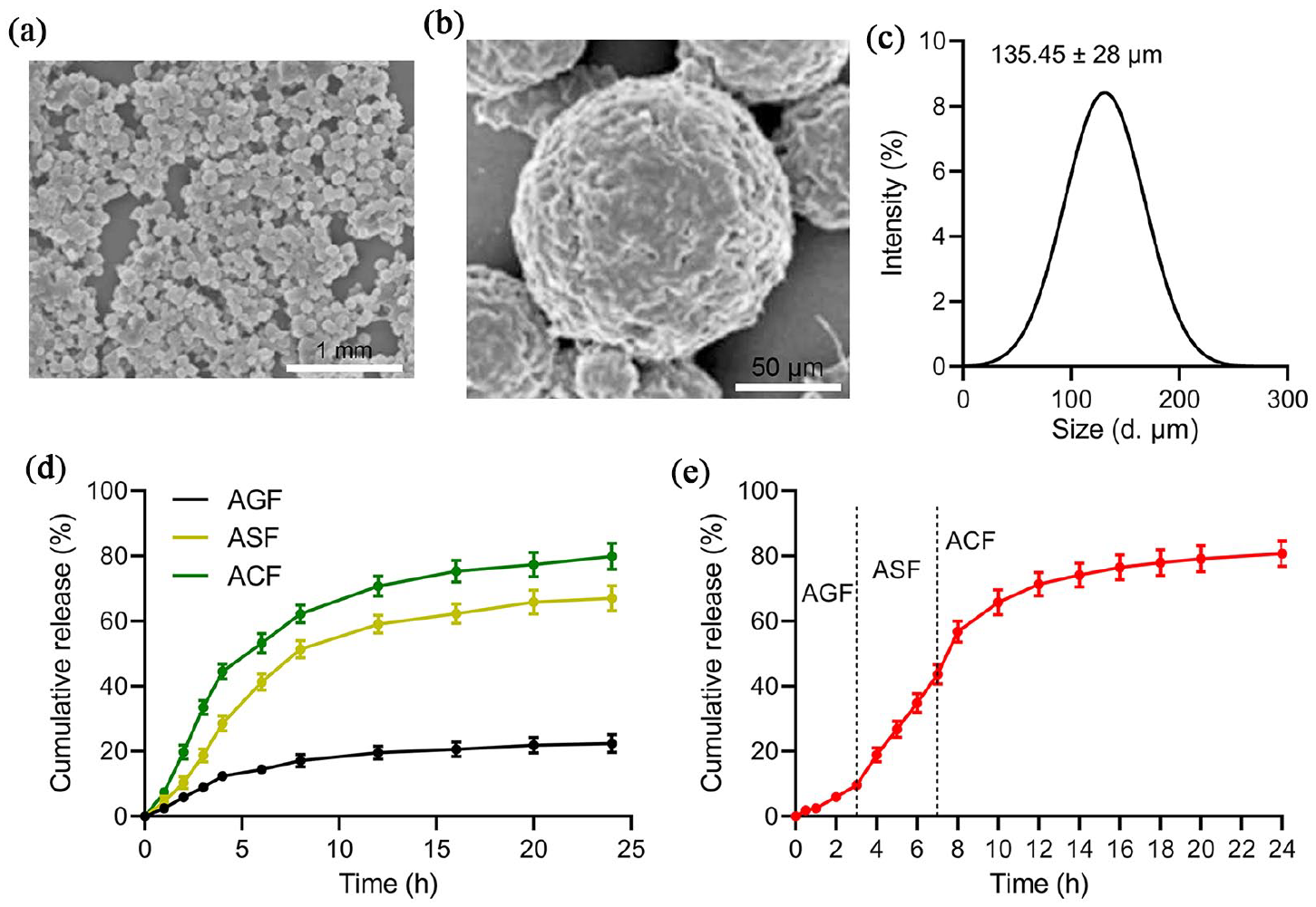
Characterization of BA-CS-SA microspheres. (a) Scanning electron microscopy images of BA-CS-SA and higher magnification shown in (b). (c) Particle size distribution of BA-CS-SA microspheres was measured by dynamic light scattering. (d) Cumulative release of betulinic acid from BA-CS-SA microspheres under artificial gastric fluid (AGF), artificial small intestinal fluids (ASF), and artificial colonic fluid (ACF). (e) BA-CS-SA continuous drug release curve in different media.
Cumulative release analysis showed that betulinic acid had the highest release in artificial colonic fluid, followed by artificial small intestinal fluid and artificial gastric fluid (Figure 1(d)). When BA-CS-SA microspheres were sequentially immersed in artificial gastric, small intestinal, and colonic fluids, betulinic acid release remained low in the first 3 h within the gastric fluid. However, the release rate significantly increased in artificial small intestinal fluid and continued to rise slightly in colonic fluid (Figure 1(e)). These findings indicate that BA-CS-SA exhibits pH-sensitive release properties.
Moreover, the results confirm that CS-SA effectively serves as a carrier for targeted betulinic acid delivery to the colon, establishing BA-CS-SA as a promising strategy for the treatment of intestinal diseases.
BA-CS-SA microspheres ameliorated DSS-induced colitis
The mouse experiment was conducted to evaluate the therapeutic effect of BA-CS-SA in DSS-induced colitis, as illustrated in Figure 2(a). Disease activity index (DAI) was assessed following treatment with different drugs. Both betulinic acid and BA-CS-SA microspheres significantly reduced the DAI in DSS-induced colitis mice, with BA-CS-SA demonstrating a more pronounced effect than free betulinic acid (Figure 2(b)).
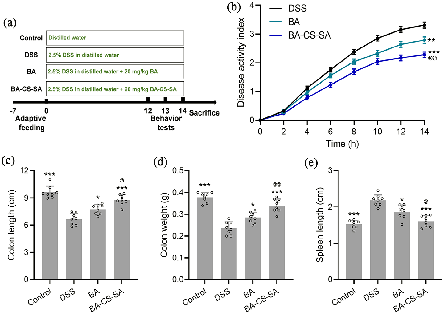
BA-CS-SA microspheres ameliorated DSS-induced colitis. (a) The scheme of the experimental design. (b) The changes of disease activity index from each group. (c–e) Comparisons of colon length, colon weight and spleen length among different groups. Data were shown with mean ± SEM for B and mean ± SD for C-E. *p < 0.05, **p < 0.01, ***p < 0.001 compared to DSS group. @p < 0.05, @@p < 0.01 compared to the betulinic acid (BA) group.
After 14 days of treatment, mice were sacrificed, and their colon length and weight were measured. DSS-induced colitis led to a significant reduction in colon length and weight compared to control mice (Figure 2(c) and (d), Supplemental Figure S2). Treatment with betulinic acid and BA-CS-SA microspheres significantly restored colon length and weight, with BA-CS-SA exhibiting a superior effect (Figure 2(c) and (d)).
Additionally, DSS-induced colitis resulted in a marked increase in spleen length compared to control mice (Figure 2(e)). Both betulinic acid and BA-CS-SA treatments significantly reduced spleen length, with BA-CS-SA demonstrating a greater suppressive effect (Figure 2(e)).
Overall, these findings highlight that BA-CS-SA provides a stronger preventive and therapeutic effect against DSS-induced colitis than free betulinic acid.
BA-CS-SA microspheres ameliorated DSS-induced colitis
It was revealed treatment with betulinic acid and BA-CS-SA microspheres reduced histological scores, inflammatory cell infiltration, and mucosal damage, with BA-CS-SA demonstrating a more pronounced protective effect (Figure 3(a)).
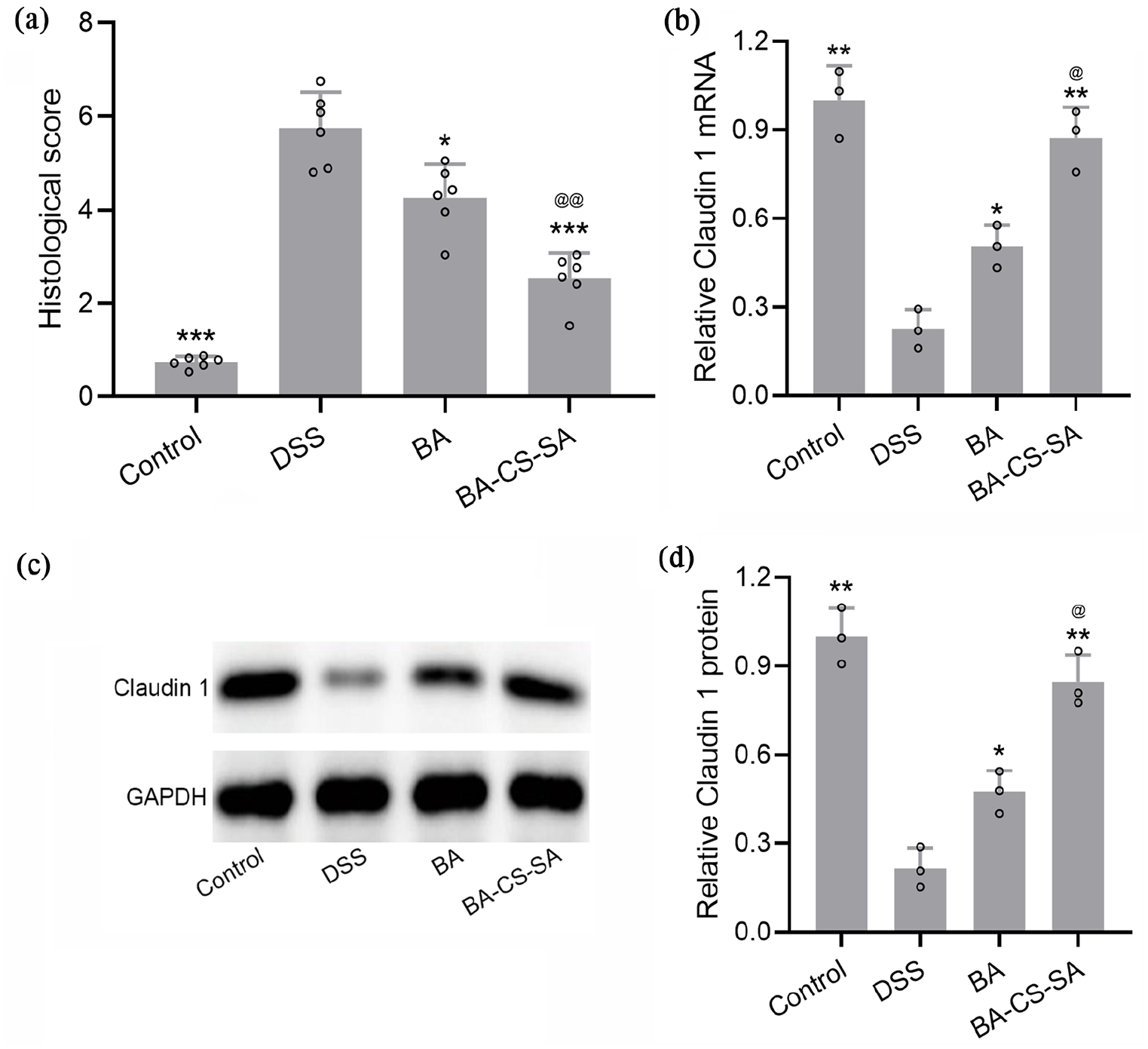
BA-CS-SA microspheres ameliorated DSS-induced colitis. (a) The comparison of histological score. (b) RT-qPCR was used to measure the mRNA expressions of Claudin 1 in the colon. (c) Western blotting was used to measure the protein expressions of Claudin 1 in the colon, and the expressions were normalized to control (d). Data were shown with mean ± SD. *p < 0.05, **p < 0.01, ***p < 0.001 compared to DSS group. @p < 0.05, @@p < 0.01 compared to the BA group.
Claudin-1 plays a crucial role in maintaining the intestinal epithelial barrier. 26 To assess mucosal barrier integrity, Claudin-1 expression was evaluated. qPCR analysis showed a significant reduction in Claudin-1 mRNA levels in DSS-induced colitis mice, whereas BA-CS-SA treatment more effectively restored Claudin-1 expression compared to free betulinic acid (Figure 3(b)).
Similarly, western blot analysis demonstrated that both betulinic acid and BA-CS-SA microspheres significantly increased Claudin-1 protein levels, which were downregulated in DSS-induced colitis mice (Figure 3(c) and (d)).
These findings suggest that BA-CS-SA more effectively protects against colonic epithelial barrier damage in DSS-induced colitis compared to free betulinic acid.
BA-CS-SA microspheres ameliorated DSS-induced inflammatory responses
To evaluate the anti-inflammatory effects of betulinic acid and BA-CS-SA, the levels of the proinflammatory cytokines TNF-α and IL-1β were analyzed. DSS-treated mice exhibited a significant increase in serum TNF-α and IL-1β levels (Figure 4(a) and (b)). Treatment with both betulinic acid and BA-CS-SA microspheres significantly reduced these cytokine levels, with BA-CS-SA showing a more pronounced effect than betulinic acid alone (Figure 4(a) and (b)).
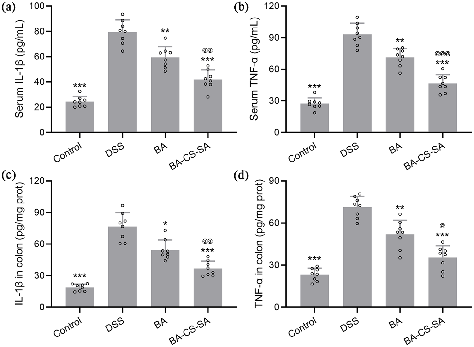
BA-CS-SA microspheres ameliorated DSS-induced inflammatory responses. Serum levels of IL-1β (a) and TNF-α (b); the contents of IL-1β (c) and TNF-α (d) in the colon were measured by ELISA. Data were shown with mean ± SD. *p < 0.05, **p < 0.01, ***p < 0.001 compared to DSS group. @p < 0.05, @@p < 0.01, @@@p < 0.001 compared to BA group.
Similarly, proinflammatory cytokine levels in colon tissues mirrored the serum findings, with BA-CS-SA treatment demonstrating superior anti-inflammatory effects compared to betulinic acid (Figure 4(c) and (d)).
These results indicate that BA-CS-SA is more effective than free betulinic acid in reducing proinflammatory cytokine levels in DSS-induced colitis.
BA-CS-SA microspheres ameliorated DSS-induced anxiety-like behaviors in mice
Patients with inflammatory bowel disease (IBD) often experience psychological disorders, including anxiety and depression. 27 To assess anxiety-like behaviors in DSS-induced colitis mice, behavioral tests were conducted following DSS treatment.
In the OFT test, DSS-treated mice showed a significant reduction in time spent in the center and total distance traveled compared to the control group (Figure 5(a) and (b)). Treatment with betulinic acid and BA-CS-SA microspheres effectively reversed these changes, with BA-CS-SA showing a stronger effect (Figure 5(a) and (b)).
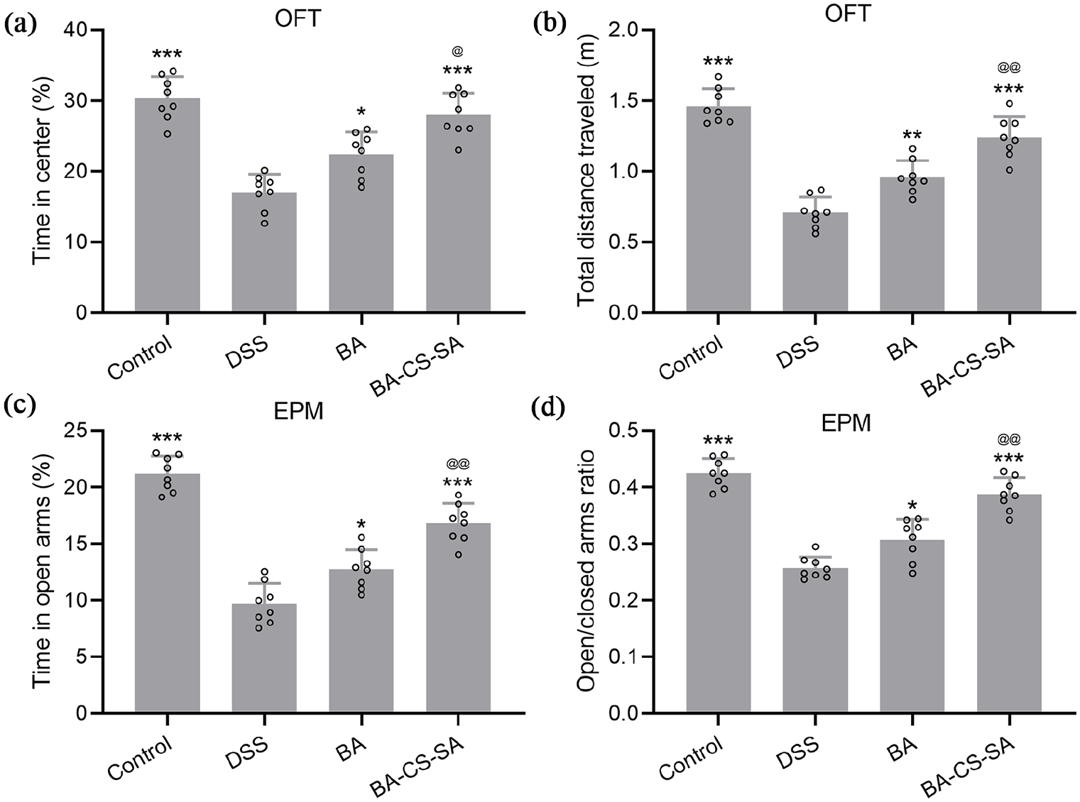
BA-CS-SA microspheres ameliorated DSS-induced anxiety-like behaviors in mice. Comparisons of time in the center (a), total distance traveled (b) in the OFT. Comparisons of time in the open arms (c), open/closed arms ratio (d) in the EPM. Data were shown with mean ± SD. *p < 0.05, **p < 0.01, ***p < 0.001 compared to DSS group. @p < 0.05, @@p < 0.01 compared to BA group.
Similarly, in the EPM test, DSS-treated mice exhibited a marked decrease in time spent in the open arms and a lower open/closed arms ratio compared to controls (Figure 5(c) and (d)). Betulinic acid and BA-CS-SA microsphere treatments significantly increased both measures, with BA-CS-SA demonstrating greater efficacy (Figure 5(c) and (d)).
These findings suggest that BA-CS-SA more effectively alleviates anxiety-like behaviors in DSS-induced colitis mice compared to free betulinic acid.
BA-CS-SA microspheres ameliorated DSS-induced neuroinflammation and improved the expressions of BDNF in the cortex
An increasing body of evidence suggests that anxiety is directly linked to inflammatory processes, characterized by elevated inflammatory cytokines and reduced BDNF levels. 28 ELISA analysis revealed a significant increase in IL-1β and TNF-α levels in the cortex of DSS-induced colitis mice compared to controls (Figure 6(a) and (b)). Notably, BA-CS-SA microsphere treatment was more effective than free betulinic acid in significantly reducing these proinflammatory cytokines (Figure 6(a) and (b)).
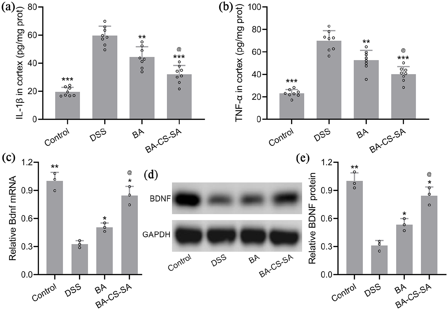
BA-CS-SA microspheres ameliorated DSS-induced neuroinflammation and improved the expressions of BDNF in the cortex. The contents of IL-1β (a) and TNF-α (b) in the colon were measured by ELISA. (c) RT-qPCR was used to measure the mRNA expressions of Bdnf in the cortex. (d) Western blotting was used to measure the protein expressions of BDNF in the cortex and the expressions were normalized to control (e). Data were shown with mean ± SD. *p < 0.05, **p < 0.01, ***p < 0.001 compared to DSS group. @p < 0.05 compared to the BA group.
BDNF loss has been closely associated with anxiety. 29 To evaluate this, we measured BDNF levels in DSS-induced colitis mice under different treatments. qPCR analysis showed a significant reduction in BDNF mRNA expression in DSS-treated mice, which was significantly restored by both betulinic acid and BA-CS-SA microspheres, with BA-CS-SA exhibiting a stronger effect (Figure 6(c)).
Similarly, western blot analysis confirmed that both treatments mitigated DSS-induced BDNF protein loss, with BA-CS-SA demonstrating greater efficacy (Figure 6(d) and (e)).
Together, these findings suggest that BA-CS-SA microspheres more effectively suppress neuroinflammation and restore BDNF levels in DSS-induced colitis mice compared to free betulinic acid.
Discussion
Betulinic acid was demonstrated to prevent colonic inflammation and fibrosis in murine models of IBD. 12 However, the poor solubility of betulinic acid limits its clinical application. Meanwhile, the traditional method of oral drug administration presents significant limitations, including intragastric degradation and poor stability due to the high acidity of gastric juice, which directly impairs the therapeutic efficacy of the drug. 30 The colon-specific drug delivery system is a specialized method of drug administration designed to prevent premature drug release in the stomach after oral administration and instead facilitate release upon reaching the colon. 31 Therefore, the preparation of nanoformulations loaded with betulinic acid to improve the bioavailability of drugs is critical for the treatment of intestinal diseases. 32 Our study successfully prepared the microsphere using the CS-SA system, which could effectively embed betulinic acid within microspheres. The in vitro drug release experiments have validated the fundamental concept that the protonation of sodium alginate resulted in particle shrinkage under artificial gastric fluid, thereby constraining the release of betulinic acid. Conversely, the alteration of buffer to artificial small intestinal fluids and artificial colonic fluid prompted the swelling and disintegration of the microspheres due to the elongation and deprotonation of the carboxyl groups in the sodium alginate and carboxymethyl chitosan, consequently facilitating the sustained release of betulinic acid, which was consistent with previous finding. 33 Notably, BA-CS-SA microspheres showed greater cumulative release of BA at higher pH levels, which contrasts with the typical behavior of calcium-crosslinked alginate gels, where acidic conditions usually promote drug release due to carboxyl group protonation. This difference likely stems from the multicomponent nature of our system. At elevated pH, both Ca²⁺ crosslinks and electrostatic interactions between CMCS and alginate may be disrupted, leading to increased matrix swelling. Additionally, BA is more soluble and mobile in neutral to alkaline environments, further enhancing diffusion. Deprotonation of CMCS may also induce electrostatic repulsion within the network, contributing to its loosening. Together, these factors explain the enhanced release at higher pH and underscore the potential of the CMCS–alginate system for pH-responsive, colon-targeted drug delivery. These findings demonstrated that BA-CS-SA microspheres could effectively deliver betulinic acid to colonic tissue. Subsequently, BA-CS-SA microspheres were better than free betulinic acid to ameliorate DSS-induced colitis by inhibiting inflammation.
While DSS-induced colitis models represent acute rather than chronic forms of intestinal inflammation, previous demonstrate that such models are sufficient to evoke anxiety- and depression-like behaviors.34 –36 These findings support the utility of DSS-induced colitis as a relevant model for investigating the neuroimmune mechanisms underlying behavioral comorbidities in IBD. DSS administration in drinking water for 7 days in mice resulted in significant weight loss and excessive neutrophil infiltration, leading to the colonic epithelium’s rupture and a marked increase in DAI.37,38 We also found similar results with decreased colon length and weight in DSS-induced colitis, and treatment with betulinic acid significantly reversed all these symptoms, consistent with Jaspreet’s findings. 39 More importantly, BA-CS-SA microspheres more greatly reversed all these symptoms, attributed to the high release of betulinic acid using the colon-specific drug delivery system. The characteristic histological changes associated with colitis include the destruction of crypts, infiltration of inflammatory cells, and denudation of the epithelium. 40 Our analysis also revealed a destroyed mucosal barrier, infiltration of inflammatory cells, and increasing histological score in DSS-induced colitis compared to normal tissues, which were significantly reversed under the treatment of betulinic acid and BA-CS-SA. The molecular biomarker of the intestinal barrier can be evaluated through the level of Claudin 1. 41 Research demonstrated that Claudin 1 levels were significantly reduced in DSS-induced colitis. However, these levels could be effectively restored through treatment with SA and BA-CS-SA, with BA-CS-SA showing an even more pronounced increase. These histopathological and biomarker findings strongly suggest the alleviation effects of betulinic acid, especially in the BA-CS-SA system.
As evidenced by clinical studies, patients with IBD have a significantly higher prevalence of psychiatric disorders, such as depression and anxiety. 27 DSS treatment in mice effectively induces colitis that closely resembles the characteristics of human UC, leading to notable depression-like and anxiety-like behaviors. 42 Our results exhibited a close relationship between colitis and behavioral performance with anxiety in DSS-induced mice, as evidenced by the OFT, EPM experiment in line with previous studies. The BA-CS-SA treatment demonstrated significantly greater efficacy in alleviating DSS-induced anxiety-like behaviors than free betulinic acid. This behavioral improvement may be explained by a combination of local anti-inflammatory effects and systemic immune modulation through the gut–brain axis, as supported by previous studies. The anxiolytic effects observed following BA-CS-SA treatment likely result from both the anti-inflammatory action of BA and the relief of DSS-induced colonic symptoms. Previous studies have shown that intestinal inflammation can trigger neuroinflammation and behavioral disturbances via the gut–brain axis. For instance, surfactin and sesamol ameliorated anxiety- and depression-like behaviors in DSS-induced colitis models by suppressing proinflammatory signaling, enhancing antioxidant defenses, and activating neuroprotective pathways such as BDNF/CREB and 5-HT signaling.43,44 Similarly, the walnut-derived peptide LP-5 improved cognitive function by restoring gut and blood–brain barrier integrity, reducing hippocampal microglial activation, and modulating the gut microbiota. 45 Consistent with these findings, our delivery system reduced both local and cortical levels of IL-1β and TNF-α, suggesting that localized immunomodulation attenuated systemic inflammation and its impact on the brain. Additionally, improved mucosal healing and reduced colitis symptoms likely lowered somatic stress, further contributing to the behavioral improvements. Together, these results highlight the dual role of BA-CS-SA microspheres in modulating both peripheral and central immune responses.
It was shown that DSS treatment increased neuroinflammatory responses and activated the TLR-4/NF-κB pathway. 42 Recent studies on animals exposed to DSS demonstrated that astrocytes and microglia were activated in the hippocampus.46,47 Furthermore, there is an increase in proinflammatory cytokines, including IL-1β, TNF-α, IL-6, iNOS, and Cox-2, within the brain. Our analysis showed an evident increase of IL-1β and TNF-α in the cortex from DSS-induced mice, which could be impaired by the betulinic acid and BA-CS-SA treatment. Meanwhile, inflammatory factors, including IL-1β and TNF-α, exhibited significant elevation in the colon tissue of mice subjected to DSS. However, these levels were notably reduced following treatment with betulinic acid and BA-CS-SA. BDNF was demonstrated to inhibit neuroinflammation and pathological pain, 48 which was significantly decreased in DSS-induced colitis and upregulated by BA-CS-SA treatment.
Accordingly, both free betulinic acid and BA-CS-SA microspheres were administered at 20 mg/kg/day, a dose consistent with established safety thresholds. Nevertheless, at higher concentrations, betulinic acid may present several risks, including poor solubility and uneven biodistribution leading to drug accumulation or carrier-associated toxicity 49 ; disruption of key metabolic pathways such as mTOR and insulin/IGF1 signaling 50 ; reduced cytoselectivity with potential damage to normal cells 51 ; impairment of DNA repair mechanisms and excessive suppression of inflammatory responses when combined with chemotherapeutic agents 52 ; nanoparticle-related hepatic or immunotoxicity 53 ; and organ-specific effects such as liver injury and enteroendocrine dysregulation via TGR5 activation. 54 Therefore, further evaluation is warranted before dose escalation, particularly with respect to pharmacokinetics, tissue distribution, and long-term safety.
This study demonstrated the high efficacy of BA-CS-SA in alleviating DSS-induced colitis and anxiety-like behaviors. The mechanistic exploration revealed that betulinic acid could significantly restore the intestinal barrier injury and attenuate the inflammation response caused by DSS treatment. However, our study still had some limitations. Initially, it is essential to assess whether the concentrations of betulinic acid in the colonic tissue are comparable between the two groups of mice subjected to treatment with free bile acids and those treated with BA-CS-SA. Furthermore, the effects of oral administration of BA-CS-SA on neuroinflammation in DSS-treated mice will be examined in future experiments. In addition, further studies are needed to distinguish whether the behavioral improvements are primarily driven by the anti-inflammatory effects of BA or by the secondary relief of DSS-induced colitis symptoms.
Conclusion
BA-CS-SA demonstrated superior efficacy in alleviating DSS-induced colitis compared to free betulinic acid. This was evidenced by greater increases in colon length and weight, higher expression of the intestinal barrier marker Claudin-1, and a more significant reduction in histological scores. Additionally, BA-CS-SA more effectively mitigated DSS-induced anxiety-like behaviors, as shown by improved performance in the OFT and EPM tests. Mechanistic analysis revealed that BA-CS-SA treatment significantly suppressed inflammatory factors, including IL-1β and TNF-α, in serum, colon, and cortex tissues.
Supplemental Material
sj-docx-1-jbc-10.1177_08839115251356266 – Supplemental material for Betulinic acid-loaded carboxymethyl chitosan/sodium alginate microspheres ameliorate dextran sodium sulfate-induced colitis and anxiety-like behaviors in mice
Supplemental material, sj-docx-1-jbc-10.1177_08839115251356266 for Betulinic acid-loaded carboxymethyl chitosan/sodium alginate microspheres ameliorate dextran sodium sulfate-induced colitis and anxiety-like behaviors in mice by Binbin Zhuang, Jinhong Zhuang, Yubin Wang and Jingyang Zeng in Journal of Bioactive and Compatible Polymers
Footnotes
Author contributions
Binbin Zhuang, Jinhong Zhuang, Yubin Wang, Jingyang Zeng: Data collection, analysis, manuscript preparation. Yubin Wang, Jingyang Zeng: Supervision, funding acquisition, and writing—review and editing.
Declaration of conflicting interests
The author(s) declared no potential conflicts of interest with respect to the research, authorship, and/or publication of this article.
Funding
The author(s) disclosed receipt of the following financial support for the research, authorship, and/or publication of this article: The study was supported by Quanzhou City Science & Technology Program of China (2021N085S).
Ethical approval
Animal studies were approved by QuanZhou Taiwanese Investment Zone Hospital. This study was performed in strict accordance with the NIH guidelines for the care and use of laboratory animals (NIH Publication No. 85-23 Rev. 1985).
Consent for publication
Not applicable.
Informed consent
N/A.
Data availability statement
The raw data could be obtained upon reasonable request to the corresponding author.
Supplemental material
Supplemental material for this article is available online.
References
Supplementary Material
Please find the following supplemental material available below.
For Open Access articles published under a Creative Commons License, all supplemental material carries the same license as the article it is associated with.
For non-Open Access articles published, all supplemental material carries a non-exclusive license, and permission requests for re-use of supplemental material or any part of supplemental material shall be sent directly to the copyright owner as specified in the copyright notice associated with the article.