
Abstract
Nanoparticles are widely recognized for their ability to cross biological membranes and modulate cellular functions. In this study, we developed and characterized nanocomposite hydrogels composed of nanochitin (NC) embedded within a PEO/PVA polymer matrix. The materials were physicochemically characterized using zetasizer analysis, FTIR–ATR spectroscopy, and SEM imaging. Their cytotoxic potential was assessed in vitro using MTT assays on normal adult human dermal fibroblast (HDFa) cells, while developmental and neuromuscular toxicities were evaluated in vivo using Drosophila melanogaster as a model organism. Our findings indicate that all NC-doped PEO/PVA nanocomposites were non-toxic to HDFa cells. In fact, 1%, 2.5% and 5% NC-doped PEO/PVA samples showed 44.6%, 20.7% and 13.7% increases compared to negative control, respectively (p
Introduction
Hydrogels are the materials used in the biomedical industry as drug delivery agents, intraocular/contact lenses, corneal prostheses, bone cements, wound dressing materials, tissue scaffolds, biosensors and hemostasis bandages. 1 They are the crosslinked hydrophilic polymeric materials that can absorb and hold large amounts of water in their specific 3D structure without self-dissolution. 2
Nanocomposite hydrogels, on the other hand, are the hybrid hydrogels that are made up of hydrated polymeric networks physically/ covalently crosslinked with each other and/or with nano-particles/structures. 3 Nanohydrogels are preferred in wound dressing applications due to their rich structural and biological properties.
The skin, being the largest and most complex organ of the human body, regulates temperature and water content while serving as a barrier against physical, chemical, and infectious stimuli. 4 However, various internal or external damages forms of damage, such as injuries, burns, and underlying diseases, impair the integrity and function of the skin and disrupt the wound healing process. Chronic wounds caused by diseases such as diabetes and venous stasis cannot complete the dynamic steps of normal wound healing, which consist of inflammation, proliferation, and remodeling.2,5 Chronic wounds that fail to heal within normal timescales and remain at these stages are a huge burden on patients and the healthcare system around the world. 6 For this reason, alternative and innovative wound dressing materials should be designed.
Providing a moist environment in wound dressings has been accepted as the gold standard since George Winter’s experiments on re-epithelialization.7,8 A moist environment allows interaction between cells involved in the wound healing process and growth factors, and protects the tissue from dehydration and cell death. 9 Hydrogels are three-dimensional structures consisting of a hydrophilic, cross-linked, polymeric meshes structure with a high water-retention capacity. 10 Hydrogels, with their ability to absorb wound exudate, facilitate the migration of fibroblasts and keratinocytes (critical cell types for the remodeling step in wound healing) to the wound bed. 11 In addition, hydrogels have become ideal for use as wound dressings due to their biocompatibility and ability to facilitate cell adhesion by mimicking the extracellular matrix layer. In order to increase their mechanical strength, hydrogels can be obtained by cross-linking multiple synthetic or natural polymers in their structure through physical or chemical interactions. 11 There are many polymers used for nanocomposite hydrogels. Among those polymers, polyvinyl alcohol (PVA) is a frequently preferred one in many biomedical applications with its biocompatible, non-toxic and non-carcinogenic properties.12–14 Polyethylene oxide (PEO) is another polymer known to enhance the mechanical properties of hydrogels, in addition to its high biocompatibility, pH-independent swelling and drug-release capabilities. 15 The reason behind the incorporation of nanoparticle/structures into the hydrogels is the fact that the studies have shown that nanocomposites obtained by incorporating nanoparticles into polymeric structures have improved water holding capacity, mechanical strength and biocompatibility.16–19
In this study, nano sized chitin was used to prepare PEO/PVA/NC nanocomposite hydrogels. Chitin is a low-cost, natural polymer that is abundant in nature and can be easily obtained from the skeleton of invertebrates or from the cell wall of fungi. 20 It is a linear polysaccharide consisting of the β-(1-4) glycosidic bond of N-acetyl-D-glucosamine units. Breakdown of chitin by enzymes such as lysozyme found in body fluids releases N-acetylglucosamine units, which leads to macrophage production and collagen storage during wound healing. 21 With its biocompatible, biodegradable, antimicrobial and hemostatic properties, it facilitates the secretion of inflammatory mediators (hormones) and fibroblast activation during wound healing.20,22
Given that nanoparticles can cross biological membranes and potentially affect cellular physiology, it is essential to assess not only cellular cytotoxicity but also reproductive and developmental toxicity for any biomaterial. 23 This study focuses on the production of biocompatible, non-toxic, cheap and easily prepared nanocomposite hydrogels. Therefore, the cytotoxicities of the PEO/PVA/NC nanocomposite hydrogels were analyzed in vitro by MTT assay after the physicochemical characterization of the materials by zetasizer, Fourier Transform Infrared Spectroscopy–Attenuated Total Reflectance (FTIR–ATR) and Scanning Electron Microscopy (SEM). In addition, the developmental and neuromuscular toxicity effects of the materials synthesized were analyzed in vivo using Drosophila melanogaster as a model organism. In literature, there isn’t any study showing the cellular, in vivo developmental and in vivo neuromuscular toxicity of the PEO/PVA/NC nanocomposite hydrogels. Therefore, this study will present the first data in this context to the literature.
Materials and methods
Materials
Poly(ethylene oxide; PEO, 99% purity, approximate M.W. 900,000) and Poly(vinyl alcohol; PVA, 98.0% purity, approximate M.W. 50,000–85,000) purchased from Acros Organics and chitin (from shrimp shells) were purchased from Merck-Sigma Aldrich, and MTT (Cell Proliferation Kit I) was purchased from Roche. Oregon-R strain was kindly provided by Department of Biology of Uludag University, Bursa, Türkiye.
Synthesis of the of PEO/PVA/NC nanocomposite hydrogels
Chitin (5 g) was ground by grinding method and reduced to nano size using Retsch PM100 planetary ball mill. Then, nanocomposites were synthesized by solution interaction method using both PEO and PVA polymers. Both of the polymer solutions were dissolved in ultra distilled water. The PEO solution was prepared at room temperature, whereas the PVA solution was prepared at 70°C. Nanochitin (NC) stock solutions were prepared in distilled water and were kept to ensure the achievement of homogeneous distribution in an ultrasonic bath for 20 min. Then, the polymer and NC solutions were separately stirred using a magnetic stirrer at room temperature for 2 h, after which the NC solution was subjected to ultrasonic treatment for an additional 20 min. Subsequently, the polymer solutions (PEO/PVA, 1/1 v/v) were combined and stirred with a magnetic stirrer at room temperature for 24 h to ensure homogeneous distribution. Three different PEO/PVA/NC solutions were prepared containing 1, 2.5, and 5 wt% NC relative to the total weight of the polymer solution. After stirring all samples on a magnetic stirrer at room temperature for 24 h, the resulting PEO/PVA/NC nanocomposites were poured into petri dishes and dried in a vacuum oven at 40°C to remove the solvents.
Characterization of PEO/PVA/NC nanocomposite hydrogels
The size and zeta potential of the nano-ground chitin were measured using a Zetasizer Malvern Nano SZ-90 particle analyzer. In addition, the backscattering angle used for particle size analysis by dynamic light scattering (DLS) was 173°. Prior to analysis, a nanochitin (NC) suspension with a concentration of 0.1 g/100 mL was prepared in ultra-distilled water by ultrasonication for 25 min at room temperature.
FTIR ATR analyses were performed using a PerkinElmer Spectrum 100 spectrophotometer. For each analysis, the FTIR ATR settings were adjusted to a resolution of 4 cm⁻¹, four scans, and a spectral range of 4000–650 cm⁻¹ in transmittance mode.
The morphology of nanocomposites was analyzed by Zeiss EVO LS 10 Scanning Electron Microscope (SEM). For SEM analysis, the samples were prepared by coating them with Au-Pd while keeping the nanocomposite materials adhered to the carbon tape under a current of 15 mA for 15 s. An acceleration voltage of 20 kV was used for SEM imaging.
The preperation of nanocomposite hydrogels for cellular, developmental and neuromuscular toxicity analyses
The hydrogel samples were cut into sizes that would fit into 24-well plates (1.5–1.9 cm2) and each sample was placed in a separate well. When cutting the materials, care was taken to ensure that each material had equal dimensions and a flat surface. The prepared plates were exposed to UV light for 24 h in a laminar cabinet with their lids open, and then the samples were used only inside the cabinet and using sterile forceps.
Cellular toxicity analysis (MTT assay)
Cytotoxicity analysis was performed according to the method of Phaechamud et al. with some modifications. 24 For this study, a normal adult human dermal fibroblast (HDF-a, ATCC® PCS-201-012™) cell line was used and cell line viability was maintained in fibroblast basal medium (FBM) containing fetal bovine serum. In addition, 0.5 mL of Penicillin-Streptomycin-Amphotericin B solution and the materials in the growth kit (L-glutamine: 7.5 mM, rh FGF basic: 5 ng/mL, rh Insulin: 5 µg/mL Hydrocortisone: 1 µg/mL, Ascorbic acid: 50 µg/mL, Fetal bovine serum: 2%) were added into FBM. Cell culture media were changed in every 3–4 days. In order to prepare nanocomposite extracts, 2 mL of completed medium was added to the sterilized hydrogel pieces and kept at 37°C for 120 h. At the end of the period, the hydrogels were removed with sterile forceps and the extracts were ready for use. From the cells that reached a sufficient number of passages and were counted with a hemocytometer, 100 μL was added to 96-well plates (7.4 × 10⁴ cells/well). Separately prepared plates for 24 and 48 h were monitored until cell attachment occurred, after the cells attached, the medium was discarded and 100 μL of the prepared extract was added. After 24 h of incubation, the medium in the wells was discarded and 100 μL of fresh medium and 10 μL of MTT reagent (Cell Proliferation Kit 1, Roche) were added to the cells and kept at 37°C for 4 h. At the end of the period, liquids were withdrawn from the wells. Then, 100 μL solubilization buffer of the MTT assay kit was added to dissolve formazan crystals and kept at 37°C for 24 h. Absorbance measurement was made at 570 nm by Thermo Fisher Scientific Multiskan GO microplate reader spectrophotometer (Finland). The same procedures were repeated for the 48-h plate. No extract was added to the negative control group. The absorbance values obtained as a result of the MTT test were calculated as a percentage by comparing them to the control group values (metabolic activity %).
In vivo analyses
Oregon-R wild type strain was used throughout the tests and the flies were kept in an incubator at 22°C. Oregon-R strains, which were removed from the incubator only during the tests, were used after being produced for several generations and inbred. All flies were fed with a semolina-based standard growth medium prepared by boiling sucrose (43 g) and agar (9 g) in 500 mL of distilled water, followed by the addition of semolina (90 g). After cooling, dry yeast (25 g), nystatin (0.3 g), and propionic acid (5 mL) were added.25,26 Subsequently, 25 g of the prepared medium was dispensed into sterile glass culture vials, and the flies were maintained in these vials throughout the experiments. In order to prepare nanocomposite hydrogel extracts for the in vivo analyses, 2 mL of sterilized distilled H2O was added to the sterilized hydrogel pieces (1.5–1.9 cm2) and kept at 22°C for 120 h. This incubation time was chosen based on preliminary optimization experiments (data not shown) which indicated that the extract composition stabilized after approximately 96 h, and extending to 120 h ensured maximal leachate release without compromising the hydrogel integrity. At the end of the period, the hydrogels were removed with sterile forceps and the extracts were ready for use. The extract solutions were incorporated into the growth medium at 50% and 100% (v/v) concentrations to assess dose-dependent biological effects. The 100% concentration represents the undiluted extract, providing maximal exposure to bioactive components, while the 50% concentration was chosen to examine submaximal effects and potential threshold responses. These concentrations were validated through pilot studies (data not shown) confirming their biological relevance and compatibility with normal fly development.
In the present study, male and female Drosophila were pooled for behavioral assays to reflect population-level responses and ensure sufficient statistical power. Although sex-specific differences have been reported in certain behaviors (e.g. courtship, aggression), previous studies have shown that for the tested endpoint (e.g. locomotion, eclosion rate, climbing) sex does not significantly influence outcomes under baseline or sublethal toxicant conditions. Furthermore, pooling sexes is a widely used and accepted approach in toxicological studies involving Drosophila, particularly when the primary focus is not on sex-differentiated traits.27,28
In vivo developmental toxicity analysis
Developmental toxicity tests were performed to monitor the rates of transformation into pupae and adults and the viability of Drosophila larvae fed with hydrogels whose toxic effects will be investigated, using the method of Liu et al. 29 In order to create larvae, 25 female and 25 male adult Drosophila were transferred to vials containing standard medium and incubated for 48 ± 4 h. At the end of this period, synchronized third instar larvae were collected from the medium and transferred in groups of 25 larvae per vial to experimental vials containing the growth medium supplemented homogeneously with hydrogel extracts at the desired concentrations, as well as to control vials without extracts. The vials were placed at 22°C for pupae and adult formation and their development was monitored. During the transformation period from larvae to pupae and from pupae to adult, which lasted approximately 10 days, the number of pupae and adults in each group were noted separately. The experiment was carried out with 3 repetitions for each group. The percentages of puparation, survival and eclosion were calculated using the following formulas;


In vivo neuromuscular toxicity analysis
In order to perform neuromuscular toxicity analysis two tests were applied to the flies. The first test, larval crawling test, was performed by modifying the method of Gautam et al. 30 In order to perform this experiment, 25 female and 25 male adult Drosophila were transferred to experimental vials treated with extracts and control groups containing no extract and incubated for 48 ± 4 h. At the end of the period, the third instar larvae were collected separately for each test group and cleaned from the media on them. For each control and test group, 2% (w/v) agar plates were prepared in Petri dishes. The larvae taken from each group were placed in the middle of the Petri dishes and their movements were marked from the moment they were placed. In order to observe the movement, millimeter paper was placed behind the Petri dishes and centered, and the movements were recorded simultaneously with a camera placed at a fixed distance. The distance covered by the larvae in 1 min was calculated in cm and the experiment was carried out with three repetitions. The crawling speeds were calculated by the total distance (cm) per min.
The second neuromuscular toxicity test was the climbing test performed by modifying the method used by Mishra et al. 31 In order to perform this experiment, 25 female and 25 male adult Drosophila were transferred to experimental vials treated with extracts and control groups containing no extract and incubated for 48 ± 4 h. At the end of the period, adult flies were transferred to a 100 mL graduated cylinder. A mark was placed on the graduated cylinder at the 80 mL line. The flies transferred into the cylinder were kept for a few minutes to adapt to the environment and the mouth of the graduated cylinder was covered with cotton so that they would not escape. A white plane was placed behind the graduated cylinder and video recording were taken from a fixed point. The flies that became accustomed to the environment were lowered to the bottom by tapping the cylinder 3–4 times and the flies that could pass the marked line within 1 min were observed. The number of flies that passed the certain line within 1 min was calculated separately for each group and the percentages of flies were given. The experiment was carried out with three repetitions. Before each repetition, care was taken to give the flies a few minutes to get used to the environment.
Statistics
Standard errors (SE) were calculated with three biological replicates. Microsoft Office Excel was used to apply the student’s t-test and the p-value < 0.05 was considered as statistically significant.
Results and discussion
Characterization results
Nanochitin can be prepared by different approaches. It is known that some of those methods that break down the starting bulk material from native chitin resources to produce nanochitin are acid hydrolysis, TEMPO oxidation, ultrasonication, grinding, high-pressure homogenization and microwave irradiation. 32 In this study, mechanical grinding method was used for nanochitin production and the size and zeta potential of the nanochitin particle was confirmed by Zetasizer Malvern Nano SZ-90 particle analyzer. Zetasizer is an advanced device used for determining some characteristic properties of nanoparticles, colloids or biomolecular particles, including size and zeta potential. 33 The size of the chitin was found 133.7 nm in this study and this showed us that the chitin particles used was nano-sized. Furthermore, the zeta potential of chitin was measured as 28.1 ± 0.2 mV at room temperature. The zeta potential of the chitin suspension was measured as +28.1 mV, indicating that the system possesses a good level of colloidal stability. This positive surface charge primarily results from the partial protonation of amino groups in the chitin chains, particularly under neutral or acidic pH conditions, which enhances electrostatic repulsive forces between particles and prevents agglomeration. Although the measurement was conducted at a single pH point, the obtained value is consistent with results reported in the literature. For instance, Jiang et al. reported zeta potential values ranging from +20 to +41 mV for nanochitin in aqueous media at neutral pH, and noted that these values are associated with stable colloidal suspensions. 34 In this context, the +28.1 mV zeta potential of chitin indicates that the structure has a suitable profile in terms of colloidal stability.
The interaction between polymer matrix and nanochitin fillers were analyzed by FTIR-ATR (Figure 1). The FTIR results of the chitin was given in Figure 1(a). According to the literature findings, the characteristic band of chitin at 3422 cm−1 can be attributed to the stretching vibrations of -NH and –OH groups. The band at 2876 cm−1 could belong to aliphatic C-H stretching vibrations. The characteristic band of chitin at 1653 cm−1 could be attributed to the carbonyl C = O stretching. The sharp band at 1375 cm−1 corresponded to the bending vibrations of the CH3 group. The band at 1552 cm−1 showed the N-H vibrations in the amide group. The vibration bands at 1067 cm−1 indicated the presence of C-O-C groups inside the chitin ring.35,36 The FTIR results of PEO, PVA and PEO/PVA/NC were given in Figure 1(b) to (d). The FTIR spectrum of the polymers were taken as a reference to explain the nanocomposite formation with FTIR-ATR. What is expected from the nanocomposite FTIR spectrum is the shift of some bands in the pure polymer or the observation of new peaks supporting the formation of new bonds. In addition, the peaks obtained from the FTIR spectra of the filler material should not be present in the nanocomposite. Because, the peak belonging to the filler material in the nanocomposite FTIR spectrum may indicate that chitin remains in bulk in the polymer matrix, is not dispersed and a homogeneous distribution does not occur. Figure 1 shows that the -OH functional group of chitin, originally observed at 3422 cm−1, shifted to 3273–3272 cm−1 in the spectra of the nanocomposites, which can be considered evidence of the interaction between the polymer matrix and the filler material (Figure 1(d)). The band at 2882 cm−1 showed the C-H stretching, the band at 1466 cm−1 showed the CH3 and CH2 bending vibrations and the band at 1279 cm−1 showed the stretching vibrations of the C-N functional groups. When the spectra of the nanocomposites were examined, it was clear that the shift occurred at the OH- functional group at 3280 cm−1. These changes mean that there is an interaction between the matrix (PEO/PVA) and the filler material (nanochitin).
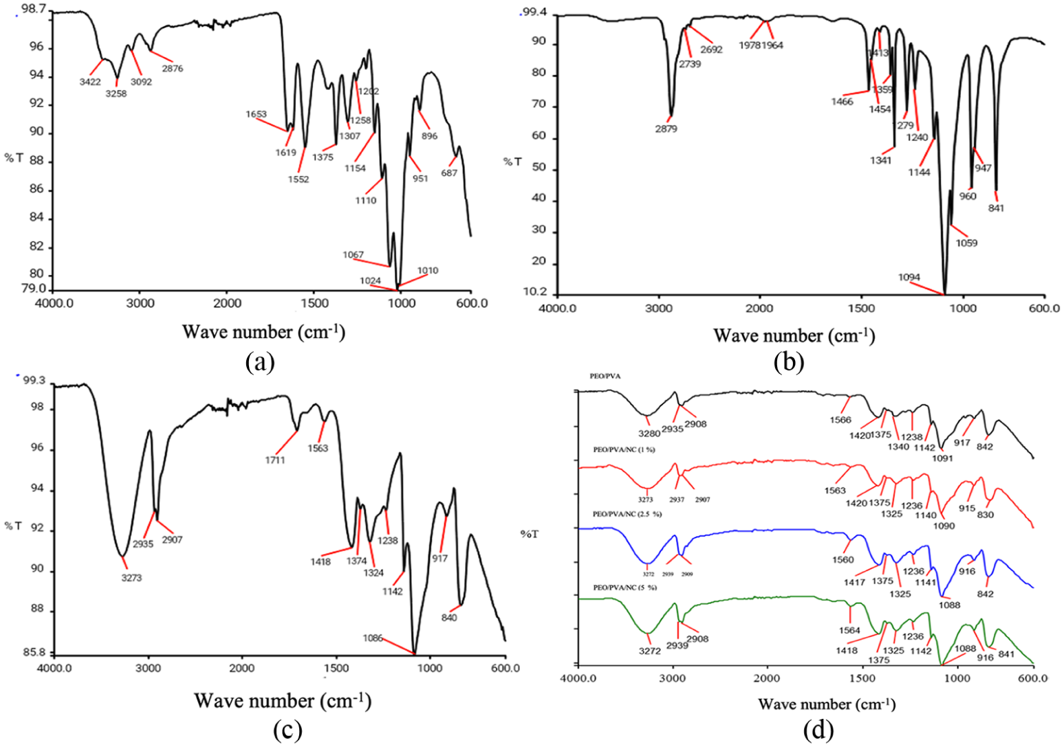
FTIR-ATR spectra of nanochitin (a), PEO (b), PVA (c) and PEO/PVA/NC nanocomposite hydrogels (d).
SEM is used to obtain the size/shape, distribution, surface morphology and purity of nanostuctural materials. 37 In this study, the morphologies of nanochitin and nanocomposite hydrogels were investigated at accelerating voltage of 10 kV and various magnifications (Figure 2). It is known that chitin has three main surface morphologies: a hard or rough surface without pores or nanofibers, a surface entirely composed of nanofibers, a surface with both pores or nanofibers and a surface with pores without nanofibers. 38 Our results showed that chitin samples used in this study has a hard or rough surface without pores or nanofibers (Figure 2(a)). The SEM image of all PEO/PVA/NC nanocomposite (Figure 2(c), (d) and (e)) hydrogel also showed that filler material (chitin) was homogeneously dispersed in the PEO/PVA matrix (Figure 2(b)).
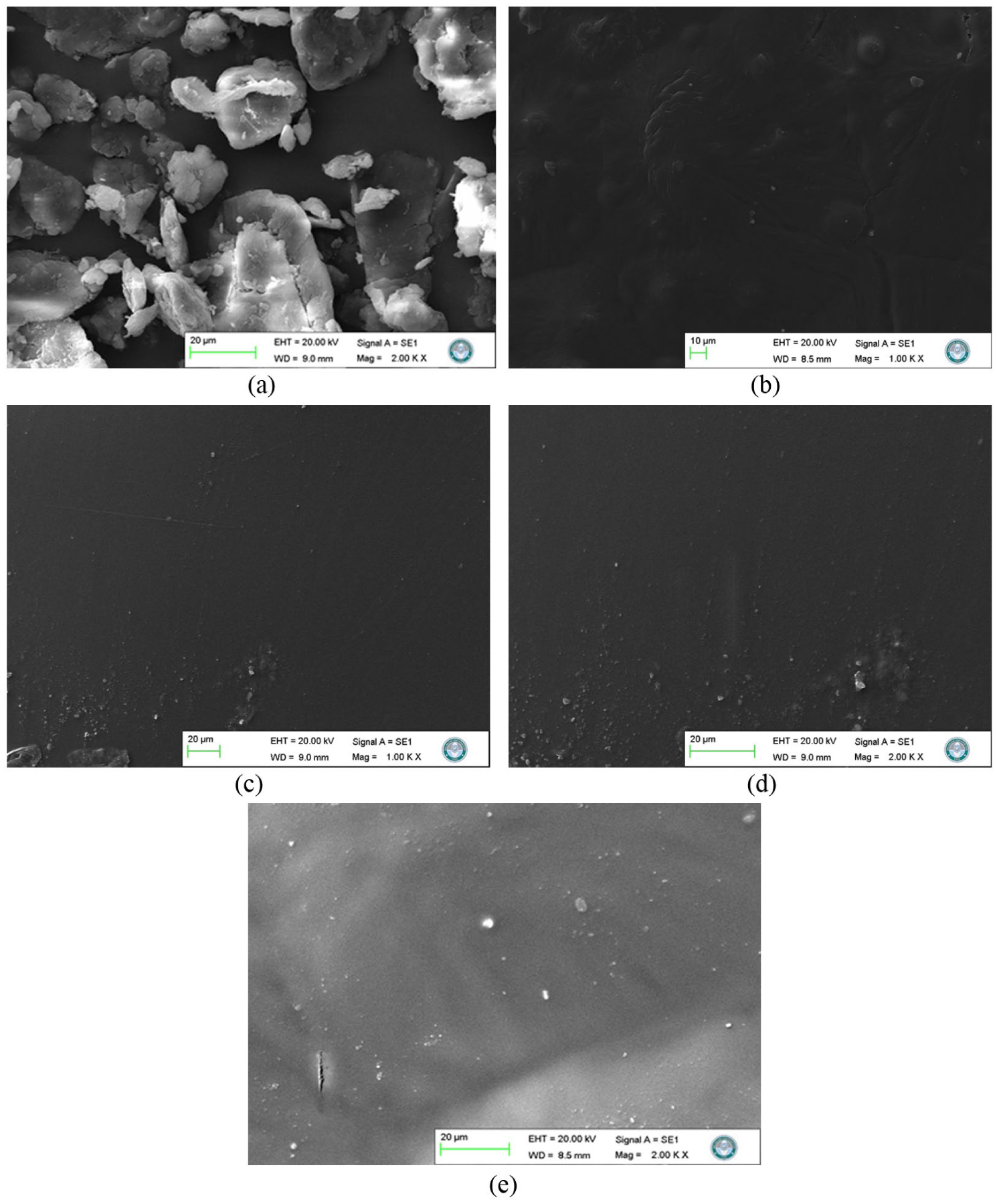
Scanning electron microscopy (SEM) images of nanochitin (a), PEO/PVA (b), PEO/PVA/NC (1%, wt.) (c), PEO/PVA/NC (2.5%, wt.) (d) and PEO/PVA/NC (5%, wt.) (e) nanocomposite hydrogels.
Cellular toxicity test results
The metabolic activity of living cells is quantitatively examined with the MTT assay which is one of the most frequently used methods in cytotoxicity tests. Metabolically active cells reduce the tetrazolium ring in tetrazolium salts by mitochondrial dehydrogenase enzymes located in the cytochrome b and cytochrome c regions of their mitochondria through electron transfers of reducing molecules. Thanks to this activity, which is only found in living cells with active mitochondria, the tetrazolium salt MTT (3-(4,5-dimethylthiazol-2-yl)-2-5-diphenyltetrazolium bromide) is used to measure the color changes obtained by the reduction of these salts by living cells. 39 In this study, the cellular toxicities were investigated by MTT assay using the HDFa cells exposed to the hydrogel extracts for 24 and 48 h and the results were given in Figure 3. According to the results, PEO/PVA/NC nanocomposite hydrogels didn’t affect the cellular viabilities after 24 h of incubation and all of the results were above 70%. However, PEO/PVA/NC (1%), PEO/PVA/NC (2.5%) and PEO/PVA/NC (5%) samples showed statistically significant increases (44.6%, 20.7% and 13.7% increases, respectively) compared to negative control (Figure 3, p < 0.05). In addition, nanochitin supported biomaterials showed higher metabolic activities in contrast to the ones observed with pure PEO, PVA and PEO/PVA samples. It is known that a material having viability in fibroblast cells above 70% as a result of the MTT test is considered non-cytotoxic and if it is below 70%, it is considered to have cytotoxic potential (DIN EN ISO 10993-5:2009 standard). 40 Therefore, it is clear that the nanocomposite hydrogels were non-toxic against HDFa cells especially when the nanochitin was used in 1% concentration (Figure 3). The results obtained in this study were compatible with the findings in literature. For example, He et al. reported that the chitin/PVA composite hydrogels were biocompatible by performing the MTT test on L929 cells and they found that the porous structure provided by chitin had a positive effect on cell viability by acting as a mechanical support for cell adhesion. 41 Islam et al. also investigated the effect of hydrogel scaffolds containing different ratios of chitosan (a linear polysaccharide obtained by extensive deacetylation of chitin) and PVA on HDFa cells using MTT test and found that all surfaces containing PVA/chitosan, regardless of chitosan ratios, increased cell metabolic activity compared to the control. They found that the PVA/chitosan composites didn’t have cytotoxic effects on fibroblast cells and they explained this situation with the fact that the chitosan could act as an ideal scaffold for cell adhesion and proliferation by mimicking the glycosaminoglycan in the ECM layer and promotes cell proliferation. 42
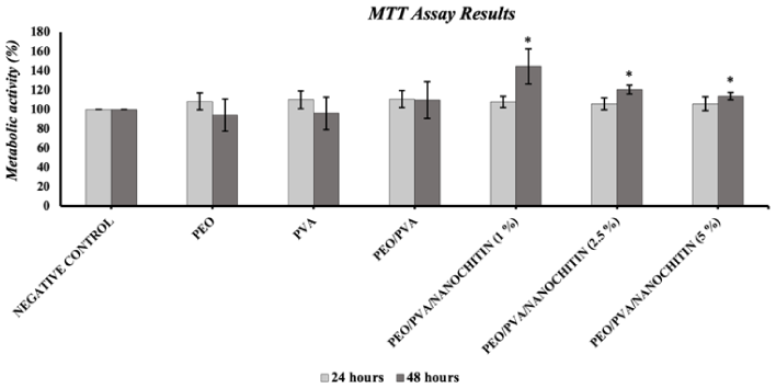
MTT assay results for cellular toxicity analysis for 24 and 48 h. Metabolic activity (%) were represented as mean ± standard error and * indicates the statistical significance (p < 0.05). Negative control is biomaterial-free.
Developmental toxicity results
The use of alternative small organism models like the fruit fly (Drosophila melanogaster) for toxicology studies has gained great importance in recent years. Although humans and fruit flieas are divergent species, Drosophila model for understanding the human condition under stress of toxicants has largely been accepted because of the abundance of highly conserved genes/ pathways controlling development, stress response and xenobiotic metabolisms. Therefore, Drosophila have been used for the toxicology analyses of the environmental contaminans/toxicants like mercury, lead, arsenic, manganese, ethanol, nanoparticles, pesticides and solvents. 43 In this study, developmental toxicity tests were performed to monitor the rates of transformation into pupae-adults and the viability of Drosophila larvae fed with PEO/PVA/NC nanocomposite hydrogel samples and the results were given in Figure 4. Results showed that all of the samples caused significant decreases in puparation and survival % (p < 0.05, Figure 4(b) and (c)). On the other hand, 50% (v/v) PEO/PVA/NC (1%) sample was non-toxic to the eclosion process (p > 0.05, Figure 4(a)). In fact, none of the materials used in this study showed eclosion rates below 50% and they can be considered as non-toxic. Because, it is known that in Drosophila, eclosion rates below 50% relative to control have been widely used as a benchmark for severe developmental toxicity, particularly in response to neurotoxicants such as methylmercury.27,28 There isn’t any similar study in literature but Vecchio et al. also investigated the in vivo toxic effects of gold nanoparticles on Drosophila via their viability/reproductive activities and observed a 50% decrease with nanoparticles compared to the control while PEG-coated nanoparticles did not create a toxic effect. 44 Mishra et al. studied the developmental toxicity of cellulose nanofibrils on Drosophila Oregon-R strains for use in drug delivery and they observed that high doses of cellulose nanofibrils prolonged the pupa formation and hatching times compared to the control and reduced the number of pupa-adult formations. However, by also measuring the levels of reactive oxygen species, it was concluded that low doses of nanofibrils were suitable for use in drug delivery and were not toxic to Drosophila. 31 In another study, Silver Key et al. added silver nanoparticles to Drosophila medium bottles and analyzed the developmental processes such as hatching from eggs, larval development, pupation and hatching from pupae. The researchers didn’t observe any significant difference between the egg-laying rate and F1 generation development of P generation adult flies placed in silver nanoparticle medium and P generation placed in normal medium. On the other hand, it was found that treatment of F1 generation with silver nanoparticles negatively affected the larval development and reduced adult fly lifespan. 45 Molecular analysis of silver nanoparticle (AgNP) exposure in Drosophila also identified dose-dependent reductions in fecundity, larval weight, pupation, and eclosion rates, along with fat body cytotoxicity, hemocyte DNA damage, and metallothionein gene induction. 46

Developmental toxicity test results. Eclosion (a), puparation (b) and survival (%) (c) were represented as mean ± standard error and * indicates the statistical significance (p < 0.05).
Neuromuscular toxicity results
As a function of their muscular and nervous systems, larvae exhibit a coordinated movement characterized by contraction and expansion. One of the important neuromuscular tests is larval crawling test which examines behaviors that include movements that are different from normal, such as twisting to the right and left, crawling straight, digging-moving slowly and monitor changes in the movements of larvae due to the effects of early neural damage caused by the toxic agent. 47 The results of the larval crawling test performed in this study were given in Figure 5(b). The results showed that none of the 50% (v/v) samples of the nanochitin added nanocomposite hydrogels caused any significant change compared to the negative control (p > 0.05), while all of the 100% (v/v) samples caused significant decreases (p < 0.05, Figure 5(b)). Another important neuromuscular test used in this study was the climbing test. The ability to fly, which has an important role in activities such as mating and food search, is a standard parameter that can be observed in adult Drosophila behavior and is a locomotor activity that can be affected as a result of the imbalance in sensory and motor activities due to the toxic agent. 48 The results of the climbing test were given in Figure 5(d). According to the results, all of the samples except 50% (v/v) of PEO/PVA/NC (1%) nanocomposite hydrogels caused significant decreases in climbing ability. In the adult climbing test, which showed similar results to the larval crawling test, the locomotor activities of adult flies were affected at high doses and the proportion of flies crossing the border decreased compared to the control. With this test, it was seen that adults exhibited positive geotropism behavior, contrary to the negative geotropism behavior they normally exhibit, due to the damage in their neural activities. 49 The dose-dependent developmental and neuromuscular toxicity observed at higher concentrations of nanomaterials might have been resulted from multiple, interacting cellular stress pathways. At elevated doses, nanoparticles can penetrate the larval gut barrier and accumulate in internal tissues, leading to oxidative stress via excessive generation of reactive oxygen species (ROS), which damages lipids, proteins, and DNA.50,51 This oxidative burden may impair mitochondrial function and ATP production, disrupting the high energy demands of developing tissues and motor circuits. Furthermore, nanoparticles have been shown to interfere with calcium homeostasis and synaptic transmission, potentially impairing neuromuscular junctions essential for coordinated movement. 52 Disruption of developmental signaling pathways, such as Notch or Wnt, has also been proposed, contributing to altered differentiation and tissue patterning during metamorphosis.53,54 Collectively, these effects can manifest as reduced pupation and eclosion rates, decreased larval motility, and impaired adult climbing behavior, particularly under chronic or high-dose exposures. Similar findings were found in literature too. For example, Ekka et al. performed a larval crawling test to examine the effects of different doses of SiO2 and TiO2 nanocomposites on Drosophila behaviors and observed larval movements in a Petri dish containing 2% agarose. A decrease in larval speed was observed compared to the control at all doses except the highest dose and it was concluded that the uptake of nanocomposites into the tissues at the early stage of development, the larval stage, had an effect on behavior by affecting sensory neurons. 55 Gautam et al. also examined the movements of larvae exposed to the h-BN and ZrO2 composites in Petri dishes containing 2% agar and calculated their speeds. While the control group larvae exhibited a straight movement toward the periphery, it was observed that the larvae treated with the test materials followed a longer movement path by stopping or changing direction several times. By comparing the observational results with the speed ratios; it was concluded that the neural activities of the larvae, whose speeds were significantly reduced compared to the control, were damaged and caused these movements. Based on the behavioral test results, the researchers stated that nano formulations of doses that showed less toxicity could be used in drug transport. 30 In addition, Bag et al. used the larval crawling test to examine the effect of guar gum nanoparticles coated with Fe3O4 on Drosophila behavior and observed the movements of larvae exposed to nanoparticles placed in petri dishes containing 2% agar. While the control group larvae exhibited a straight movement, it was observed that the test larvae exposed to the nanoparticles moved by stopping at many points and their speed was calculated to decrease compared to the control. The researchers observed that the speed of the larvae increased as the amount of nanocomposite increased and stated that this test provides control of neural functions in the early stages of development. 56 On the other hand, Sundararajan et al. performed an adult climbing test to examine the in vivo toxicity of CeO2 nanoparticles. The researchers calculated the percentage of flies that could climb the 3 cm limit in 15 s by placing adult flies in 10 cm tubes after treating them with nanoparticles prepared at different concentrations for 1 month. No significant difference was found in the control and test groups, and it was concluded that the nanoparticles did not cause neurodegenerative damage and did not have a toxic effect on adult flies. 57 Sabat et al. investigated the effects of TiO2 nanoparticles on Drosophila behavior and sensory organs, they applied a climbing test to adult flies exposed to nanoparticles. According to the results of the test, climbing activities decreased compared to the control with increasing concentrations, and a decrease of more than 20% in climbing speed was observed at high concentrations. The decreased climbing activity was explained by the damage occurring in the larval period not being repaired by metamorphosis, causing abnormal adult behavior. It was stated that the abnormal behaviors observed in flies as a result of the toxic effect of nanoparticles on neurons are an important indicator and a parameter that should be taken into consideration before using nanoparticles in the medical field. 53 Matthews et al. also demonstrated that chronic dietary exposure to 1 µm and 20 nm polystyrene plastics did not affect mortality or developmental endpoints in Drosophila but significantly altered adult locomotion and daily activity patterns. 58 In vivo testing of a ZnFe₂O₄-based nanocomposite in Drosophila revealed gut barrier penetration, developmental delays, reduced pupation and eclosion rates, weight loss, sensory organ defects, and impaired larval crawling and adult climbing. 59 Another study revealed that dietary ingestion of carbon nanomaterials (C₆₀, CB, SWNTs, MWNTs) during the larval stage did not affect egg-to-adult survival, but adult exposure to dry particulate CB/SWNTs severely impaired locomotion and increased mortality. 60
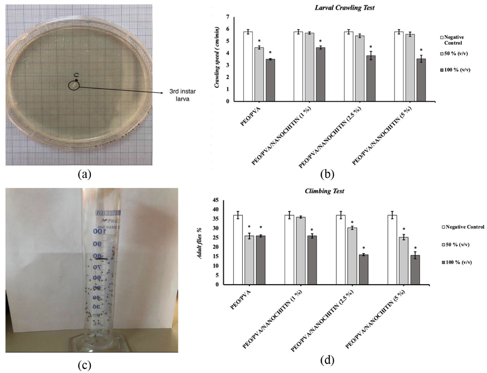
Results of the neuromuscular toxicity tests: (a) petri dishes used for larval crawling assay with grid background; (b) Crawling speed (cm/min) of third instar larvae exposed to 50% and 100% (v/v) concentration of PEO/PVA and NC loaded (1, 2.5 and 5% wt.) nanocomposites; (c) 100 mL graduated cylinder with 80 mL mark used for adult climbing test; (d) percentage of adult flies climbing above the 80 mL mark on a 100 mL graduated cylinder with 1 min. (p < 0.05 for 100% vs control). * indicates the statistical significance (p < 0.05).
Mechanistic discussion
The findings obtained in this study demonstrate the cellular, developmental, and neuromuscular toxicity of nanocomposite hydrogels. At the cellular level, it is known that embedded nanoparticles, particularly metal or oxide types, frequently provoke excessive reactive oxygen species (ROS) generation, leading to oxidative stress, mitochondrial damage, DNA strand breaks, and inflammatory responses in cultured cells and stem–cell models.61–63 During developmental stages, these materials may cross biological barriers (including the placenta or yolk sac), accumulate in embryonic tissues, and dysregulate morphogenetic signaling pathways such as Wnt, Hedgehog, and neurogenic gene cascades; this disruption impairs neurogenesis, synaptogenesis, and vascular development, evident in organoid models and zebrafish embryos.64,65 Neuromuscular toxicity mechanisms manifest through ROS-driven apoptosis in motor neurons, interference with axon outgrowth and motor nerve formation, altered expression of key neurodevelopmental genes (e.g. elavl3, gap-43, syn2a, neurog1), and consequent behavioral deficits such as inhibited locomotion, delayed escape responses, and reduced spontaneous movement in models like zebrafish larvae. 62 Collectively, these findings suggest a hierarchical mechanistic framework in which initial oxidative stress and transcriptional dysregulation at the cellular level may perturb essential developmental signaling pathways, culminating in structural and functional impairments of the neuromuscular system.
Conclusion
This study focused on the production of biocompatible, non-toxic, cheap and easily prepared nanocomposite hydrogels. Therefore, the cytotoxicities of the PEO/PVA/NC nanocomposite hydrogels were analyzed in vitro by MTT assay in this study after the physico-chemical characterization of the materials by zetasizer, Fourier transform infrared spectroscopy – attenuated total reflectance (FTIR–ATR) and Scanning Electron Microscope (SEM). In addition, the developmental and neuromuscular toxicity effects of the materials synthesized in this study were analyzed in vivo using Drosophila melanogaster as a model organism. According to the results, PEO/PVA/NC (1%), PEO/PVA/NC (2.5%) and PEO/PVA/NC (5%) samples showed statistically significant increases in the metabolic activities of HDFa cells compared to negative control and it was clear that the nanocomposite hydrogels were non-toxic against HDFa cells especially when the nanochitin was used in 1% concentration. Developmental toxicity test results showed that all of the samples caused significant decreases in puparation and survival % (p < 0.05), but 50% (v/v) PEO/PVA/NC (1%) sample was non-toxic to the eclosion process. The larval crawling test results showed that none of the 50% (v/v) samples of the nanochitin added nanocomposite hydrogels caused any significant change compared to the negative control (p > 0.05), while all of the 100% (v/v) samples caused significant decreases. The climbing test results also showed that all of the samples except 50% (v/v) of PEO/PVA/NC (1% wt.) nanocomposite hydrogels caused significant decreases in climbing ability. In literature there isn’t any study showing the cellular, in vivo developmental and in vivo neuromuscular toxicity of the PEO/PVA/NC nanocomposite hydrogels. Additionally, future research could focus on optimizing polymer-to-nanochitin ratios to balance mechanical strength, biocompatibility, and toxicity profiles more effectively. Expanding analyses to include detailed molecular mechanisms of interaction with cellular and developmental pathways would also provide deeper insights into the safety and functionality of these hydrogels. In addition, although Drosophila offers a genetically tractable and sensitive model, its physiological differences from mammals may limit direct translational relevance. Future studies should include chronic exposure paradigms, assessment of reproductive fitness and lifespan, and validation in vertebrate models to fully elucidate systemic and long-term toxicity. Moreover, exploring the effects of varying nanocomposite formulations such as different polymer-to-metal ratios or surface modifications may offer insights into structure toxicity relationships and aid in the design of safer nanomaterials.
Footnotes
Acknowledgements
This work produced from a part of Sedef Kirdar’s and Ulas Kumral’s MSc Theses. The authors thank Assoc. Prof. Dr. Berna Kocer Kizilduman, Assoc. Prof. Dr. Emel Tamahkar Irmak, Prof. Dr. Yasemin Turhan and Prof. Dr. Mehmet Dogan for the chemical characterization analyses and the scientific ideas that the authors benefited from during the progress of the study.
Author contributions
Funding
The authors disclosed receipt of the following financial support for the research, authorship, and/or publication of this article: The Scientific Research Projects Commission of Balikesir University (BAP 2018/178 and BAP 2019/114).
Declaration of conflicting interests
The authors declared no potential conflicts of interest with respect to the research, authorship, and/or publication of this article.
Data availability statement
Data Availability is not applicable. All findings is presented in the work.