






Abstract
Polymer/DNA hybrid hydrogels represent a novel class of biomaterials that integrate the programmable nature of DNA with the mechanical strength of synthetic polymers. In this study, we present a simple and adaptable method to fabricate DNA-crosslinked polyacrylamide hydrogels using two 5′-acrydite-modified DNA strands and a partially complementary DNA crosslinker. The hydrogels were synthesized via radical-initiated polymerization using APS and TEMED, allowing the DNA strands to become covalently incorporated into the polymer network. Subsequent hybridization with the crosslinker strand formed a three-dimensional hydrogel structure. A systematic investigation of key parameters—such as DNA concentration, molar ratios, reaction temperature, and complementary sequence length—was conducted to optimize gel formation. The most rigid and stable hydrogel was achieved using 60 µM concentrations of each DNA strand in a 1:1:1 molar ratio. Gelation was optimal at lower temperatures, while higher temperatures resulted in losing hydrogen bonding and inhibited the formation of a stable gel network. Moreover, hydrogels fabricated using shorter complementary regions exhibited enhanced stability, likely due to a reduction in undesired intramolecular interactions. Morphological analysis confirmed the successful formation of a porous, interconnected structure. The developed DNA/polymer hybrid hydrogels exhibit promising properties for potential applications in bioengineering.
Introduction
Hydrogels are hydrophilic polymers widely utilized in various fields such as tissue engineering, purification, and detection techniques due to their high-water retention capacity, flexibility, biocompatibility, and responsiveness to environmental stimuli such as pH, temperature, and magnetic fields.1–4 In recent years, the development of hydrogels incorporating antibodies, 5 polypeptides 6 or enzymes 7 has gained attention to enhance specificity and biocompatibility. DNA-based hydrogels particularly have attracted considerable interest due to their high stability and flexibility, offering promising applications in detection,8–10 environmental analysis11,12 controlled drug release13,14 cell adhesion,15–17 tissue engineering18,19 and cancer therapy.20,21 DNA hydrogels are distinguished not only by their ability to maintain structural integrity, but also by their unique ability to be programed based on regulation of polymer chain interactions. DNA hydrogels combine the robustness of polymeric scaffolds with the information coding capacity of nucleic acids. Functional DNA motifs—such as aptamers for target-specific binding, i-motifs for pH sensitivity, G-quadruplexes for ion-mediated folding, and DNAzymes for catalytic activity—have been integrated into polymer frameworks to confer sensing, actuation, and catalytic functions.8,22–25 Through Watson–Crick base pairing, DNA crosslinks can be programed to undergo reversible gel–sol transitions or strand displacement reactions, enabling stimulus responsive behavior for applications in biosensing, controlled drug release, cell adhesion modulation, tissue engineering, and even cancer therapy.
Three principal strategies exist for DNA hydrogel synthesis. In the first method, DNA forms self-crosslinks in the presence of enzymes or specific motifs, leading to hydrogel formation. The second method involves the entrapment of DNA within polymer matrices via physical interactions such as electrostatic forces. The third method involves the synthesis of DNA hybrid hydrogels by incorporating DNA strands into hydrophobic polymers, allowing side-chain formation. 26 Pioneering work by Nagahara and Matsuda demonstrated one pot co polymerization of acrylamide with two distinct acrydite tagged single stranded DNAs, followed by hybridization with a complementary crosslinker to yield a DNA-polyacrylamide hydrogel. 27 These two polymers were subsequently combined via two different approaches to complete the hydrogel formation. In the first approach, a complementary crosslinking oligonucleotide was used to bind the two DNA-polyacrylamide polymers, resulting in hydrogel formation. In the second approach, the hydrogel was formed directly by binding the two DNA-polyacrylamide polymers together. 27 Subsequent studies have extended this methodology to various polymer/DNA combinations, yet a systematic exploration of fabrication parameters and thorough structure–function characterization remain to be realized.28–32
Conventional polyacrylamide (PAAm) hydrogels employ N,N′-methylenebisacrylamide to establish irreversible covalent junctions via free-radical polymerization, producing mechanically stable but static networks. They use N,N′-methylenebisacrylamide for covalent crosslinking, yielding mechanically robust but static networks. When N,N’-methylenebisacrylamide is used as a crosslinker, the addition of free radicals during polymerization facilitates the formation of covalent bonds between the carbon atom of the bisacrylamide molecule and the carbon atom on the polyacrylamide chain. As a result, an irreversible and highly stable gel structure is synthesized. However, in the present study, DNA molecules served as crosslinking agents for the synthesis of polyacrylamide hydrogels. Replacing bisacrylamide with DNA crosslinkers introduces reversible, sequence-defined junctions and adjustable crosslinker lengths for dynamic tunability. 27 There have been certain advantages of DNA crosslinked polymer as compared with acrylamide-crosslinked polymer. For example, the length of the crosslinker could be adjusted in DNA crosslinked polymer fabrication which in turn provides flexibility. DNA crosslinks exhibit lower dissociation energies—arising from hydrogen bonding—than carbon–carbon covalent bonds in PAAm, facilitating stimuli triggered network remodeling under mild conditions.33,34
This study presents an optimized platform for fabricating DNA–polyacrylamide hybrid hydrogels via radical polymerization of 5′-acrydite–modified DNA strands, followed by hybridization with a partially complementary DNA crosslinker. The acrydite-modified DNAs exhibited a crosslinking activity like that of free acrylamide monomers, enabling their efficient integration into the hydrogel network by facilitating the formation of side branches on the backbone of acrylamide polymer. Two acrydite-functionalized DNA strands, each containing sequences partially complementary to a crosslinking DNA strand, were employed to mediate the formation of linear polymeric structures, thereby completing the DNA polymerization process by hybridization between DNA crosslinker. The influence of key parameters—such as DNA concentration, the molar ratio among the three DNA components, and the length of the complementary regions between the strands and the crosslinker—was systematically assessed via qualitative visual analysis. Furthermore, the effect of temperature on the gelation process was investigated by changing both the assembly temperature and incubation time during hydrogel fabrication. The resulting DNA hydrogel, synthesized under optimized conditions, was subsequently characterized by Fourier Transform Infrared (FTIR) spectroscopy and Scanning Electron Microscopy (SEM) to evaluate its structural and morphological properties. By integrating the molecular programmability of DNA with the structural robustness of polyacrylamide, our study provides fundamental insights into the design rules governing DNA–polymer hybrid hydrogels. This work lays the groundwork for the rational development of the next generation, stimuli responsive materials for applications in biosensing, controlled release, and regenerative medicine.
Materials and methods
Materials
Acrylamide (⩾99%), ammonium persulfate (APS), N,N,N′,N′-tetramethylethylenediamine (TEMED; ⩾99%), tris(hydroxymethyl)aminomethane hydrochloride (Tris·HCl), and disodium ethylenediaminetetraacetate (EDTA·2Na) were purchased from Sigma-Aldrich (St. Louis, MO, USA). The 5′-acrydite-modified oligonucleotides (two DNA strand monomers and a partially complementary crosslinker) used in this study were synthesized and HPLC-purified by Integrated DNA Technologies (Coralville, IA, USA). These strands were modified at their 5′ ends with acrydite groups, enabling their covalent incorporation into the polyacrylamide network. This modification ensures the efficient integration of DNA into the polymer network during radical-initiated polymerization. All aqueous solutions were prepared using nuclease-free deionized water (dH₂O).
Methods
Fabrication of DNA hydrogels
Linear polyacrylamide–DNA conjugates were generated in two separate reactions, each containing one acrydite modified DNA strand (strand 1 or strand 2). In a typical protocol, DNA strand 1 (60 µM) were mixed with acrylamide monomer solution (10%), TE buffer (pH 7.2), and dH2O to a total volume of 100 µL. Freshly prepared APS/TEMED initiator (0.14% (v/v); comprising 10% (w/v) APS and 10% (v/v) TEMED) was added to trigger free radical polymerization. The reaction mixture was incubated at room temperature for 30 min to yield linear polyacrylamide–DNA strand 1 conjugates. An identical procedure was used to prepare the polyacrylamide–DNA strand 2 conjugate. Both linear polymer DNA strand 1 and 2 were then mixed with crosslinker DNA solution (60 µM) to achieve a final volume of 300 µL, which incubated at room temperature for 90 min to form a DNA hydrogel. To assemble the hybrid hydrogel, the two linear polymer–DNA conjugates were combined with the DNA crosslinker (60 µM) and brought to a final volume of 300 µL with TE buffer. The mixture was incubated at ambient temperature (25°C) for 90 min to allow sequence specific hybridization and network formation.
The efficient fabrication of DNA hydrogels was achieved through the optimization of key parameters, including the concentration of each DNA component (50, 60, and 70 µM), the molar ratios between DNA components (strand 1 : strand 2 : crosslinker at 1:1:0.5, 1:1:0.8, 1:1:1, and 1:1:1.5), temperature (4°C, 25°C, 37°C, and 50°C), and incubation time (0–90 min). Gelation was assessed visually and by inversion tests at defined time points.
Characterization of DNA hydrogels
A DNA hydrogel sample was prepared, and time-dependent visual analysis was performed for the gelation control by periodic inversion of reaction tubes. For the morphological analysis, a 300 μL of the prepared DNA hydrogels were subjected to heat dry at 60°C for 2 h. Dried samples were then sputter coated with gold layer and imaged using a Hitachi SU-5500 SEM at an accelerating voltage of 5 kV for SEM imaging. FTIR spectra of the dried hybrid hydrogels were recorded on a Bruker Vertex 70 spectrometer over the range 4000–400 cm⁻¹ to confirm incorporation of DNA within the polyacrylamide network.
Results and discussion
Synthesis of DNA hydrogels
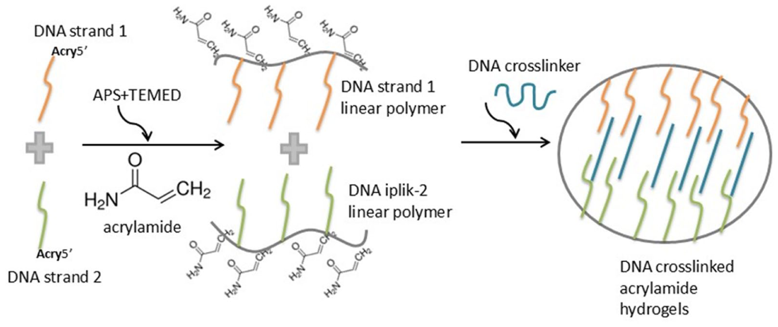
The synthesis principle of hybrid DNA hydrogel in this study is as follows (Scheme 1):
DNA strand 1 monomer solution, modified at the 5′-end with an acrylate group and partially complementary to DNA crosslinker, is used to form linear polymer solution using the polymerization initiator APS and the accelerator TEMED.
Similarly, DNA strand 2 monomer solution, also modified at the 5′-end with an acrylate group and partially complementary to DNA crosslinker, is used to form a linear polymer solution using APS and TEMED. Through this process, the two acrylate-modified DNA strands form side chains on the acrylamide polymer structure.
DNA crosslinker, possessing regions partially complementary to these two DNA strands, is mixed with linear polymer solutions of DNA strands to facilitate the hybridization of the linear polymer strands, thereby completing polymerization. In this synthesis principle, crosslinker brings together the two DNA strands that are incorporated as side chains into the acrylamide polymer, thereby enabling gelation.
Scheme 1
Schematic illustration of the DNA-crosslinked polyacrylamide hydrogel fabrication process. The synthesis proceeds in two main steps: First, 5′-acrydite-modified DNA strands (Strand 1 and Strand 2), each partially complementary to a DNA crosslinker, are separately polymerized with acrylamide monomers using APS and TEMED as initiator and accelerator, respectively. This radical-initiated polymerization covalently incorporates the acrydite-modified DNA strands as side chains onto the growing polyacrylamide backbone, forming linear polymer-DNA conjugates. Second, these linear polymer-DNA conjugates (containing both Strand 1 and Strand 2) are mixed with the partially complementary DNA crosslinker. The DNA crosslinker then facilitates the hybridization of the two linear polymer-DNA conjugates through sequence-specific Watson-Crick base pairing. This hybridization step effectively crosslinks the linear polymer chains, leading to the formation of a stable, three-dimensional DNA-polyacrylamide hybrid hydrogel network.
DNA crosslinker used in this study corresponded to the sequence (CZ13) generated by Eissa and Zourob study. 35 The two DNA strands are 18 base long- short DNA sequences which are modified at the 5′ end by acrydite molecule. The 59 base long-DNA crosslinker, along with the two complementary DNA strands have been designed accordingly for this study. Their properties are summarized in Table 1.
Properties of oligonucleotides used in hydrogel synthesis in this study.

DNA strand-1 and DNA strand-2 are 18-base DNA strands that contain 15-base regions complementary to the DNA crosslinker sequence.
Initial assessment of gelation was primarily conducted using the Sydansk 36 bottle test method, which provided immediate and valuable insights into the sol-gel transition and the relative rigidity of the synthesized hydrogels. This method, widely recognized in polymer and gel science for its simplicity and effectiveness in preliminary evaluations, involves observing the flow behavior of the gel in an inverted vial.37,38 The gel strength is then assigned an alphabetic code from A (indicating no discernible gel formation) to J (representing a highly rigid, non-flowing gel), providing a qualitative yet systematic measure of rigidity. For instance, a gel that does not flow at all when inverted would receive a higher code (e.g. J), indicating greater rigidity. While various interpretations exist, gelation onset is typically marked when the solution transitions from a non-gelled state (A) to a minimally flowing gel (e.g. Code C, as per some studies, 39 or Code B, as defined by others 40 ). This bottle test methodology remains a preferred choice due to its efficiency and economic viability, enabling the simultaneous evaluation of numerous samples and facilitating long-term monitoring of thermal stability, which is often impractical with more complex analytical techniques. In our study, gelation time was defined as the point at which the solution transitioned from code A (no gel detected) to a slightly deformable non-flowing gel of code H, as observed through the bottle test method. This same criterion was consistently applied as our gel acceptance standard throughout all optimization studies discussed below.
Effect of DNA concentration on hydrogel fabrication
The effect of DNA concentration on hydrogel synthesis was evaluated with the use of DNA strand 1, DNA strand 2, and DNA crosslinker at the initial concentration of 50, 60, and 70 µM. Linear polymer solutions were formed by adding the polymerization initiator APS and the accelerator TEMED to the monomer solutions of DNA strands 1 and 2. Subsequently, hydrogel synthesis was carried out by mixing the linear solutions of DNA strand 1 and DNA strand 2 with DNA crosslinker at a molar ratio of 1:1:1, and the hydrogel synthesis process was monitored visually at 25°C. Time-dependent gelation images of the hydrogels at the initial concentration of 50, 60, and 70 µM DNA are shown in Figure 1. Time-dependent gelation was assessed using the Sydansk bottle test method, 36 and gelation was specifically defined as the transition of the sample from a liquid state (A) to a rigid, non-flowing gel (H).
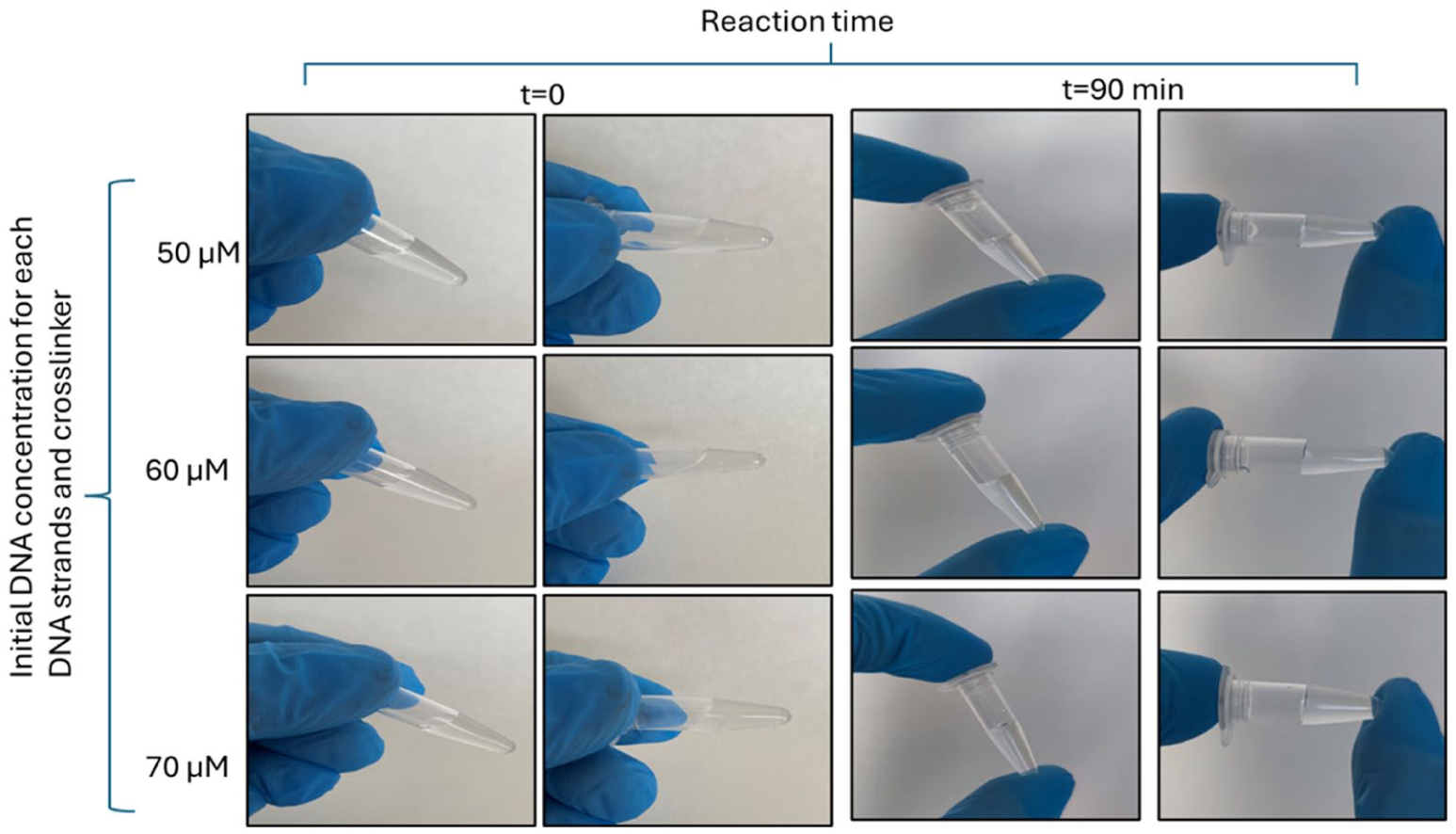
Gelation of DNA based hydrogels synthesized with linear polymer solutions of DNA at initial concentration of 50, 60, and 70 µM at 0 and 90 min.
At the beginning of polymerization, all DNA hydrogels appeared in liquid form consistent with the pre-gelation phase as gelation had not yet started; however, a gradual transition from liquid to gel form was observed over time. After 90 min of polymerization, the hydrogel obtained from the 1:1:1 molar mixture of DNA strand 1, DNA strand 2, crosslinker with 60 µM of each DNA formed a more rigid gel exhibiting characteristics indicative of a higher gel strength code (approaching H), whereas the sample prepared with 50 µM of each DNA sample exhibited a more fluid gel form suggesting a less efficient network formation and a lower gel strength code. Similarly, the hydrogel obtained from the 1:1:1 molar mixture of linear polymer solutions with the highest DNA content (70 µM) also formed a more rigid gel, comparable to the one obtained with 60 µM of each DNA. At this stage, linear polymer solutions with 60 µM of initial DNA concentration for each DNA molecules were determined to be the optimum formulation for hydrogel synthesis due to their ability to form a firmer gel while being more cost-effective than DNA formulation by 70 µM DNA concentration. In subsequent studies, the DNA concentration for each DNA strands and crosslinker was set at 60 µM in the synthesis of DNA based hydrogels.
Effect of the molar ratios of DNA strands and aptamer on hydrogel synthesis
It has been known from the previous studies that the appropriate ratio of DNA building blocks in hydrogel network was critical for the structural integrity of hydrogel. The excess of either the DNA strands or the crosslinker in hybrid DNA hydrogels resulted in insufficient DNA assembly formation, thereby preventing the establishment of a continuous network.41,42 Therefore, following the determination of the optimal DNA concentration (60 µM for each component), the effect of molar ratios of the three DNA components of hydrogel was evaluated on gelation efficiency. DNA strand 1: DNA strand 2: DNA crosslinker solutions were prepared at molar ratios of 1:1:0.5, 1:1:0.8, 1:1:1, and 1:1:1.5. Given that gel fabrication fundamentally relies on the sequence-specific hybridization between the crosslinker and the two DNA strands, it was expected that the fabrication solution prepared at a 1:1:1 molar ratio (DNA strand1: DNA strand 2: DNA crosslinker) resulted in faster and more rigid gelation while gels prepared by mixing the crosslinker DNA or DNA strand solutions in lower molar ratios were expected to form softer gels. As visually represented in Figure 2, gelation was slower in the samples with ratios other than 1:1:1, and softer gels were obtained at the end of 90 min, as expected. The formulations with a crosslinker deficit (e.g. 1:1:0.5 and 1:1:0.8) took considerably longer to transition from a liquid (A) to a non-flowing gel state (H), and even after 90 min, they typically resulted in softer gels (lower gel strength codes) compared to the 1:1:1 ratio. These findings are consistent with previous studies in the literature. Tang et al. 43 synthesized DNA–poly(phenylenevinylene) hybrid hydrogels and demonstrated that when the ratio of the monomer to DNA was less than 0.5, the resulting hydrogels were unstable. In the same study, it was also observed that decreasing the molar ratio led to the formation of softer gels and an increase in swelling capacity. In another study conducted in 2010, a DNA hydrogel responsive to heat and enzymes was synthesized, and the effect of varying the molar ratio between the main DNA structure and the cross-linking DNA strand on the structural properties of the hydrogel was investigated. 41 Hydrogels synthesized using 2:1 and 1:3 molar ratios of Y-scaffold to cross-linker behaved like liquids, suggesting that insufficient DNA content prevented proper network formation. Conversely, hydrogels synthesized with 1:1 and 1:1.5 molar ratios exhibited higher mechanical strength and resulted in the formation of solid hydrogels. 41 In another study, it was reported that lower molar ratios between monomers reduced the efficiency of cross-linking and led to decreased hydrogel stiffness. 44 Furthermore, a recent study demonstrated that an equimolar use of components was optimal for a DNA hydrogel designed against the SARS-CoV-2 cDNA target. 45

Time-dependent gelation of the hydrogels obtained by DNA strand linear polymers and DNA crosslinker at different molar ratios at 25°C.
Effect of reaction temperature
Temperature is another parameter that alters the mechanical properties and structural stability of DNA based hydrogels. Fabrication of DNA based hydrogel depends on the design of the crosslinker or the hydrogel motifs which precisely control the melting temperature and thus the structural properties of hydrogels. Therefore, to evaluate the effect of polymerization temperature on gelation, four different reaction temperatures were tested in the synthesis process. As given in Table 1, crosslinker DNA exhibited a higher Tm compared to the DNA strands due to its longer sequence. The calculated melting temperatures of the most stable secondary structures of the crosslinker, DNA strand 1, and DNA strand 2 were 39.0°C, 53.4°C, and 16.2°C, respectively. To assess the effect of temperature, DNA based hydrogels were fabricated at optimal DNA concentration and molar ratios previously determined at 4°C, 25°C, 37°C, and 50°C. Figure 3 illustrated the time-dependent gelation results at each temperature.

Time-dependent gelation of the hydrogels obtained by DNA strand linear polymers and DNA crosslinker (60 µM each and 1:1:1 molar ratio) at different temperatures.
Visual examination of the samples at 90 min of reaction time revealed that hydrogel synthesized at 50°C remained in a liquid state (A), indicating no gelation. This observation is consistent with the melting temperature of the crosslinker (39.0°C), suggesting that at 50°C, the hydrogen bonds essential for DNA hybridization and subsequent crosslinking were largely destabilized, preventing the formation of a stable network. Hydrogel synthesized at 37°C exhibited more pronounced gelation than the one at 50°C, while DNA gels synthesized at 4°C and 25°C formed completely solid gels. DNA hydrogels synthesized at 4°C and 25°C formed completely solid and rigid gels (H), demonstrating optimal network formation and stability. The results established that temperature was one of the key factors influencing the gel–sol phase transition of DNA hydrogels. The temperature sensitivity of DNA-based crosslinked hydrogels was attributed to the thermal stability of base pairing, which decreased with the increase in temperature. When the temperature exceeds the melting point, hydrogen bonds between hybridized DNA molecules in hydrogel structure broke down, causing the DNA strands to dissociate. Based on these results, the optimum temperature for DNA hydrogel synthesis was determined as 4°C and 25°C. To ensure the single strand structure of DNA sequences prior to reaction, DNA linear polymer solutions and crosslinker were pre-incubated at 50°C for 5 min and then hydrogel was synthesized at a 1:1:1 molar ratio to either 4°C or 25°C.
Effect of complementary base pair length
Two sets of DNA strands with different lengths of complementary region to crosslinker DNA were designed to evaluate the effect of the length of the complementary region between DNA strands and crosslinker on gel formation. The designed DNA strands with complementary region (bold nucleotides) were shown in Figure 4(A) and hydrogels synthesized using these strands at 4°C and 25°C were shown in Figure 4(B). At 90 min-reaction time, the gel synthesized using the 15-base complementary sequences was observed to be more solid and stable than the gel synthesized with the 25-base complementary sequences at both temperatures. It was found that increasing the length of the complementary region resulted in slower gelation and a more liquid-like form in the structure of hydrogels.

(A) The sequences of DNA components of hydrogel (complementary regions were shown in bold) with their possible secondary structures (drawn by RNAfold software http://rna.tbi.univie.ac.at//cgi-bin/RNAWebSuite/RNAfold.cgi); (B) Time-dependent gelation of the hydrogels obtained by DNA strand linear polymers and DNA crosslinker (60 µM each and 1:1:1 molar ratio) at 4°C and 25°C (upper and lower images were of DNA strands with 15 and 25 base-long complementary regions to DNA crosslinker, respectively).
Previous studies also have evaluated the effect of complementary region length between DNA sequences on hydrogel stability and mechanical properties. For instance, Pan et al. 46 demonstrated that the DNA hydrogels synthesized by flexible chains had better stability and mechanical properties as compared to the ones having rigid chains in their structure. In fact, computational analyses revealed that, under identical conditions, the crosslinking density of the hydrogel composed of flexible polymer chains was approximately twice that of the hydrogel composed of rigid polymer chains. 41 There have been certain studies proposing opposing results related to the length of complementary regions between DNA components of DNA hydrogels. Xing et al. demonstrated that the proper ratio of DNA building blocks is critical for the mechanical strength of the hydrogel, and that an excess of cross-linkers may result in insufficient DNA content for the formation of a continuous network. They also emphasized that the length of base pairing in the DNA structure plays a vital role in determining the mechanical strength of the hydrogel. While longer terminal regions can contribute to stronger DNA hydrogels, mismatched regions at the termini may reduce stability and lead to decreased mechanical strength. 41 As opposed to the expectation that longer complementary regions yield stronger gels, the results of this study indicated that hydrogels synthesized using shorter complementary sequences formed more rigid and stable gels which could be explained by the increased probability of secondary structure formation due to intramolecular base pairing in longer DNA strands.
As a result, the systematic optimization of key parameters, including DNA concentration, molar ratios, reaction temperature, and complementary sequence length, was paramount in this study. This approach allowed to achieve the most rigid and stable DNA-crosslinked polyacrylamide hydrogels. Without such optimization, the formation of a robust and reliable network would be compromised, leading to inconsistent material properties and limiting potential applications. Furthermore, understanding the impact of these parameters, particularly the enhanced stability observed with shorter complementary regions, provides crucial design rules for developing next-generation stimuli-responsive materials with tailored properties. While a 90-min gelation time might seem long for some applications requiring rapid gelation, this duration was determined through systematic optimization to ensure stable network formation. This optimization ensures the reproducibility and practical utility of our DNA-hydrogel system. The DNA crosslinkers used in this study offer reversible, sequence-defined junctions and adjustable crosslinker lengths instead of the irreversible covalent linkages found in conventional polyacrylamide (PAAm) hydrogels that use N,N′-methylenebisacrylamide. This provides dynamic tunability to the hydrogels. 27 The lower dissociation energies of DNA crosslinks, arising from hydrogen bonding, facilitate stimuli-triggered network remodeling under mild conditions.33,34 This feature offers a significant advantage, especially in applications such as biosensors, controlled drug release, and regenerative medicine, where the controlled modification or dissolution of the network structure is required. This system also combines the molecular programmability of DNA with the structural robustness of polyacrylamide. This integration endows the hydrogels with information coding capacity. For instance, functional DNA motifs such as aptamers, i-motifs, or DNAzymes can be integrated into the polymer frameworks to confer sensing, actuation, and catalytic functions.8,22–25 This enables the hydrogels to respond specifically and programmably to environmental stimuli (e.g. pH, temperature). Finally, the method presented in this study represents a simple and adaptable synthesis method for DNA-polyacrylamide hybrid hydrogels. This offers a platform that can be easily modified and scaled for future research and various applications. Compared to conventional methods, the unique programmability and dynamic tunability of this system make it a promising candidate for the development of next-generation stimuli-responsive materials.
Characterization of DNA based polyacrylamide hydrogels
The photographs of pure DNA hydrogel and ethidium bromide-entrapped DNA hydrogel under UV light showed also the hybrid polymer/DNA structure of DNA hydrogel (Figure 5(A)). Upon synthesis of DNA hydrogel by formation of the hydrogen bonding between DNA crosslinker and DNA strands incorporated into the growing polyacrylamide network as side chains; the resulting hydrogels sank to the bottom of the tube. By inversing the tube, the hydrogels with a compact structure remained at the bottom of the tube. After the entrapment of ethidium bromide, the UV exposure revealed the compact gel structure of DNA hydrogel. This visual evidence served as a primary confirmation of successful hydrogel formation and, more importantly, the effective integration of DNA within the polymer matrix. The observation that the synthesized hydrogels sank to the bottom of the tube and maintained a compact structure upon inversion indicated the formation of a stable, macroscopic gel network. This macroscopic stability was a fundamental requirement for most hydrogel applications, demonstrating that the chosen synthesis parameters effectively led to a robust material. The use of ethidium bromide, a well-known intercalating agent for DNA, followed by UV exposure, provided direct evidence of the presence and accessibility of DNA within the hydrogel structure. Ethidium bromide fluoresced strongly when bound to DNA, and its uniform distribution throughout the gel, as implied by the compact UV-exposed image, suggested that the DNA strands were indeed crosslinked within the polyacrylamide network rather than merely being encapsulated or aggregated. This confirmed the hybrid nature of the hydrogel, where DNA acted as an integral structural component rather than a simple additive. The compact structure observed under UV light further reinforced the idea of a well-formed, interconnected DNA-polymer scaffold.
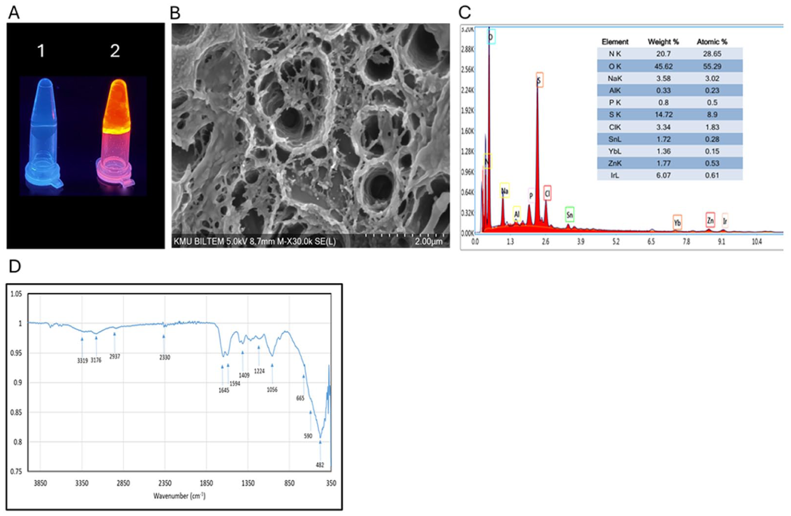
Characterization of DNA based hydrogels. (A) Photographs of DNA hydrogel (1) and ethidium bromide-entrapped DNA hydrogel (2) under UV light; (B) Scanning electron microscopy morphology; (C) EDX analysis; (D) FTIR spectrum of DNA hydrogel.
Scanning electron microscopy (SEM) was employed to investigate the morphology of the DNA hydrogels. As shown in Figure 5(B), SEM image revealed interconnected microstructures likely stabilized via hydrogen bonding to form porous sheet structures. The image clearly shows interconnected microstructures, which are characteristic of a porous network. This observation aligned with the proposed mechanism of hydrogel formation, where DNA crosslinkers formed hydrogen bonds with the acrylamide polymer chains, creating a stable, three-dimensional network. The interconnected nature of the pores is crucial for efficient transport phenomena within the hydrogel, which is vital for its functionality in biological and chemical systems. Furthermore, the distribution of the elements in the structure of DNA hydrogel was characterized by energy-dispersive X-ray (EDX) elemental mapping and the result was given in Figure 5(C). The elemental analysis of the DNA hydrogel revealed the predominant presence of Oxygen (O), Nitrogen (N), Sulfur (S), Carbon (C), and Phosphorus (P). The detection of phosphorus was particularly significant as it is a unique and defining element of the DNA backbone, unequivocally confirming the successful integration of DNA within the polyacrylamide matrix. Nitrogen and carbon were abundant in both DNA and the polyacrylamide structure, while oxygen was present in both components and water within the hydrogel. The presence of sulfur could potentially originate from residual initiators (e.g. ammonium persulfate) or other minor components used during synthesis, providing further evidence of the chemical environment within the hydrogel. The mapping of these elements (as shown in Figure 5(C)) further corroborated their uniform distribution throughout the interconnected network, indicating a homogeneous hybrid material formation.
FTIR spectroscopy further supported the structural composition of DNA hydrogels (Figure 5(D)). The characteristic peaks of -OH, C = O stretching, C-O-C stretching and C-O stretching at 3319, 1645, 1224, and 1056 cm−1 were exhibited by DNA hydrogels. The peak at 3319 cm⁻¹ was corresponding to the stretching vibration of -OH groups which are present in both the polyacrylamide backbone (from residual water or hydroxyl groups) and, more significantly, in the deoxyribose sugar of the DNA molecule. The presence of a sharp band at 3623 cm⁻¹ further specifically proves the integration of DNA due to the -OH group in the DNA molecule, distinguishing it from general hydroxyl presence. The strong absorption at 1645 cm⁻¹ is attributed to the C=O stretching vibration, primarily from the amide I band of the polyacrylamide which confirmed the successful polymerization of acrylamide monomers into the polymer network. The peaks at 1224 and 1056 cm⁻¹ are associated with C-O-C stretching and C-O stretching vibrations, respectively, and they could arise from the polyacrylamide backbone and also from the sugar-phosphate backbone of DNA. A broad band in the range of 2997–2883 cm−1 was attributed to the stretching vibration of C–H bonds in −CH2 group, which confirmed the presence of DNA and the macromolecular chains of polyacrylamide within the structure of DNA hydrogels. Moreover, the peaks at lower wave number at 590, 821, and 1281 cm−1 and in the region of 1108–1224 cm−1 were attributed to stretching vibration of the phosphate groups in deoxyribose of DNA. The presence of these peaks strongly indicated the successful incorporation of DNA into the hydrogel structure.
Conclusion
In conclusion, this study successfully demonstrated the fabrication and characterization of DNA-crosslinked acrylamide hydrogels. The synthesis principle was through the incorporation of 5′-acrydite modified DNA strands into a polyacrylamide network and hybridization of modified DNA strands with partially complementary DNA crosslinker. The methodology facilitated the efficient integration of DNA strands as side-chain components, subsequently hybridized with a partially complementary DNA crosslinker to form a three-dimensional network. Optimization study in synthesis process revealed that a DNA concentration of 60 µM for each component, combined in a 1:1:1 molar ratio, yielded a stable hydrogel which could be easily visualized by naked eye. Furthermore, temperature was found to be a critical factor, with optimal gelation observed at 4°C and 25°C due to the preservation of hydrogen-bond interactions essential for network stability. Notably, hydrogels incorporating shorter complementary regions exhibited superior stability property. Characterization techniques, including FTIR spectroscopy and SEM, confirmed the successful incorporation of DNA strands within the polymer matrix, as well as the development of porous, interconnected morphology in the network structure of DNA hydrogel. Overall, the developed hybrid DNA hydrogels have the potential to offer a stable matrix for bioengineering applications by combining the programmability of DNA with the structural advantages of synthetic polymers.
Footnotes

Ethical considerations
No approval from the Board of Ethics is required.
Author contributions
This paper is derived from the first author’s doctoral dissertation supervised by the second author. They all read and approved the final version of the paper.
Funding
The authors disclosed receipt of the following financial support for the research, authorship, and/or publication of this article: This work was supported by the Scientific and Technological Research Council of Türkiye (TÜBİTAK), Grant number: 123Z498.
Declaration of conflicting interests
The authors declared no potential conflicts of interest with respect to the research, authorship, and/or publication of this article.