





Abstract
Hemophagocytic syndrome, also known as hemophagocytic lymphohistiocytosis (HLH), is a rare and frequently fatal disorder caused by an uncontrollable and ineffective systemic immune response. Patients initially present with fever, cytopenia, and hepatosplenomegaly, and subsequently develop multiorgan failure (MOF). Hemophagocytosis can be found on biopsy specimen but is not required. Acquired forms of HLH can occur in apparently healthy adults, while children present more often with an inherited form of the disease. Since HLH often presents with sepsis-like symptoms and organ dysfunction, patients are usually treated for presumed sepsis, which inevitably leads to delayed diagnosis and treatment. Intensivists need to have a low threshold for suspecting this disorder when previously healthy individuals present with a fulminant sepsis-like syndrome, which are unresponsive to conventional treatment. We present 3 patients with HLH who were admitted to our adult medical intensive care unit (MICU) over a 2-year period with fatal outcomes and emphasize the diagnostic importance of markedly elevated serum ferritin levels and the need for tissue biopsy in making an accurate diagnosis in a timely manner.
Keywords
Introduction
Hemophagocytic lymphohistiocytosis (HLH) is characterized by a life-threatening unregulated activation of the immune system, resulting in a systemic inflammatory response syndrome (SIRS). Clinically, HLH presents with prolonged malaise, fever, cytopenia, and hepatosplenomegaly, leading to multiorgan failure (MOF) and fatal outcomes if left untreated. Hemophagocytic lymphohistiocytosis is a heterogeneous group of disorders, including familial or primary HLH, acquired or secondary HLH, and macrophage-activation syndrome (MAS). Familial HLH (FHLH) is the most thoroughly investigated HLH disorder being first described by Farquhar and Claireux in 1952 as “familial hemophagocytic reticulosis.” 1 Onset of the disease is under the age of 1 year in 70% to 80% of patients, although there have been some reports of FHLH first manifesting in older children and even adults.2,3 Acquired forms of HLH are associated with infections (viral, most commonly Epstein-Barr virus [EBV], bacterial, parasitic, and fungal), malignancy, autoimmune disorders such as lupus and rheumatoid arthritis and medications such as chemotherapy and immunosuppressants, although the etiology of secondary HLH often remains unknown despite extensive antemortem and postmortem investigations. Macrophage-activation syndrome is a distinct form of HLH associated with autoimmune disorders, particularly systemic onset juvenile inflammatory arthritis, although whether it is truly a unique disease entity different from secondary HLH is a point of contention. 4
The Histiocyte Society recently revised their diagnostic guidelines to include molecular diagnosis, fever, splenomegaly, cytopenia, hypertriglyceridemia, hypofibrinogenemia, and hyperferritinemia, low serum natural killer (NK) cell activity and elevated serum soluble interleukin 2 (IL-2) receptor (CD25; Table 1 ). 5 If a genetic mutation known to be associated with familial HLH is found by mutation analysis, a patient does not need to satisfy the diagnostic guidelines to be diagnosed with HLH. Examples of known mutations are those found in perforin, UNC13D and STX11 genes. The diagnosis of HLH remains elusive, primarily due to a nonspecific presentation mimicking that of sepsis and the lack of specific laboratory findings. In addition, the pathologic finding of hemophagocytosis can be seen in other conditions such as hemolytic anemia and malignancy without meeting the diagnostic criteria of HLH.
Histiocyte Society Diagnostic Guidelines for HLH
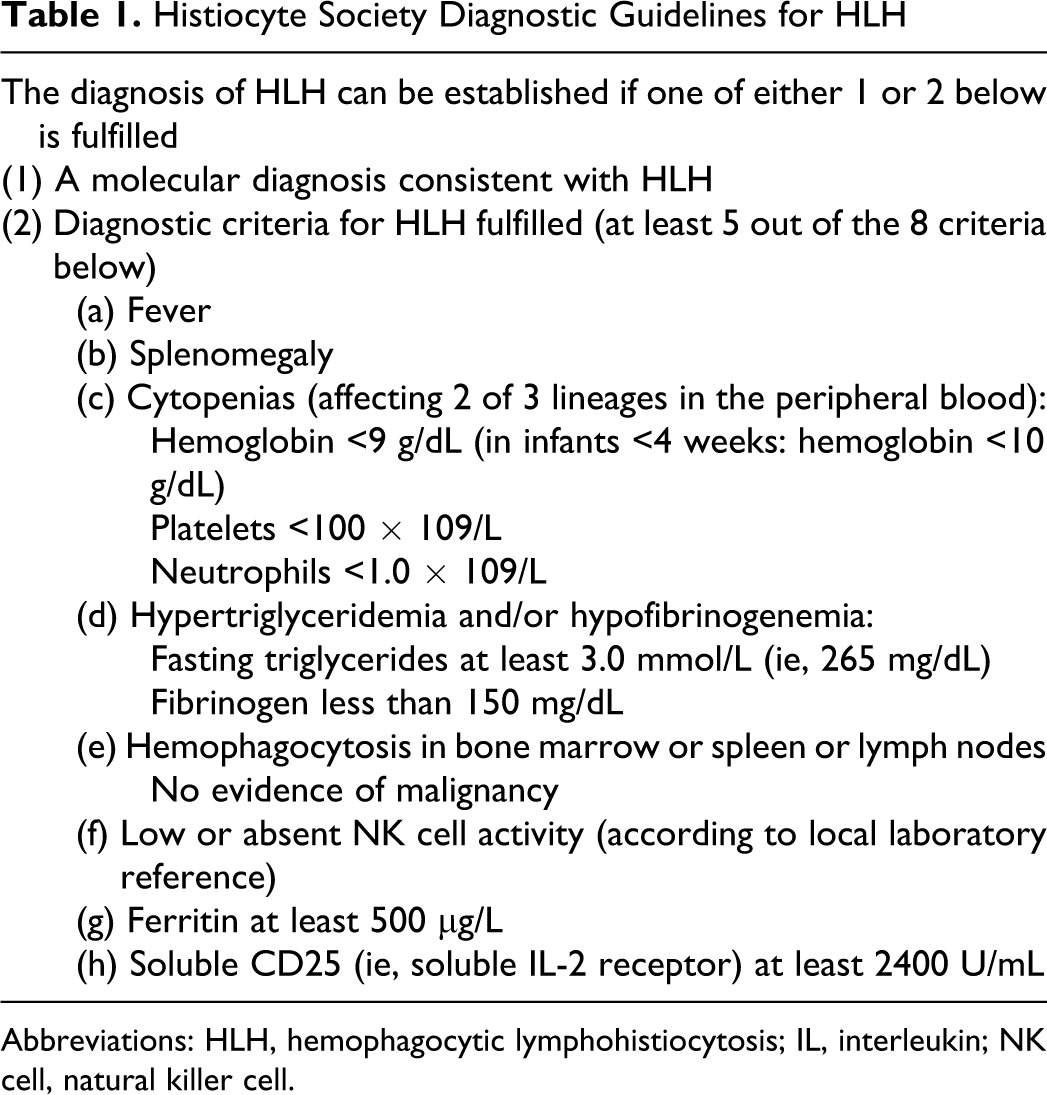
Abbreviations: HLH, hemophagocytic lymphohistiocytosis; IL, interleukin; NK cell, natural killer cell.
We describe 3 cases of acquired HLH with fatal outcomes in our adult medical intensive care unit (MICU) from 2008 to 2009 (Table 2). The purpose of our case series is to raise awareness of the diagnosis of acquired HLH as a cause of what appears to be fulminant sepsis in the adult MICU, as well as to demonstrate the importance of hyperferritinemia and repeating tissue examination in making a prompt and accurate diagnosis.
Characteristics of 3 Cases
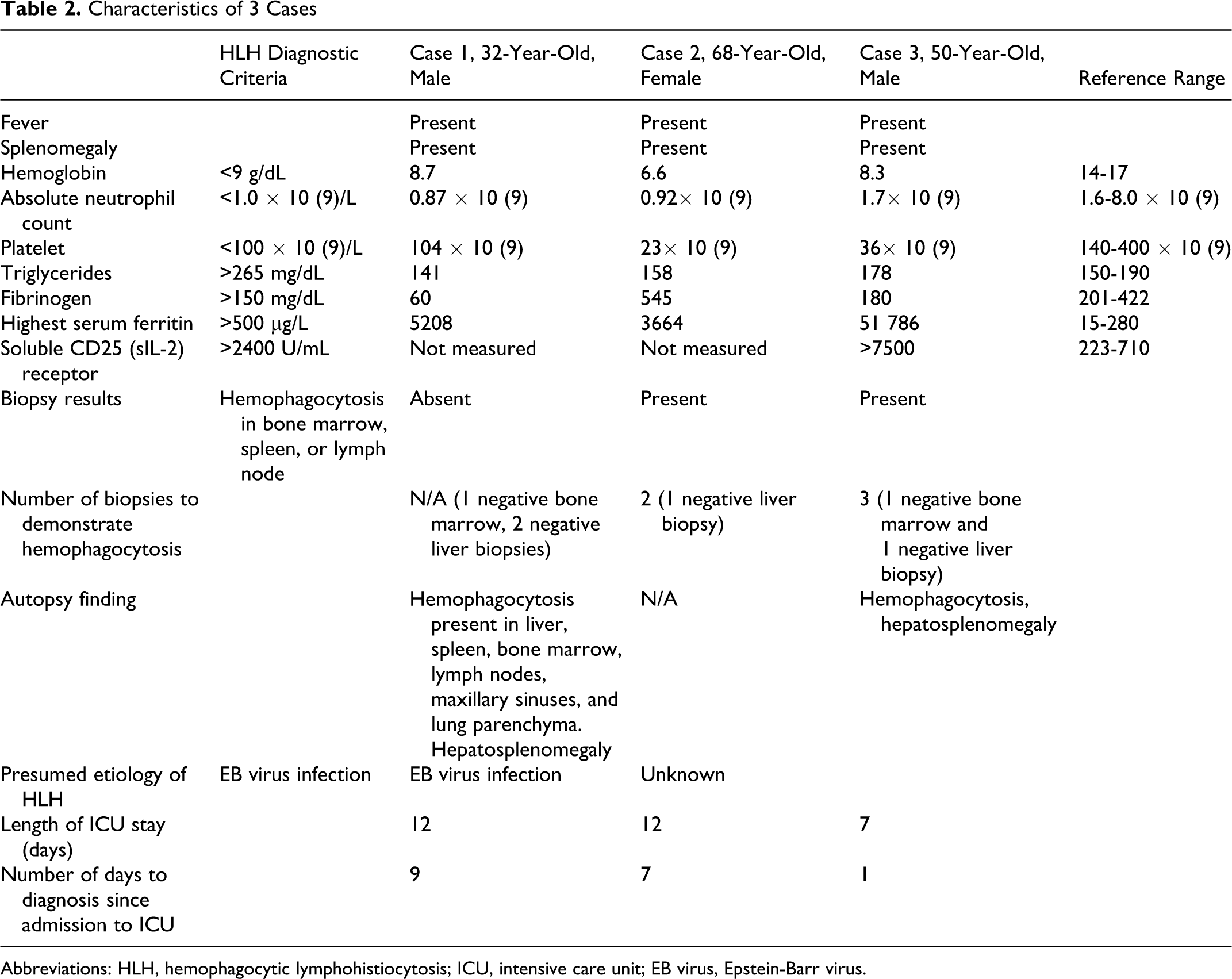
Abbreviations: HLH, hemophagocytic lymphohistiocytosis; ICU, intensive care unit; EB virus, Epstein-Barr virus.
Case Reports
Case 1 (Diagnosed in January 2008 ) 6
The patient was a 32-year-old male with a new diagnosis of “hepatic sarcoidosis,” who was admitted to our hospital with persistent fever, body aches, rhinorrhea, jaundice, and pruritus beginning 2 weeks prior to admission. A liver biopsy performed at an outside hospital 2 months earlier revealed scattered noncaseating granulomas within the parenchyma and portal tracts with a negative stain and culture for mycobacterium. He was subsequently started on oral prednisone at 20 mg daily for hepatic sarcoidosis.
His medications included alprazolam, benadryl, omeprazole, guaifenesin, hydroxyzine, and prednisone. The patient denied any significant family history. He gave a history of heavy tobacco and alcohol use in the past and worked as a correctional officer.
On transfer to our hospital, he was febrile, tachycardic, and hypotensive. He was alert and oriented but markedly jaundiced with small right axillary lymphadenopathy and hepatosplenomegaly. The remainder of his examination was unremarkable. Initial laboratory studies found him to be pancytopenic (see Table 2), with a bilirubin of 33 mg/dL (normal: 0.2-1.2 mg/dL), asparate aminotransferase (AST) of 25 IU/L (normal: 7-42 IU/L), alanine aminotransferase (ALT) of 135 IU/L (normal: 1-45 IU/L), alkaline phosphatase (ALP) of 1431 IU/L (normal: 25-120 IU/L), triglycerides (TG) of 161 mg/dL (normal: 150-190 mg/dL), and a serum ferritin of 5208 ng/mL (normal: 15-280 ng/mL). Epstein-Barr virus immunoglobulin G (IgG) was positive, which was consistent with previous exposure. Workup for autoimmune disorders was negative. Computed tomographic (CT) scans of the head, chest, and abdomen demonstrated a pansinusitis, innumerable scattered bilateral lung nodules measuring up to 1 cm in diameter, right axillary lymphadenopathy, and hepatosplenomegaly. His bone marrow biopsy exhibited a cellular marrow containing all normal hematopoietic elements with left-shifted myelopoiesis, but no evidence of hemophagocytosis, malignancy, or granulomatous disease. A stain for acid-fast bacilli was negative. He was admitted to the ICU and was empirically treated with broad-spectrum antibiotics for presumed severe sepsis.
The patient’s hospital course was complicated by persistent fevers, with progressive pancytopenia and recurrent episodes of hypotension, requiring vasopressors, as well as mechanical ventilation for severe hypoxemia. He required frequent red cell and platelet transfusions, as well as granulocyte colony-stimulating factor (G-CSF). As there was no clinical improvement, a repeat liver biopsy was performed revealing ductopenia with no granulomas or malignancy. On the basis of his clinical picture of SIRS and MOF in the absence of any positive cultures that point to a specific infection and unresponsiveness to 2 weeks of empiric broad-spectrum antibiotics and perplexing liver pathology, the possibility of HLH was considered. Once it was established that he met the diagnostic criteria for HLH (fever, splenomegaly, cytopenia, hypofibrinogenemia, and hyperferritinemia), the patient was treated with high-dose methylprednisolone and immunoglobulins. His deteriorating condition precluded the use of chemotherapy, and he subsequently expired. The autopsy confirmed the diagnosis of HLH and revealed marked hepatomegaly (4800 g) and splenomegaly (2930 g). Antemortem serum EBV polymerase chain reaction (PCR) was later found to be markedly elevated at 692 000 copies/mL. In addition, molecular studies for EBV on the postmortem bone marrow, liver, spleen, and lung were positive (>1 copy/cell), confirming EBV-associated HLH.
Case 2 (Diagnosed in December 2008 )
The patient was a 68-year-old female with a past medical history of atrial fibrillation, breast cancer, ovarian cancer, and sarcoidosis, who presented with 3 weeks of persistent fatigue and liver lesions seen on a previous abdominal CT. She also complained of chronic dry cough, daily fevers, and night sweats for the past year since her diagnosis of sarcoidosis. Her medications included metoprolol, digoxin, warfarin, esomeprazole, cholestyramine powder packet, and prednisone. Upon admission, she was afebrile and had an unremarkable physical examination. A repeat CT scan of the abdomen revealed a 1.7 cm low-attenuation enhancing solid focal lesion at the inferior aspect of the right lobe of the liver, a 0.8 cm lesion in left lateral segment of liver, multiple low-attenuation focal splenic lesions with perisplenic fluid and mild splenomegaly. A CT of the thorax revealed irregularly marginated and spiculated nodules in the right lower lobe and a right fissural nodule, as well as mild mediastinal hilar lymphadenopathy. A core biopsy of the liver revealed a diffuse fibrohistiocytic infiltrate consistent with sarcoidosis. Initial laboratory tests revealed hemoglobin of 6.6 g/dL, platelet count 23 × 10 9 /L, white blood cell 0.92 × 10 9 /L (see Table 2), ALP of 189 IU/L, AST of 38 IU/L, ALT 43 of IU/L, ferritin of 2920 ng/mL, and a fibrinogen of 805 mg/dL (normal: 201-422 mcg/dL).
Over the next few weeks of her hospital stay, she continued to have fevers and developed worsening coagulopathy and liver dysfunction, as well as persistent pancytopenia. Despite pulse dose steroids for presumed sarcoidosis, her general condition progressively worsened, and she was eventually transferred to the ICU. Broad-spectrum antibiotics were empirically started for presumed sepsis. In the ICU, her hepatitis panel and cytomegalovirus (CMV) PCR were negative, however, she was noted to have a markedly elevated EBV viral load (210 353 copies/mL). All cultures remained negative. Her fever was refractory to antibiotics, and eventually, the possibility of HLH was raised. A bone marrow biopsy revealed occasional macrophages engulfing hematopoietic cells, suggestive of hemophagocytosis. No granulomas or evidence of malignancy was noted. Immunohistochemical stain demonstrated CD25 cells scattered throughout the marrow core, consistent with the diagnosis of HLH.
The patient was started on infliximab and cyclosporine A. Her profound pancytopenia required multiple blood transfusions with marrow support with G-CSF and erythropoietin. Fungemia and severe MOF ensued, and the patient succumbed to her illness. Her family declined autopsy. The etiology of her HLH was considered due to EBV infection.
Case 3 (Diagnosed in May 2009 )
The patient was a 50-year-old male with a past medical history of insulin-dependent diabetes mellitus and hyperlipidemia, who 4 months prior to admission to our hospital developed fever, chills, fatigue, and a severe headache following a tick bite for which he was treated for presumed Lyme disease and recovered uneventfully. Two weeks prior to admission to our hospital, he went to the same outside hospital with fever and chills where he was found to have a mild pancytopenia (WBC 4.1 ×10 9 /L, Hgb 8.9 g/dL, platelet count 100 000/mm 3 ) and elevated creatinine (2.1 mg/dL). Over the hospital stay, he continued to spike fevers up to 39.4°C with rigors despite negative cultures and broad-spectrum antibiotics. His blood work revealed gradually worsening pancytopenia and increasing creatinine and hepatic enzymes. His bone marrow biopsy revealed normocellular marrow with adequate megakaryocytes and matured cell lines. A CT scan of the abdomen and pelvis showed significant hepatosplenomegaly. He was transferred to our hospital for further workup of fever of unknown origin.
On the day following admission, he was transferred to the ICU due to progressively worsening respiratory distress, acute kidney injury, and hyperkalemia. On physical examination, he was afebrile, tachycardic, and tachypneic requiring 5 L of supplemental oxygen by nasal cannulae to maintain an oxygen saturation greater than 90%. The patient appeared confused and diaphoretic. His lungs were clear to auscultation bilaterally, however, his breathing was labored. The remainder of the physical examination was unremarkable. He was subsequently intubated for acute respiratory distress and underwent emergent hemodialysis for hyperkalemia. Laboratory results revealed pancytopenia (see Table 2), abnormal hepatic enzymes (AST 416 U/L, ALT 159 U/L, and ALP 1500 U/L), and a remarkably elevated ferritin of 56 000 ng/mL.
A transjugular liver biopsy yielded inconclusive result of nonspecific inflammation. Along with the repeat bone marrow biopsy revealing increased macrophages, some of which showed evidence of erythrophagocytosis, the patient had fever, splenomegaly, pancytopenia, hyperferritinemia, and a soluble CD25 receptor greater than 7500 U/mL; thus, the diagnosis of HLH was made. Despite aggressive supportive care including mechanical ventilation, continuous hemodialysis, and treatment with intravenous IgG, infliximab, daclizumab, dexamethasone, and cyclosporine, he succumbed to the illness. Autopsy uncovered marked erythrophagocytosis in bone marrow (Figure 1), significant hepatomegaly (3550 g), and splenomegaly (1000 g) with red pulp congestion (Figure 2). Serologies for EBV, CMV, herpes viruses, HIV, hepatitis viruses, and Lyme disease were all nonreactive. The etiology of HLH in his case remained unknown.

Hematoxylin-eosin stain of antemortem bone marrow sample showing numerous macrophages engulfing red blood cells.

Autopsy revealed significant hepatomegaly (below: 3550 g) and splenomegaly (above: 1000 g).
Discussion
Epidemiology
The incidence of HLH in adults is unknown; however, in children, it is estimated to be 1.2 per million per year in a Swedish study, but the true incidence is probably higher as the disorder is believed to be underdiagnosed.7,8 In addition, most studies reported are relevant to children than adults. Lack of specific biomarkers and the presence of similar conditions that mimic the presentation of HLH, such as sepsis, make antemortem diagnosis particularly challenging.
Pathophysiology
Putative pathophysiology of HLH is an uncontrolled mutual stimulation of histiocytes (macrophages and dendritic cells), NK cells, cytotoxic T-cells (CTL), which leads to persistent hypercytokinemia and SIRS. 9 Secondary HLH is a rare disorder with an undetermined incidence. Conditions associated with secondary HLH include infections, particularly viral, malignancy, and autoimmune disorders. 10 Notably, EBV-associated HLH has been frequently reported in Asian countries, particularly Japan, and has a notoriously relentless clinical course unless treatment is initiated promptly. 11 The estimated incidence of EBV-associated HLH in Japanese children is at least 25 cases annually. 11 Tabata et al suggest a possible link between the higher incidence of EBV-associated HLH and the prevalence of unique and virulent strains of EBV in Asia, but this hypothesis needs to be further elucidated in the future. 12 Furthermore, it is less clear how apparently immunocompetent patients develop dysfunction of NK cells and CTLs following triggering conditions, such as viral infections. The mechanism by which EBV causes HLH has been extensively investigated. It is believed that while primary infection with EBV usually affects B lymphocytes, in patients with EBV-associated HLH, the virus infects NK-T cells and causes proliferation of more T cells with subsequent production of proinflammatory cytokines, particularly tumor necrosis factor-α (TNF-α) and interferon-γ, by stimulation by EBV latent membrane protein 1 (LMP-1). This hypercytokinemia stimulates macrophages and is thought to be the pathophysiologic mechanism for EBV-associated HLH. Epstein-Barr virus has also been implicated in the pathogenesis of lymphoma-associated hemophagocytic syndrome (LAHS). In fact, it is proposed that EBV-associated HLH and LAHS are part of a continuum that represents the natural progression of T-cell infection with EBV. Some patients diagnosed with EBV-associated HLH who survived have been observed to have relapsing disease or progression to T-cell lymphoma. Latent membrane protein 1 is believed to cause activation of TNF receptor and nuclear factor-κB. This process subsequently causes EBV-infected T-cells to escape TNF-α-induced apoptosis. These cells are thought to survive and can later cause disease relapse. Furthermore, chromosomal analysis of EBV-infected T-cells in patients with HLH showed clonality early in the course of the disease, which may further account for progression to lymphoma.13–15
Diagnosis
In order to achieve a rapid diagnosis of HLH, the Histiocyte Society proposed diagnostic guidelines in 2004 (Table 1). 5 The provisional diagnosis of secondary HLH requires 5 or more of the 8 diagnostic criteria in the absence of a positive family history or known gene defects associated with primary HLH such as PFR1, UNC13D, and STX11.16–19 Among the diagnostic criteria, a pronounced hyperferritinemia should alert physicians to the possibility of HLH. 20 Since Esumi et al first proposed the potential value of hyperferritinemia in assisting the diagnosis of HLH in 1989, 21 the validity of hyperferritinemia as a biomarker for HLH has been widely studied in multiple observational studies. In the review of 1093 pediatric blood samples with elevated ferritin greater than 500 mcg/L, Allen et al described that the 10 patients diagnosed with HLH had the highest maximum ferritin levels ranging between 994 and 189 721 mcg/L (median 15 830 mcg/L). 22
Ferritin is an iron storage protein that plays a key role in iron homeostasis. Serum ferritin is frequently measured in patients with suspected iron-deficiency anemia, iron overload, and hemochromatosis. More recently, the role of elevated serum ferritin levels in other medical conditions, such as neurodegenerative disorders, cardiovascular diseases, and malignancy, has been increasingly recognized.23–25 Although the exact cause of the profound hyperferritinemia in HLH has not been fully elucidated, several factors appear to play a role, including the increased release of ferritin from cellular damage in the liver and spleen, increased secretion by macrophages and hepatocytes, and decreased serum clearance due to reduced glycosylation or downregulation of ferritin receptors. 26
Ferritin is an iron storage protein that plays a key role in iron homeostasis. Serum ferritin is frequently measured in patients with suspected iron-deficiency anemia, iron overload, and hemochromatosis. More recently, the role of elevated serum ferritin levels in other medical conditions, such as neurodegenerative disorders, cardiovascular diseases, and malignancy, has been increasingly recognized.23–25 Although the exact cause of the profound hyperferritinemia in HLH has not been fully elucidated, several factors appear to play a role, including the increased release of ferritin from cellular damage in the liver and spleen, increased secretion by macrophages and hepatocytes, and decreased serum clearance due to reduced glycosylation or downregulation of ferritin receptors. 26
Another diagnostic criterion is the presence of elevated triglyceride level (at least 265 mg /dL) in the blood. The exact mechanism for hypertriglyceridemia in HLH is unknown, but hypercytokinemia resulting in lipoprotein lipase inhibition probably plays a central role. 27 It is interesting to note that none of our patients had serum triglyceride level above the diagnostic criterion. The incidence of hypertriglyceridemia in HLH has been estimated to be 60% to 70%. 10 In a small series of Japanese children with EBV-associated HLH, all the patients had elevated triglyceride levels. 28 In another report on patients with familial HLH, 77% had hypertriglceridemia. 29 A recent analysis of 28 patients diagnosed with secondary HLH in Japan reported that only 32% of cases had triglyceride levels greater than 265 mg/dL. 30 Thirty-six percent had levels between 150 mg/dL and 265 mg/dL, and another 32% had levels within the normal laboratory range of less than 150 mg/dL. The authors of this study further reported that triglyceride level was positively correlated with the ferritin level, degree of hepatosplenomegaly, as well as the need for chemotherapy. They also showed that mean triglyceride levels significantly decreased in those patients that showed clinical improvement after treatment. The trend toward improvement of triglyceride levels after treatment seems to hold true, regardless of initial triglyceride level; however, they failed to report a subgroup analysis to show that even those with normal initial triglyceride levels had statistically significant decline in triglyceride levels after treatment. The authors propose that triglyceride level might be an important predictor of severity and response to treatment. In our series, 1 patient had triglyceride level less than 150 mg/dL and 2 patients had levels between 150 mg/dL and 265 mg/dL. All 3 patients expired and none of the patients received the recommended standard chemotherapeutic regimen secondary to severe deterioration and MOF. Repeat triglyceride levels were also not available.
Although hemophagocytosis in biopsy specimens is the hallmark of HLH, it is not the sine qua non for the diagnosis, and the absence of this finding should not hinder a treating physician from initiating treatment if other diagnostic criteria are adequately met. It is not uncommon to require more than 1 biopsy specimens to demonstrate microscopic evidence of hemophagocytosis. 7 Conversely, the mere presence of hemophagocytosis in the reticuloendothelial system does not automatically imply the diagnosis, as it also occurs in other hematological disorders such as hemolytic diseases and malignancy. Ductopenia, or bile duct paucity, is a rare and nonspecific finding in liver histology but may help clinch the diagnosis of HLH in the appropriate clinical context. Vanishing bile duct syndrome (VBDS) loosely refers to the group of acquired disorders characterized by progressive destruction and disappearance of the intrahepatic bile ducts. Vanishing bile duct syndrome represents a final common pathological pathway, resulting from a whole host of causes including autoimmune disorders such as primary biliary cirrhosis (PBC) and primary sclerosing cholangitis (PSC), medications, genetic abnormalities, infectious diseases, and malignancy. 31 Hepatitis B virus (HBV), hepatitis C virus (HCV), CMV, EBV, as well as other viral infections, have been linked to VBDS, and sporadic cases of VBDS associated with EBV-associated HLH have been reported. 32 In our case 1, the finding of ductopenia in the liver biopsy prompted a literature search, and eventually raised the possibility of EBV-associated HLH as the diagnosis.
Treatment
The standard immunochemotherapy protocol proposed by the Histiocyte Society (HLH-2004), includes therapy with dexamethasone, etoposide (VP-16), cyclosporine, and intrathecal methotrexate. 5 In our 3 cases, advanced organ dysfunctions, particularly renal and hepatic dysfunction at the time of diagnosis, precluded aggressive treatment in accordance with the HLH-2004 protocol. Early treatment is vital to prevent progressive organ failure and death. Imashuku et al illustrated a statistically significant mortality benefit in adult patients with EBV-associated HLH treated with etoposide within 4 weeks of presentation compared to those not treated or treated late. In his study, the Kaplan-Meier analysis showed the 2.5-year survival of 85.7% in the early etoposide-treated patients, compared to 10.3% in the remaining patients (log rank test, P = .0141). 33 Case reports of successful treatments with antimonoclonal antibodies, such as daclizumab, an anti-CD25 antibody, and infliximab, an anti-TNF-α antibody, have been reported in refractory cases, but the effectiveness of these therapeutic approaches warrant rigorous exploration in the future.34,35
Organ dysfunction, affecting the central nervous system, liver, kidney, lung, and bone marrow, occurs at various degrees resulting in a high mortality from HLH. 27 Close collaboration by the intensivist with a hematologist and pathologist experienced in diagnosing HLH is required, for the early diagnosis and treatment of this disorder. The principal role of the treating intensivist is to ensure that patients with HLH receive early and comprehensive supportive care often involving mechanical ventilation, hemodynamic support, renal replacement therapy, blood transfusions, and antibiotics. Lastly, since HLH can quickly inflict serious consequences on previously healthy adults, the importance of multidisciplinary support cannot be overemphasized.
Conclusion
Patients with HLH often present to the adult ICU with sepsis-like signs and symptoms. Early recognition and prompt treatment of the disorder in collaboration with hematologists and pathologists is the key to preventing fatal outcomes. While we recognize that ferritin is an acute phase reactant, and can be elevated in many conditions, measurement of serum ferritin levels is an inexpensive, yet critical test in suggesting the diagnosis in patients with SIRS and MOF after infectious cause is thoroughly excluded. Initial tissue biopsy may not reveal hemophagocytosis and needs to be repeated if the suspicion remains high. Hemophagocytic lymphohistiocytosis is a remarkably elusive diagnosis unless intensivists carry a high index of suspicion and vigilance whenever a patient presents with prolonged fever, cytopenia, and organ dysfunction refractory to antibiotics.
Footnotes
The author(s) declared no conflicts of interest with respect to the authorship and/or publication of this article.
The author(s) received no financial support for the research and/or authorship of this article.