






Abstract
Gastric reactance has been proposed as a measure of mucosal ischemic injury in the critically ill. The purpose of this study was to evaluate the incidence of gastric mucosal injury as measured by gastric reactance in different subgroups of critical patients. We studied 100 adult patients admitted to 7 different hospital intensive care units, requiring a nasogastric tube. Gastric impedance measurements were continuously obtained from each patient for 24 hours. Patients were managed based on conventional protocols by hospital staff, blinded to the changes in gastric impedance parameters. The low-frequency central reactance (X L) reflects tissue edema caused by prolonged ischemia. The previously reported threshold of X L ≥ 13 − jΩ was used to classify injured mucosa; 80% of all patients had mean X L above this threshold. No significant differences were found in the incidence of mucosal ischemia between medical versus surgical, hemodynamic versus respiratory or neurological patients. Significant lower urine output was found in patients with X L above threshold (P < .01); also, there was a significant effect of fluid balance in those patients (P < .05). More complicated patients had higher average reactance. This study shows that gastric ischemia as estimated by gastric reactance has a very high incidence in the critically ill, independently of the reason for admission. High reactance is related with higher morbidity in agreement with other reports using different methods of assessing splanchnic hypoperfusion in this patient population.
Introduction
Shock is characterized by acute systemic hypoperfusion, leading to tissue hypoxia and vital organ failure.1,2 An association between abnormal gastrointestinal (GI) perfusion and critical illness has been suggested for many years; and largely circumstantial evidence continues to implicate the GI tract in the pathogenesis of the systemic inflammatory response syndrome (SIRS) and multiple organ dysfunction syndrome (MODS). 3 Inadequately oxygenated, the splanchnic tissues may become prone to ischemia-related complications. 4 However, at present, there is no clinically useful method to directly monitor the level of mucosal ischemia. In particular, it may be of benefit not only to measure the adequacy of perfusion but also to detect the onset and monitor the level of ischemic injury.
Ischemic damage of the GI tissue is characterized by alterations in cellular function and energy metabolism. After a certain period of ischemia, the GI tissue becomes vulnerable to damage due to reperfusion, in which restored oxygen supply produces free radical species, which lead to further tissue injury. 5 The mucosal injury increases as ischemia persists. 6 For an effective and correctly timed therapy, it may be very useful to know the ischemic level and the tissue damage in a continuous and simple manner. 7
Impedance spectroscopy, the study of the passive electrical properties of biological tissues as a function of frequency, has been proposed as a tool for monitoring mucosal tissue dysoxia and damage in different organs.8–11 On a conceptual level, the cell cytoplasm and extracellular space act as conductive media isolated from each other by the cell membrane. The conductivity of the extra- and intracellular space contributes to the overall resistance of the tissue, while the cell membrane contributes to the capacitive effect. 9,12 Impedance measurements, tissue reactance in particular, have been shown to reflect changes in a tissue structure; namely, the intracellular to extracellular volume ratio and cell membrane ion permeability, both of which are directly affected during ischemia. A specific device and system has been developed and tested for this specific application13–15 (Critical Perfusion Inc, California). Along with the technique, a Gastric Impedance Spectroscopy Probe (GISP) (Critical PerfusionInc, California) 16 allows the direct acquisition of an electric impedance spectrum of the mucosa, which can be used to identify and continuously monitor the level of tissue damage.
The impedance of biological tissues results from the interaction of an electrical current with the tissue at cellular and molecular level. Impedance has 2 components: the electrical resistance which restricts the flow of electrons and dissipates energy, and the electrical reactance which is related to the capacity to store and release energy. 17 All the impedance parameters increase with duration of ischemia as ion pumps fail, cells swell, and ion membrane permeabilities decrease, allowing cellular membranes to store greater amounts of electrical energy, and reducing the available extracellular current paths. Reactance has been shown to be directly proportional to intracellular to extracellular volume ratio, and therefore an increase in reactance is relatively specific to ischemia. Other forms of injury such as apoptosis, fluid overload, or tissue breakdown actually decrease reactance. An algorithm was developed to calculate the characteristic electrical values that best describe human gastric impedance measurements and simplify the information obtained with this method. 18 It was reported that hypoperfusion and ischemia cause changes in the impedance measurements of the gastric wall in cardiovascular surgery patients. 19 The value of these measurements as a prognostic index of postoperative outcome in cardiovascular surgery patients was validated by Beltran et al. 20
The objective of this study is to describe the incidence of abnormal gastric reactance in different subgroups of critically ill patients. A secondary objective is to evaluate the effects of highly positive fluid balance and increased intraabdominal pressure (IAP) in gastric reactance.
Methods
The target population of this study was general critical care patients that required nasogastric tube for gastric decompression or feeding. All the patients were onTotal Parenteral Nutrition (TPN) and some patients were on Proton-PumpInhibitor (PPI) therapy, following standard procedures for each hospital. We did a previous unpublished study in healthy volunteers to demonstrate that suction and feeding did not significantly affect the measurements of gastric impedance spectroscopy. The study was approved and supervised by the Ethical Research Committee of 7 different hospitals in Mexico City. Informed consent from the patient or his next of kin was obtained in all cases. Patients with massive GI bleeding, esophageal obstruction, or nasopharyngeal obstruction were excluded from the study. GISP was positioned in the stomach to measure tissue reactance. For all cases, GISP positioning was confirmed by chest or abdominal radiography. Exclusion criteria included improper GISP positioning or less than 12 hours of continuous monitoring. Patients entered in the study were followed up prospectively until intensive care unit (ICU) discharge or after 28 days of follow-up during the study period. Age, sex, primary diagnosis, Acute Physiology and Chronic Heath Evaluation II (APACHE II) score, and Sequential Organ Failure Assessment (SOFA) score were recorded upon admission.
During ICU stay, patients were managed based on the conventional hemodynamic and respiratory protocols by the health care staff, blinded to the changes in gastric impedance parameters. All patients were mechanically ventilated and sedated. Patients were monitored until they no longer required a nasogastric tube, after 3 days of monitoring or death. Impedance measurements obtained from these patients, automatically recorded every 15 minutes, were processed to calculate the central reactance at low frequencies (X L), as the characteristic gastric impedance parameter of interest, as reported by Beltran et al. 20 To analyze differences in X L between the different groups, we calculated the average reactance over the first 24 hours of monitoring for each patient (mean X L).
Mean arterial pressure (MAP), central venous pressure, lactate, base excess, arterial blood gases, and urine output were obtained hourly to evaluate disturbances in hemodynamics and organ function. The study protocol prospectively defined SIRS, sepsis, and shock patients according to the Society of Critical Care Medicine (SCCM) guidelines 21,22 after ICU admission by medical staff, during the first day of impedance monitoring. The SIRS was manifested by 2 or more of the following conditions: (1) temperature >38°C or <36°C; (2) heart rate >90 bpm; (3) respiratory rate >20 bpm or PaCO2 <32 mm Hg; and (4) white blood cell count >12 000/mm3 or <4000/mm3. Sepsis was manifested by 2 or more of the previous conditions as a result of infection. Septic shock was manifested when sepsis was induced with hypotension (systolic blood pressure <90 mm Hg) despite adequate fluid resuscitation along with the presence of perfusion abnormalities that may include lactic acidosis or oliguria. We compared patients with SIRS, sepsis, and septic shock.
The subgroups of critically ill patients by reasons of ICU admission were classified as surgical or medical patients with a classification of hemodynamic, respiratory, or neurological, according with their principal problem and monitoring necessity upon ICU admission. Patients with cardiovascular disorders (arrhythmia, tachycardia, unstable angina, etc) or hemodynamic instability were classified as hemodynamic; patients with low oxygen saturation, respiratory insufficiency or failure were classified as respiratory; and patients with cerebrovascular diseases or any neurological problem were classified as neurological.
Because a large proportion of patients in the ICU have highly positive fluid balance as part of their therapy, we evaluated the effect of large positive fluid balance on mean X L, comparing patients with highly positive fluid balance (>20 mL/kg in 24 hours) against patients with negative fluid balance (<0 mL/kg in 24 hours). Some studies23–25 have demonstrated the hemodynamic effects of increasing IAP and its correlation with low gastric tonometry pHi and lactic acidosis. We also evaluated the effect of high IAP on gastric reactance measured indirectly using a pressure transducer to the bladder. To determine the IAP transvesiclely, 50 mL of sterile saline was instilled into the empty bladder through a Foley catheter. The tubing of the drainage bag was clamped and a needle was advanced through the aspiration port and connected to a pressure transducer.
Changes in gastric reactance were evaluated according to the thresholds proposed previously to predict postoperative complications in 55 higher risk cardiovascular surgery patients with cardiopulmonary bypass. 20 X L < 13 is considered as normal tissue and X L > 26 as very injured tissue. 20 We sought to use impedance parameters as early indicators of splanchnic hypoperfusion. According to the receiver–operating characteristics (ROC) curves, setting X L threshold at 13, the sensitivity was 0.9. This value better identified patients at risk. Below this level, all healthy volunteers from a previous unpublished study would be classified as normal. Setting the XL threshold at 26, the specificity was 0.9, allowing more accurately to identify patients who developed complications.
The Student t test was used to evaluate the differences in continuous variables. Analysis of variance controlling for the effect of shock was used to evaluate the effect of positive fluid balance in X L. A P value <.05 was considered statistically significant. Data are presented as mean ± standard deviation (SD).
Results
A total of 100 patients were included in the study and monitored prospectively. Patient characteristics are shown in Table 1. There were 58 men and 42 women, mean age 60 ± 19 years. Among these 60 patients survived, 26 died and 14 were discharged to continue their treatment in other hospitals. Forty patients were surgical patients and 60 were medical. For the surgical group, 30 had hemodynamic, 4 respiratory, and 6 neurological problems. In all, 21 of the medical patients developed hemodynamic instability, 27 respiratory problems, 7 neurological impediments, and 5 other complications. Some physiological variables as well as gastric reactance are also shown in Table 1.
Patient Characteristics, Physiological Variables, and Impedance Spectroscopy Parameters.a

Abbreviations: SOFA, Sequential Organ Failure Assessment; APACHE, Acute Physiology and Chronic Health Evaluation; R, risk of hospital mortality; ICU, intensive care unit; MAP, mean arterial pressure; CVP, central venous pressure; BE, base excess; RL, central resistance at low frequencies; X L, central reactance at low frequencies; XH, central reactance at high frequencies.
a Data are represented as mean ± SD.
b Survivors.
Incidence of normal and abnormal mean X L measurements in the population included in the study is shown in Table 2. At the time of measurement, 92 patients were diagnosed with SIRS, 50 were confirmed septic, and 25 were in shock (septic or nonseptic). Eighty percent of all patients had a mean reactance X L ≥13 − jΩ, which is considered above normal. No significant differences were found in the incidence of X L > 13 − jΩ between medical versus surgical patients (P = .61); between patients with hemodynamic versus respiratory or versus other complications (P = .45); or between patients with or without SIRS (P = .06), sepsis (P = .29), or shock (P = .51). More complicated patients had higher average reactance, though not significantly (X L of 20.4 ± 7 − jΩ for 21 patients with septic shock versus X L of 16.4 ± 4.6 − jΩ for 8 patients without SIRS). Figure 1 shows histograms of mean X L incidence including the thresholds for X L proposed in a previous study 20 for SIRS, sepsis, and septic shock subgroups. The lowest incidence of abnormal reactance was in patients admitted with neurological problems (62%, n = 13); whereas the highest incidence was in patients admitted with hemodynamic instability (80.4%, n = 51) and in patients admitted with respiratory problems (83.9%, n = 31).
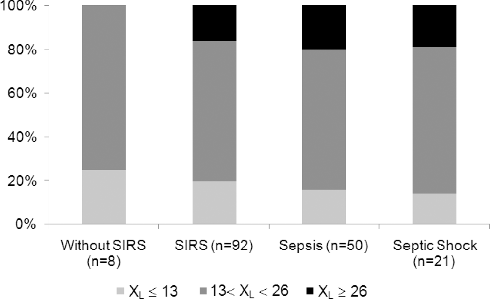
Mean central reactance at low-frequency (X L) distribution during monitoring day according to X L thresholds for patients with SIRS, sepsis, and septic shock. SIRS indicates systemic inflammatory response syndrome.
Mean Central Reactance at Low-Frequency (X L) Distribution During Monitoring Day According to Impedance Parameter Thresholds for Subgroups of Patients by Reasons for ICU Admission.

Abbreviation: ICU, intensive care unit.
From a total of 15 patients with a highly abnormal mean X L > 26 − jΩ during monitoring, 8 were admitted for medical reasons: 1 was admitted for hemodynamic instability and developed septic shock; 4 were admitted for respiratory problems, 2 of those developed sepsis and the others developed septic shock during the monitoring period; 1 was admitted for neurological problems, with sepsis during the monitoring period, 1 was admitted for acute hepatic encephalopathy with sepsis during the study, and 1 was admitted with acute pancreatitis, metabolic acidosis, acute renal insufficiency, and SIRS. Of the 15 patients with highly abnormal reactance, 7 were surgical patients, 1 neurological, and 7 abdominal surgeries with hemodynamic instability. Main complications that these 15 patients developed during their ICU stay included sepsis (10 cases), hydroelectrolytic imbalance (5), renal failures (4), reinterventions (4), respiratory failures (4), and death (4). It is important to mention that 67% of those 15 patients developed sepsis and 20% septic shock. We had 1 particular case of hypoperfusion and ischemia of proximal and distal segments of the intestine with a mean X L of 31.2 − jΩ during the observation period that shows that high values of X L are related with tissue damage.
Twenty patients had mean X L < 13 − jΩ (considered as healthy tissue). 20 Of the 20 patients, 11 were medical and 9 were surgical. One of those patients subsequently died after 25 days in ICU with multiple organ failure (MOF). Four of those patients developed neurological complications and 4 respiratory problems after pneumonia, but all were discharged from ICU later. The other 55% of the 20 patients were discharged from ICU without further complications.
Table 3 shows Student t test results evaluating differences in some continuous variables between patients with normal and abnormal reactance. Significant lower urine output was found in patients with mean X L ≥ 13 (P < .01). Both SOFA and APACHE scores were significantly higher in the same group. We found significant effect (P < .05) on mean X L comparing patients with highly positive fluid balance (>20 mL/kg in 24 hours) against patients with negative fluid balance (<0 mL/kg in 24 hours). Table 4 shows a comparison of the overall mortality and morbidity (as measured by an ICU length of stay >5 days), between patients with normal and abnormal reactance. Patients with an abnormal reactance had a significantly higher mortality, but no significant difference between groups was found for the proportion of patients with an extended ICU length of stay. We had 5 patients with an IAP >15 mm Hg, and in all cases mean X L values were greater than 26 − jΩ (very injured tissue).
T Test for Some Variables According to Mean Central Reactance at Low-Frequency (X L) Thresholds for Healthy (<13) and Unhealthy (≥13) Tissues.

Abbreviations: SOFA, Sequential Organ Failure Assessment; APACHE, Acute Physiology and Chronic Health Evaluation; MAP, mean arterial pressure; CVP, central venous pressure; *Statistically significant.
Indicators of Patient Outcome.a

Abbreviation: ICU, intensive care unit; LOS, length of stay; * Statisticallysignificant.
a Patients are classified by normal and abnormal mean X L. Mortality analysis excludes patients transferred to other hospitals. ICU length of stay (LOS) analysis excludes patients transferred and patients that died.
Discussion
Gastric reactance was conceived under the assumptions that GI hypoperfusion is common, is an early indicator of hypoperfusion (the Canary of the body), is directly related to further complications in the ICU (the gut is the motor of MOF), and no other generally available measurement provides specific information about the adequacy of mucosal perfusion. Gastric reactance is easy to use: Catheters are placed in the same way as standard naso gas trictube (NGT), with no additional training required. The measurement is reliable and robust, does not require calibration, and is an absolute measurement, and if the sensor is not in a good contact with the mucosa, the device will give an error message but will not give false measurements (very much like a pulse oximeter). The technology is not yet commercially available but cost could be in the same order as other monitoring technologies such as pulse oximetry or mixed venou soxygensaturation (SvO2). However, before we may advocate the clinical use of this technology, we need to first answer the following questions: (1) Is the measurement clinically relevant and independent of other available indicators? (2) What patients are at risk and may benefit from gastric reactance monitoring? and (3) Can we use this information to improve patient management and treatment? This last question is the hardest to answer and will require further research and experience. This study was primarily designed to give a first answer to question no. 2, but we also hoped to corroborate our previous findings that the measurement is relevant and independent of other indicators of perfusion, in a broader population of patients.
This is the first observational study designed to obtain a database of human gastric reactance under varied conditions and pathologies to identify the incidence of gastric mucosal injury in critically ill patients. The target population of this study was the general critically ill with a broad range of diagnoses including trauma, sepsis, and respiratory distress, because we want to identify subgroups of patients who may benefit from this technology. Overall, we found that 80% of all critically ill patients had a mean reactance X L above the normal values for healthy tissue (X L ≥ 13 − jΩ) and 15% of the patients had very high measurements (X L > 26 − jΩ), indicating significant level of tissue damage. This incidence matches the previously reported incidence of mucosal ischemia in the ICU as measured by gastric tonometry. 26 As expected, patients with cardiovascular and respiratory problems had the highest incidence of abnormal mean reactance. However, no significant difference was found in any of the patient subgroups, and all had very high incidence suggesting splanchnic hypoperfusion is very common, and any intubated ICU patient may be at risk.
Patients with a mean X L ≥ 13 − jΩ had significantly lower urine output and lower MAP, though not quite significant (as patients were under standard treatment protocols to control blood pressure). This is what we would expect of patients with compromised splanchnic perfusion. Accordingly, these patients also had significantly higher SOFA and APACHE scores as compared to normal X L patients. Most importantly, patients with abnormal mean reactance had a significantly higher mortality in the ICU. We did not see a difference in ICU length of stay. This is probably due to the very long average stay seen in this sample of Mexican ICUs (as compared, eg, to US hospital averages).
Other measurements are used as indicators of hypoperfusion such as lactate, base deficit, or SvO2. These measurements are very specific indicators of systemic hypoperfusion but are very insensitive to splanchnic hypoperfusion and already have been shown in our previous animal and clinical studies to correlate very poorly with gastric reactance. As expected, in this study, lactate did not correlate with gastric reactance: X L is identifying patients at risk well before significant changes in lactate are observed; this means we may be able to identify patients and adjust treatment earlier.
Patients with sepsis or septic shock also showed higher mean reactance. However, this study was designed to provide a 1-day snapshot of ICU patients, and as can be seen from Figure 1, reactance is a poor diagnostic marker for sepsis and septic shock. But gastric reactance is proposed not as a diagnostic but as a monitoring tool for these patients. We would expect septic patients with normal mean X L to develop higher reactance in subsequent days. (Some studies have shown that the GI microvasculature is injured in the development of sepsis, causing hypoxia and cell necrosis. 27 ) By the same token, we could expect, according to the theory that the gut is the motor of MOF, that nonseptic patients with high reactance may subsequently develop sepsis. Our study did show that patients with mean reactance X L > 26 − jΩ had hemodynamic instability or sepsis, and the others developed sepsis or septic shock during their ICU stay; but this study was not designed to test these hypotheses. Indeed, based on these and other earlier results, a longitudinal study of changes in reactance over the length of an ICU stay as related to patient outcomes would be an excellent next research step.
We observed a strong relationship between increased IAP and high mean reactance X L (greater than 26 − jΩ). Some studies23–25 demonstrated the hemodynamic effects of increasing IAP and its correlation with low gastric tonometry phi and lactic acidosis. Elevated IAP may collapse major veins within the abdominal cavity and induce a decrease in venous return from the abdomen and the lower extremities to the heart. 28 Moreover, experimental and clinical studies have shown that an acute increase in IAP on the normovolemic model has territorial circulatory repercussions, such as decreased splanchnic blood flow.29,30 If gastric blood flow is adversely affected with IAP >15 mm Hg, gastric reactance should be a useful tool for monitoring tissue damage, which can be used in combination with other measures capable of identifying regional ischemia. The interpretation of individual values of any parameter should be avoided, as suggested by Vincent, 31 and an evaluation of the trend in values should be better.
Conclusions
This study showed a very high incidence of abnormal gastric reactance, which is a measure of mucosal ischemic injury, in all medical and surgical critically ill patients, independent of the reason for ICU admission. Overall incidence of abnormal reactance agrees with previously reported incidence of gastric ischemia in 80% of intubated patients in the ICU. These patients had significantly worse indicators of perfusion and prognosis and had a significantly higher mortality. These results are in agreement with our understanding of the measurement and the physiology behind it and show that gastric ischemic damage is highly prevalent and relevant to ICU patient outcomes. The measurement itself is easy and reliable and provides information currently not available to the physician. Further research is recommended to understand how these measurements change with the evolution of disease and treatment in order to understand whether and how this measurement may be used as a useful clinical monitoring tool.
Footnotes
Acknowledgments
The study was performed at the ICUs of the Hospital Español, Instituto Nacional de Cardiologia “Ignacio Chavez” (INCICh), Centro Medico ABC, Medica Sur, Hospital Angeles del Pedregal, Hospital Juarez de Mexico, and Centro Medico Nacional “La Raza”, Mexico City. The research was supported by a grant from Innovamedica S.A. de C.V. Critical Perfusion Inc (CPI), of Palo Alto California, now owns all commercial rights to gastric reactance technology. Dr Sacristan is the inventor of this technology and founder of both Innovamedica and CPI but is no longer affiliated with either company. None of the authors are affiliated with CPI. The authors wish to thank Ursina Diaz for her help with data collection and analysis, as well as Drs Juan Esponda, Manuel Poblano, and Pedro Ambriz for their help with patient recruitment, as well as the intensive care departments and nursing staff of the participating hospitals for their help in collecting data.
Declaration of Conflicting Interests
The authors declared no potential conflicts of interest with respect to the research, authorship, and/or publication of this article.
Funding
The authors received no financial support for the research, authorship, and/or publication of this article.