
Abstract
Background:
Atrial fibrillation and atrial flutter are atrial tachycardias associated with embolic strokes. To date, there have only been a few reports highlighting the incidence of these atrial tachycardias due to mechanical compression of myocardial structures and the pulmonary vasculature in certain mediastinal masses and cysts.
Case:
We present a case of a 75-year-old gentleman who is a nonsmoker with a history of hypertension who presents with an acute embolic stroke due to atrial flutter likely from mechanical compression from an underlying squamous cell carcinoma of the lung.
Conclusion:
This case represents, to the best of our knowledge, a rare case of squamous cell carcinoma of the lung in a nonsmoker likely leading to mechanical compression and a resultant atrial tachycardia with an embolic stroke.
Background
Atrial fibrillation and atrial flutter are atrial tachycardias widely known to be linked to embolic strokes. To date, there have been few cases highlighting the incidence of atrial fibrillation and atrial flutter in certain mediastinal masses 1 –3 and lung cysts 4,5 due to mechanical compression of myocardial structures and pulmonary vessels. Here, we present a case of squamous cell lung carcinoma (SQCLC) associated with new-onset atrial flutter leading to subsequent embolic strokes.
Case
A 75-year-old gentleman with hypertension presented with an episode of right facial drooping and aphasia. He noticed that when he stood up, he was leaning toward his right side. He also exhibited garbled and slurred speech with some right-sided drooling and was found to be unresponsive. The entire episode lasted for 5 minutes. He denied any focal weakness or vision changes. He also denies any muscle contractions or seizure activity and denied any incontinence. His wife then called for an ambulance, and the patient was brought to the emergency department. The patient did report a nonproductive cough but otherwise no chest pain, dyspnea, or hemoptysis. He also served as a volunteer firefighter. On presentation, he was afebrile with a blood pressure of 108/72, heart rate of 160, respiratory rate of 16, and oxygen saturation of 97% on room air. On auscultation, his heart demonstrated regular rhythm with no murmurs, and his lungs revealed decreased breath sounds bilaterally. Neurological examination demonstrated normal language and fluency and context, intact cranial nerves II to XII, preserved motor strength 5/5 throughout, and normal finger-to-nose testing and heel-to-shin testing. His deep tendon reflexes were 2+ and symmetric in the patella, biceps, and Achilles tendon. His electrocardiogram demonstrated atrial flutter with rapid ventricular response.
Given his concerning history, a computed tomographic angiogram of his head and neck was obtained, which showed no acute intracranial abnormality but incidentally captured a partially visualized 5- to 7-cm solid mass in the left apex of the lung that was further confirmed on computed tomography (CT) of the chest (Figure 1). A subsequent magnetic resonance imaging of the brain revealed 2 small acute infarcts within the right cerebellum and left frontal lobe, likely embolic in etiology. An expedited bronchoscopy was performed on the following day of presentation with findings of a fungating mass eroding from the posterior aspect of the left upper lobe takeoff obliterating everything except the lungula. A staging CT of the abdomen and pelvis was also obtained, which additionally demonstrated a lytic lesion in the T10 vertebral body, a 2-cm hypodense lesion involving the lateral aspect of the interlobar left kidney, and a stable 1.1-cm hypodense lesion in the lateral aspect of the interlobar right kidney with an indeterminate hepatic hypodense lesion.
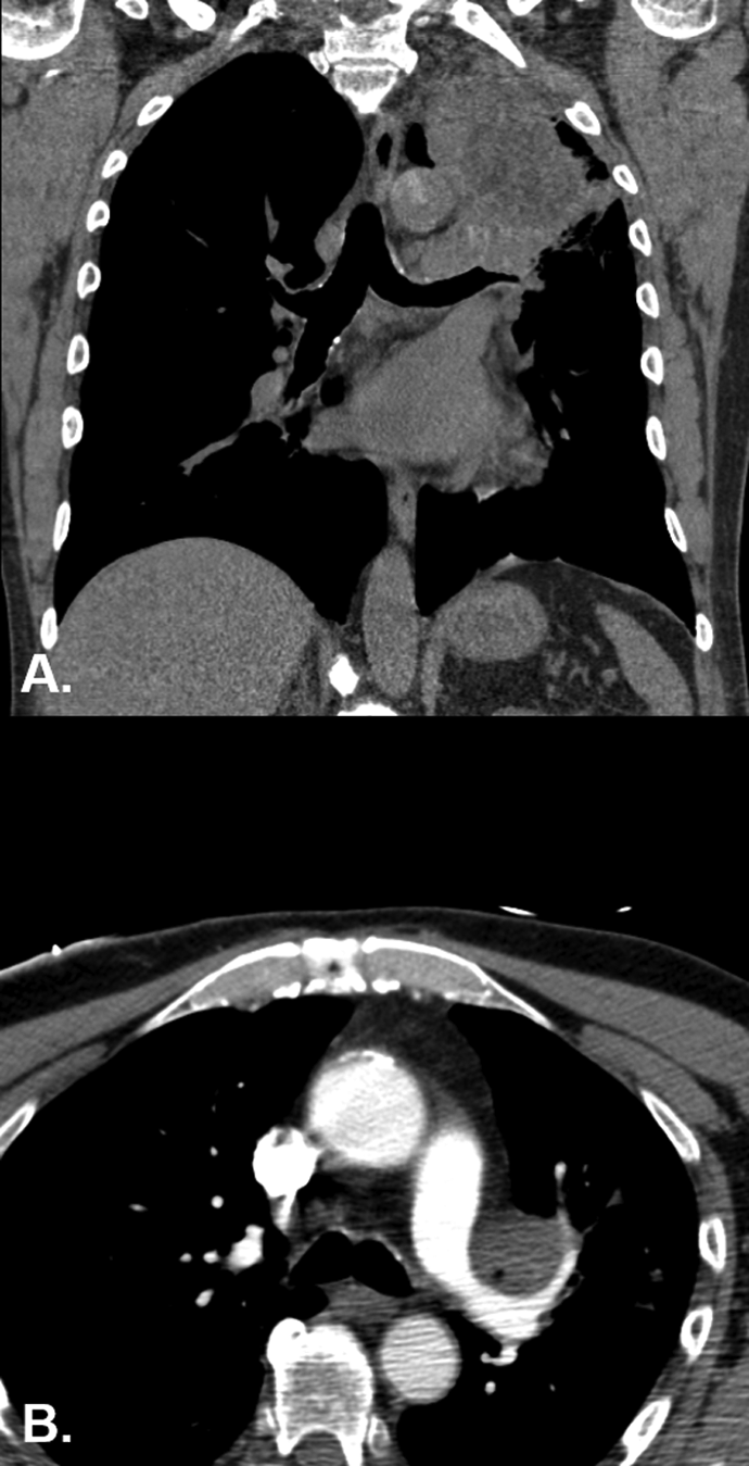
A, Computed tomography (CT) of the chest showing a 6.8 × 6.1 × 9.2 cm soft tissue mass in the left upper lobe extending to the left hilum and abutting the left main pulmonary artery. B, Axial image from a contrast-enhanced CT angiogram of the neck demonstrating encasement and narrowing of the left main pulmonary artery by the left hilar mass.
Subsequent hematoxylin and eosin staining of the biopsied section revealed the presence of keratin and was positive for tumor protein p63 immunostaining and negative for thyroid transcription factor-1 (TTF-1). Four hours after biopsy, the patient was started on a continuous intravenous infusion of heparin. A transthoracic echocardiogram demonstrated severe diffuse hypokinesis, moderately dilated right atrium, and severe tricuspid regurgitation with an estimated left ventricular ejection fraction of 25%. A transesophageal echocardiogram identified no thrombus in the left and right atrium. He subsequently underwent a successful cardioversion to normal sinus rhythm. He was transitioned to therapeutic subcutaneous enoxaparin upon discharge from the hospital. Based on the pathologic findings from the fungating mass visualized on bronchoscopy and imaging findings involving a T10 vertebral body and suspicious liver and kidney lesions, he was diagnosed with metastatic SQCLC with plans to initiate chemotherapy with carboplatin and gemcitabine following a scheduled fluorine 18-labeled fluorodeoxyglucose positron emission tomographic scan to complete his staging workup.
Discussion
SQCLC is a subtype of non–small cell lung cancers that is strongly associated with cigarette smoking. Other risk factors include genetic and familial predisposition, occupational and environmental exposures, and diet. 6 Cough, hemoptysis, dyspnea, and, on occasions, fevers in the setting of postobstructive pneumonia comprise some of the clinical features. 7 Our patient otherwise did not demonstrate any respiratory symptoms aside from a chronic productive cough. Although our patient was not a cigarette smoker, he was exposed to second-hand smoking by his parents while growing up. Second-hand smoking accounts for about 1.6% of this type of cancer. 8 In addition, he also served as a volunteer firefighter at a younger age and was exposed to many fires as well as ambient air pollution that can include emissions rich in various polycyclic aromatic hydrocarbon compounds.
To date, there have been incidences of atrial fibrillation and atrial flutter in certain mediastinal/lung tumors and cysts due to possible mechanical compression of myocardial structures and pulmonary vessels. 1 –5 In this case, echocardiographic findings of biventricular heart failure with dilated right atrium and severe tricuspid regurgitation may be due to the compression of the pulmonary artery (Figure 1). Acute biventricular heart failure can certainly lead to increased risk of developing atrial arrhythmias. Furthermore, recently, new-onset atrial fibrillation in and of itself has been found to be a possible marker of occult malignancy. 9 Our patient presented with new-onset atrial flutter, which was likely related to mechanical effect from the underlying left upper lung mass. The new-onset atrial flutter subsequently manifested as cerebral embolic infarcts in the right cerebellum and left frontal lobes. In addition, malignancy, by itself, can lead to hypercoagulability, an underestimated but another important risk factor for stroke. 10,11 Of the common cancer types, lung cancer was found to be the sixth most common in ischemic strokes with a prevalence of 4.5%. 11 Although the majority of ischemic strokes related to lung cancer have been found to be metastatic, of primary lung cancers, SQCLC was found only in 31% of patients. 10 Moreover, atrial fibrillation was found in only 16% of patients. 10
The relationship between cancer and atrial fibrillation is a complex bidirectional one that is still incompletely understood. 12 Treatment of atrial fibrillation and treatment of cancer can individually lead to multiple complications. While the prognosis in the case of metastatic SQCLC is poor with a median survival of 4 months and a 1-year and 5-year survival of 14.6% and 1.6%, respectively, 13 it is likely even further compounded by the presence of atrial fibrillation. Furthermore, progression of cancer leading to mass effect or mechanical compression, in this case, of cardiac structures, likely suggests advanced disease. Currently as it stands, cancer screening is not recommended beyond standard routine health care with a new diagnosis of atrial fibrillation. 12 In our patient, the left upper lung was a purely incidental finding in the setting of an embolic stroke. To the best of our knowledge, this case represents a rare occurrence of SQCLC in a nonsmoker leading to new-onset atrial tachycardia with a subsequent embolic stroke due to possible mechanical compression.
Footnotes
Declaration of Conflicting Interests
The author(s) declared no potential conflicts of interest with respect to the research, authorship, and/or publication of this article.
Funding
The author(s) received no financial support for the research, authorship, and/or publication of this article.