
Abstract
Right heart (RH) failure carries a high rate of morbidity and mortality. Patients who present with RH failure often exhibit complex aberrant cardio-pulmonary physiology with varying presentations. The treatment of RH failure almost always requires care and management from an intensivist. Treatment options for RH failure patients continue to evolve rapidly with multiple options available, including different pharmacotherapies and mechanical circulatory support devices that target various components of the RH circulatory system. An understanding of the normal RH circulatory physiology, treatment, and support options for the RH failure patients is necessary for all intensivists to improve outcomes. The purpose of this review is to provide clinical guidance on the diagnosis and management of RH failure within the intensive care unit setting, and to highlight the different pathophysiological manifestations of RH failure, its hemodynamics, and treatment options available at the disposal of the intensivist.
Introduction
Right heart (RH) failure is a disease process characterized by a complex web of dynamic physiologic and hemodynamic pathways with diverse etiologies. The RH circulatory system includes the systemic veins, right atrium (RA), coronary sinus, tricuspid valve (TV), RV free wall, RV outflow tract (RVOT), pulmonic valve, the main pulmonary artery (PA) (post-pulmonic valve), and the secondary and tertiary branches of the PA. 1 RH failure can represent disruption of any of the aforementioned components of the RH circulatory system, not simply the RV, leading to suboptimal delivery of blood flow or oxygen to the pulmonary circulation with or without elevated venous pressures. 1
In the acute setting, RH failure presents challenges across a range of presentations including acute ischemic heart failure, non-ischemic cardiomyopathies with right ventricle (RV) involvement, myocardial infarction (MI), valvular disease, pulmonary hypertension (PH) from any cause, pulmonary embolism, congenital heart disease, primary respiratory failure, and sepsis. 2 The purpose of this review is to provide clinical guidance regarding the diagnosis and management of RH failure in the intensive care unit and to provide a framework to optimize understanding of RH hemodynamics and physiology to ensure better outcomes in states of critical illness.
The Physiological Right Ventricle
The RV and the left ventricle (LV) are embryologically, morphologically, and physiologically distinct from one another. Compared to the LV, the RV volume is up to 15% larger and its mass is up to one-sixth smaller. 3 This slightly larger RV volume in conjunction with the high capacitance-low resistance feature of the pulmonary circulation, ensures that the RV and LV have an almost equal cardiac output (CO). 4 With exercise, depending on the imposed workload, there are varying degrees of elevation in CO, left atrial (LA) pressure, mean pulmonary artery pressure (mPAP) and RV systolic pressure (RVSP).5,6 The increase in CO is accompanied by a decrease in pulmonary vascular resistance (PVR), attributable to the initial recruitment and subsequent distension of the small pulmonary resistive vessels in healthy subjects.6,7 In this setting, the RV afterload increases from an associated decrease in PA compliance and is accompanied by a concomitant increase in RV contractility in order to maintain RV-arterial coupling.7–10
The orientation of RV muscle fibers is a foundational component of its distinct physiology. The LV's muscle fibers are organized in a helical continuum between the sub-endocardium and sub-epicardium with circumferential constrictor fibers in the middle layer. 11 In the RV, subendocardial muscle fibers are arranged circumferentially in continuity with the LV and parallel to the atrioventricular groove and encircling the sub-pulmonary infundibulum, while at the apex the superficial myofibers are organized in a spiral fashion to form the subendocardial fibers. 12 In the interventricular septum, a longitudinal contractile pattern predominates due to the helical orientation of the fibers1,13 (Figure 1). The continuity of the circumferential fibers with the LV fibers is implicated in RV free wall movement towards the intraventricular septum in systole 4 and contributes to systolic ventricular interdependence, a fundamental property in RV pathophysiology. 14 In fact, it is estimated that 20% to 40% of RV contractile force is generated by LV contraction.2,11,14 Systolic interventricular dependence explains why in part, following insertion of a LV assist device (LVAD) in a patient with combined acute LV failure and mild PH can precipitate acute RV failure. 15
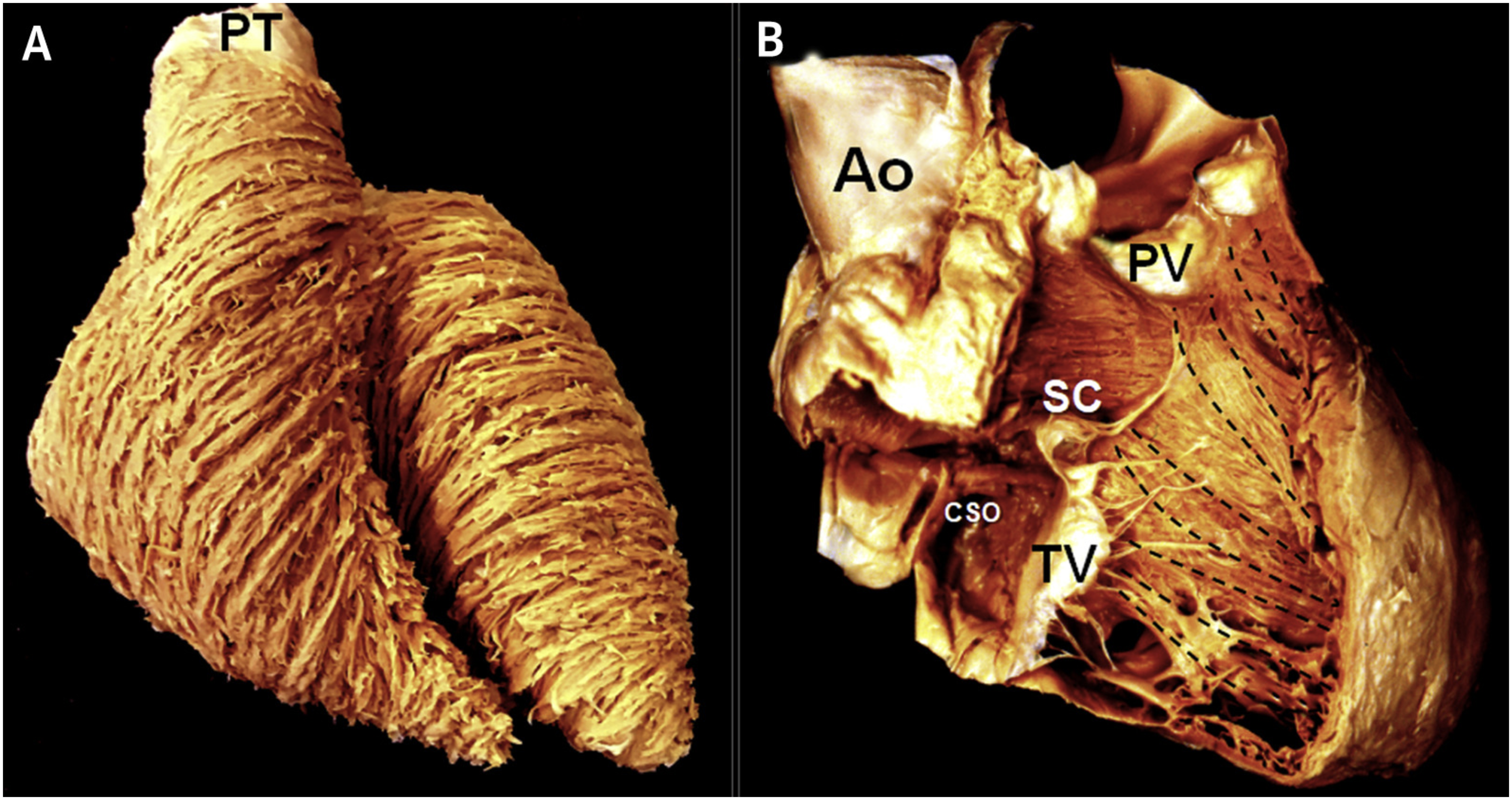
RV myoarchitecture. (A) Normal heart viewed from the front shows the circumferential arrangement of superficial RV and middle LV layers of aggregated cardiomyocytes. (B) Opened normal RV showing longitudinally arranged subendocardial myocyte aggregates (black dashed lines).
The muscle fiber orientation of the RV is integral to understanding its unique means of contraction. 16 In the LV, circumferential fiber shortening and twisting secondary to oblique fiber shortening and longitudinal axis shortening leads to concentric contraction with a wringing type movement.4,15,17 The RV, however, contracts in sequential fashion with a peristalsis-like movement via longitudinal shortening. 3 The bellows-like movement allows the RV to expel high volumes with little free wall surface area change; this helps to explain how the RV can achieve a greater end-diastolic volume and surface area per volume of blood in order to reach an equal stroke volume (SV) to the LV with lower energy expenditure and with low contribution to RV wall stretch.4,17
A major determinant of RV function is ventriculoarterial coupling, which is the ratio of end-systolic elastance (Ees) to arterial elastance (Ea, the vascular load) and helps to explain RV contractility in the context of varying loading state, otherwise understood as efficient RV adaptation to afterload.2,3 In optimal physiologic states, RV efficiency is maintained when end-systolic elastance (Ees) is equal to or greater than Ea, which reflects complete transference of potential energy from the ventricle to the arterial system; in the RV, this ratio (Ees/Ea) is optimal at 1.5 to 2.0, and uncoupling occurs when the ratio is decreased; for instance, when PVR acutely increases, Ea increases out of proportion to the Ees and as ventriculoarterial uncoupling occurs, the RV must expend additional energy to maintain its SV.2,3
RV Response to Preload and Afterload
The function of the RV depends on multiple factors including RV preload, RV afterload, RV contractility (including the RV free wall and intraventricular septum), and pericardial compliance. 18
RV preload refers to the load prior to contraction, the end-diastolic volume, or wall stretch, and RV filling is influenced by intravascular volume, ventricular relaxation, RV chamber compliance, heart rate, passive and active arterial characteristics, and LV filling.3,14 RV afterload is variably characterized as maximum wall tension, external RV work or power, arterial elastance, or the change in pressure for a given change in volume. 3 The RV afterload is dependent upon PVR, resistance, compliance, and wave reflection. 19 The RV wall is thin and compliant with a low volume-to-wall surface area ratio and thus while it is able to accommodate significant preload or venous return, it has difficultly accommodating increased afterload, and even minor increases in afterload can lead to comparatively large decreases in SV. 3 This is largely due to the fact that the RV's end-systolic pressure volume slope is shallower than the LV.2,3 This is interestingly illustrated by patients with Fontan circulation, or cavo-pulmonary anastomosis that bypass the RV as these patients can rapidly deteriorate with any increase in PA pressure such as with high altitude or acute thrombosis of the Fontan's circulation. 20 This is in contrast to the LV which can generally tolerate increases in afterload, but not preload, quite well. 17 To compensate for increases in afterload, the RV will first dilate to preserve its SV and ventriculo-arterial coupling (ie, the Frank-Starling response) to preserve its outflow.2,21 With continued increase in afterload, the RV then uses a homeometric, or functional contractility adaptation described by Anrep's law to sustain its stroke output. 22
Ultimately this adaptation becomes maladaptive as the reduced RV SV results in impaired antegrade filling of the LV, and as the RV continues to distend, the RV end-diastolic pressure of the now pressure and volume overloaded RV, exceeds that of the LV, shifting the inter-ventricular septum leftward, which further impedes LV filling.17,23 Additionally, the increasing RV transmural pressure along with reduced systemic arterial pressure results in reduced coronary artery to RV perfusion gradient with subsequent RV ischemia and infarction.2,24
What are the Scenarios in the ICU Setting That RH Failure is Encountered?
The underlying hemodynamic cause of RH failure is generally due to a pressure or volume overloaded RH system and is encountered in the setting of excessive RH preload (as can be seen with increased venous congestion), afterload (as can be seen in the setting of intermediate and high risk pulmonary embolism, acute hypoxia, acutely decompensated PH, and acidemia), and insufficient contractility (as can be seen in RV ischemia, myocarditis, and pericardial disease). While each of these hemodynamic dyscrasias represents a unique conundrum for the cardiopulmonary system, it is usually an amalgamation of these 3 properties that leads to hemodynamic collapse and is often seen in the acute illness phase in patients with underlying pulmonary vascular disease from any cause. Indeed, RH failure is a common complication of PH especially in patients with pulmonary arterial hypertension (PAH) or chronic thromboembolic PH (CTEPH), though patients with PH due to left heart disease and/or lung disease and hypoxia can also present with this issue. Patients with RH failure due to PH often present in the setting of advanced underlying illness refractory to treatment or due to the presence of concomitant disease such as sepsis, pregnancy, surgery, pulmonary embolism, arrythmias or myocardial ischemia. 25 Previous reviews have been published detailing specific evaluation and management of acute RH failure states including high-risk PE, 26 RV MI, 27 pregnancy, 28 and RV failure after LVAD placement. 29
In the ICU, there are common precipitating factors that can be detrimental to RV function and lead to RH failure in the appropriate clinical context. Some of these conditions include acute hypoxia from any cause including exacerbations of chronic lung disease and acute respiratory distress syndrome (ARDS), acidemia secondary to shock from any cause, left heart dysfunction due to ischemia, arrythmias, valvulopathies, and cardiac surgery. 30 Certain therapies in an ICU may also lead to decreased RV function such as positive pressure ventilation, fluid overload, and certain medications such as propofol and dexmedetomidine.25,31,32 Sepsis has been shown to be independently associated with RV myocardial dysfunction potentially due to cytokine mediated myocardial depression.23,33
RV Monitoring in the ICU
In patients with suspected RH failure in the ICU, initial assessment should begin with a thorough physical exam and a detailed history pertaining specifically to cardiopulmonary, hematologic, vascular, and rheumatologic disease. Patients with RH failure often present with typical signs of RV dysfunction, systemic congestion, and a low output cardiac state: cool and diaphoretic/clammy extremities, relative hypotension, signs of end-organ hypoperfusion, peripheral edema, and hypoxemia. 30 Sometimes, patients can present with clinical signs of RH failure but with elevated CO as can be seen in patients with large arteriovenous malformations, chronic hemolytic anemia (eg, sickle cell disease), and sepsis. 25 Jugular venous distension is an important sign of elevated right-sided filling pressures. Other clinical signs include a RV heave on chest palpation, a prominent pulmonic component of the second heart sound, a tricuspid regurgitation murmur over the right sternal border, a palpable/pulsatile liver, and hepatojugular reflux. 13 Of note, these physical exam signs should only serve to complement more objective markers of disease as below and should not be relied upon to make or discard a diagnosis.
Pertinent labs should include N-terminal pro-brain natriuretic peptide levels, lactic acid, and markers of kidney and hepatic function (none of which are specific for RH disease but can help to characterize this syndrome in clinical context). Evaluation of plasma troponin can be considered in the right setting. For instance, elevated plasma troponin may be associated with worse prognosis in patients with acute pulmonary embolism. 34 An electrocardiogram should routinely be obtained, which may show evidence of RA dilatation, rightward axis, RV hypertrophy or a right bundle branch block. Chest x-ray can be obtained to evaluate for pulmonary congestion, but lung ultrasound has been demonstrated to have higher sensitivity for identifying patients with pulmonary congestion due to heart failure than clinical assessment or chest x-ray. 35 Further assessment of RH failure requires additional advanced imaging and hemodynamic monitoring devices to evaluate etiology and guide treatment.
Computed Tomography (CT) Imaging
CT imaging is a commonly obtained imaging test for ICU patients and when completed with contrast can help determine if there is underlying RH dysfunction. Findings on CT scan that are associated with RH dysfunction include an enlarged PA of greater than or equal to 30 mm, a ratio of main PA diameter compared to aorta diameter of 1.0 or greater, reflux of contrast in the IVC and/or hepatic veins, septal bowing, and an increase in volumetric RV to LV ratios. 36 CT imaging may also be helpful to diagnose acute or chronic pulmonary embolism and abnormal lung parenchyma as seen in settings such as interstitial lung disease.36,37
Echocardiography
Two-dimensional (2D) echocardiography is generally one of the first exams to be performed to assess RV morphology, function, and load, and is one of the most widely available diagnostic tools. In the ICU setting, however, it is important to recognize that there are limitations of echocardiographic measurements in those with lung disease, and especially in patients undergoing positive-pressure ventilation or mechanical ventilation. 17
Due to the intricate shape of the RV, it is important to image the RV in multiple views, including in the parasternal long axis, the short axis, and the apical 4 chamber. 12 The parasternal long axis plane allows primarily for visualization of the RVOT, RV free wall, interventricular septum, and the moderator band; but it can also assess tricuspid valve (TV) morphology and function and the RV outlet. 12 In the short axis view, the shape of the intraventricular septum can be assessed throughout the cardiac cycle at the level of the LV papillary muscles; this view also facilitates visualization of the RVOT, main PA and the right and left main pulmonary arteries allowing for assessment of possible proximal pulmonary emboli. 38 In the apical 4 chamber view, there is the opportunity to appreciate RA dimensions and RV morphology, and to assess the TV. 12 RV dilatation and enlargement can be best appreciated here, with a diameter >42 mm at the base and >35 mm at the mid-level indicating RV dilatation and a longitudinal dimension >86 mm indicating RV enlargement. 38 Finally, the subcostal view is important because it allows assessment of the diaphragmatic wall of the RV, RA, and IVC to be visualized and provides information about the RV inlet; importantly, the IVC should be measured immediately proximal to the entrance of the hepatic vein with a sniff test to assess collapsibility, measurements of which can help to approximate RA pressures.12,38 This view in diastole also allows for measurement of RV wall thickness with M-mode or 2D imaging (RV wall thickness >5 mm indicates RV hypertrophy in the appropriate clinical context). 38
RVSP can be measured with the peak systolic gradient of the tricuspid regurgitant jet added to the RA pressure estimated from IVC collapsibility 38 using the modified Bernoulli equation. 19 RVSP approximates PA systolic pressure in the absence of pulmonary valve stenosis or RVOT obstruction. 39 Of note, it is important to recognize that there can be significant variance of up to 10 mm Hg in up to 50% of patients between echocardiographic estimates of RVSP and RVSP measured during RH catheterization. 40 Additionally, direct estimations of PA systolic pressures are poor given difficulties in measuring pulmonic valve insufficiency jet velocity during early diastole. 19
RV systolic function can be evaluated with the RV index of myocardial performance (RIMP), tricuspid annular plane systolic excursion (TAPSE), 2D fractional area change (FAC), and Doppler tissue velocity or S’ velocity of the tricuspid annulus. 38 RIMP and 2D FAC provide an index of global RV function (RIMP >0.40 by pulsed Doppler and >0.55 by tissue Doppler and 2D FAC <35% indicate RV dysfunction). 38 TAPSE and S’ velocity are both measures of longitudinal RV systolic function measured from the tricuspid lateral annulus, and a TAPSE value of <16 mm has been shown to be an adverse prognostic indicator in PAH. 41 In addition, using TAPSE and the estimated PA or RVSP, the TAPSE:PASP ratio, a noninvasive measure of RV-PA coupling can also be determined. 42 TAPSE however does not account for other measures of RV systolic function (eg, the circumferential or anterior-posterior contractile pattern of the RV). Additionally, TAPSE is angle and load dependent, and it can be pseudo-normal or even be overestimated in PH patients with clockwise apical longitudinal rotation of the RV. 43 RV diastolic function can be indirectly estimated with echocardiography using pulsed Doppler of the tricuspid inflow, tissue Doppler of the lateral tricuspid annulus, pulsed Doppler of the hepatic vein, and measurements of the IVC 38 (Figure 2).
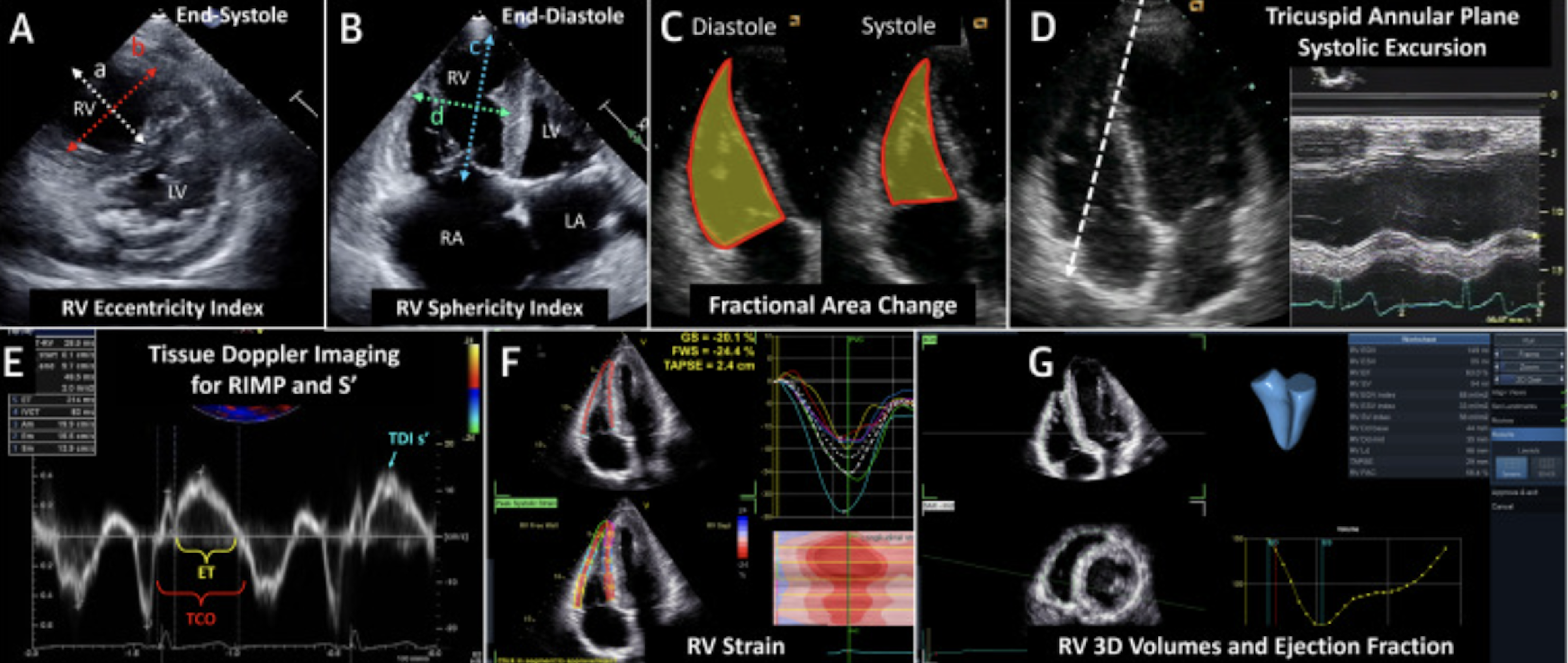
Echocardiographic parameters for the assessment of right ventricular function. (A) A short-axis view of the right ventricle (RV) with the eccentricity index as the ratio of a to b. (B) An apical 4-chamber view with RV sphericity index as the ratio of c to d. (C) The diastolic and systolic measurements of the RV area for calculation of fractional area change. (D) The M-mode cursor (white dashed line) aligned with the tricuspid annulus, allowing measurement of the tricuspid annular plane systolic excursion (TAPSE). (E) Tissue Doppler imaging (TDI) profile of the tricuspid annulus with the measurement of ejection time (ET) and tricuspid closure opening time (TCO) used for calculation of the right ventricular index of myocardial performance (RIMP), and measurement of systolic velocities (S’, blue arrow). (F) The measurement of strain imaging. (G) An example of a fully automated quantitation of RV volumes using 3-dimensional (3D) volume acquisitions.
Non-Invasive CO Monitors
In critically ill patients, invasive monitoring may either not be feasible or felt to represent an unacceptable risk. 45 This becomes particularly problematic for those with RH dysfunction in whom advanced hemodynamic data are necessary both to guide choice of therapy and decisions regarding weaning therapy. In this subset of patients, it may be beneficial to use non-invasive or minimally invasive devices to measure CO and other hemodynamic parameters. A wide variety of alternatives to invasive measurement of CO are available. Recent reviews describing these devices in detail are available.46–48 Devices in current use are broadly based upon 4 methodologic approaches: (a) analysis of an arterial pulse wave, which is primarily predicated upon the established relationship between SV and the area under the systolic portion of the arterial pressure waveform; (b) acquisition of SV surrogates using sensors placed on the thorax or imbedded in an endotracheal tube, which are primarily based on updated bioimpedance methodology and the related technologies of electrical velocimetry and bioreactance; (c) ultrasound based estimations of SV and CO, which incorporates conventional echocardiography as well as external and internal (esophageal) probes specifically designed to measure aortic blood flow velocity; and (d) CO2 rebreathing for estimation of SV and CO representing a permutation of the Fick equation for calculating blood flow.
Despite the value of monitoring CO in the setting of RH failure and the variety of commercially available devices that are less invasive than a PA catheter (PAC), few studies have specifically focused on comparative accuracy and precision in shock states and there are little data to clearly support efficacy much less superiority of any technology. That said, there appear to be data suggesting inferiority; while a previous study indicated acceptable performance of a non-invasive, surface sensor-based bioreactance device in patients with compensated PH, 49 recent data indicate relatively poor performance in the setting of cardiogenic shock. 45 Overall, when considering non-PA catheter options for CO monitoring it is worthwhile taking into account the effect of accompanying factors such as atrial fibrillation, valve dysfunction, and high-dose pressor use on both the less invasive method and the validation predicate to which it has been compared. 50 For patients with RH failure in particular, the high prevalence of tricuspid regurgitation takes on added importance because of potential impact on PA catheter-based thermodilution measurement of CO, 51 the most common reference standard for validating minimally and non-invasive CO technologies. Ultimately, it seems prudent to limit these devices to short-term use as a bridge to minimally invasive methods in the setting of RH failure and if used in this setting, CO data should be referenced to other clinical metrics such as systemic blood pressure, central venous pressure (CVP), mixed venous oxygen saturation, and intermittent transthoracic echocardiography.
Catheter-Based Hemodynamic Monitoring
The CVP typically approximates the RA pressure in the absence of superior vena cava stenosis. The CVP and RA pressure is commonly used as a surrogate of the RV end diastolic pressure particularly in the absence of severe tricuspid regurgitation. The CVP and RA pressure is relatively easy to obtain and can be recorded from the superior vena cava or RA, respectively, using a central venous catheter. While often utilized in decision making regarding volume management in patients, CVP or RA pressure as a static marker has not been shown to be associated with circulating blood volume, and has not been shown to predict fluid responsiveness in various clinical settings. 52 In fact, prior studies have demonstrated that there is no relationship between the trans-mural filling pressure of the RA (ie, RA pressure–pericardial pressure) and either the RVEDP or SV.53,54 This is because the normal RV fills at or below its unstressed volume, such that any increase in RV end diastolic volume can occur without corresponding change in RV diastolic wall stretch. 15 Additionally, these measurements are often skewed in the various clinical scenarios encountered in the ICU or operating room setting and this value should be interpreted with caution. For example, an increase in RA pressure with decreasing RV dilatation (ie, without RV dysfunction) from reduced venous return can occur in the context of increased intra-thoracic pressure from positive end expiratory pressure application (PEEP). With that said, in patients with known PH and associated RV dysfunction, not only does an elevated RA pressure portend poor outcomes in PH, 55 but an acute rise in this pressure in response to fluid resuscitation can be an indicator of impending RH failure in the right clinical context.
In general, the use of PA catheters in the medical ICU has declined over time due to literature suggesting that its use does not improve outcomes.56,57 However, often it is necessary to pursue more invasive hemodynamic monitoring with a PA catheter in the case of RH failure, which is the cornerstone method of evaluating the RV, and has been shown to improve survival in patients with cardiogenic shock. 58 PA catheters are able to directly measure pulmonary and intracardiac pressures as well as mixed-venous oxygen saturation, and the data from these measurements are used to calculate important hemodynamic measurements like CO/index and pulmonary and systemic vascular resistances; these data are important to understand a patient's individual hemodynamics and can help both to guide choice of therapeutic interventions and to dynamically assess response to therapy; they can help optimize volume status, adjust vasoactive medications, and facilitate the weaning of life support. 58 PA catheter measurements can also be uniquely helpful for managing RH failure in the setting of LVAD placement. For instance, the PA pulsatility index, which is the ratio of PA pulse pressure and CVP and has been shown to predict RH failure following LVAD placement. 59 On a practical note, recent literature has highlighted the impact of respirophasic variation on pulmonary vascular pressures, especially in conditions like obesity or scenarios when positive pressure is applied (eg, during mechanical ventilation). Recently, Khirfan and colleagues demonstrated that correction for respirophasic changes through simultaneous measurement of esophageal pressure led to changes in PH diagnosis and classification. 60 However, while esophageal pressure corrections are ideal, given the technical complexities associated with esophageal balloon use in the ICU setting, particularly in awake individuals, an acceptable compromise may be to obtain average pressure readings over several respiratory cycles.61–63
More recently, the use of continuous RV pressure monitoring has been gaining notoriety in cardiac surgery, critical care, and in the peri-operative setting of liver transplantation to help diagnose dynamic RV dysfunction and RVOT obstruction.64,65 For RV monitoring, a thermodilution paceport catheter (Paceport 5 Lumen TD Catheter, 7.5 Fr, 110 cm, reference model 931F75; Edwards Lifesciences Corp, Irvine, CA) allows for continuous right atrial, RV, and PA pressure monitoring (Figure 3). The thermodilution paceport catheter is placed similarly to a conventional PAC with the tip located in the PA to allow for continuous PA monitoring. In that position, the RV pacing lumen port typically is located within the RV allowing for continuous RV pressure monitoring by connecting it to a pressure transducer. Our group has been using continuous RV pressure waveforms during invasive cardio-pulmonary exercise testing (iCPET).7–9,66–68 and in the peri-operative setting for surgical pulmonary thromboendarterectomy for CTEPH. In the latter context, should the patient develop intra- or post-operative RH failure, while the PA pressure may pseudo-normalize from diminished RV SV, the coincident rise in RV pressure (and RA pressure/CVP) can herald impending circulatory collapse. Additionally, an instantaneous intra- or peri-operative diagnosis of dynamic RVOT obstruction of a hypertrophied RVOT or infundibulum following normalization or near normalization of PA pressure following cardiac surgery for non-congenital heart disease, 69 lung transplantation, 70 and balloon pulmonary valvuloplasty 71 can be made with continuous RV pressure monitoring. 69 In this clinical scenario (often referred to as the “suicide RV”), a significant systolic RV-PA gradient of >25 mm Hg can develop in the setting of markedly reduced PA pressure. If dynamic RVOT obstruction is suspected, immediate echocardiographic evaluation is warranted to help confirm dynamic RVOT obstruction or exclude other potential source of hemodynamic instability. 69 In dynamic RVOT obstruction, inotropic agents are contraindicated as they would reduce RV filling and further exacerbate the obstruction, while volume and beta-adrenergic blockade can help improve or normalize the systolic RV-PA gradient. 69
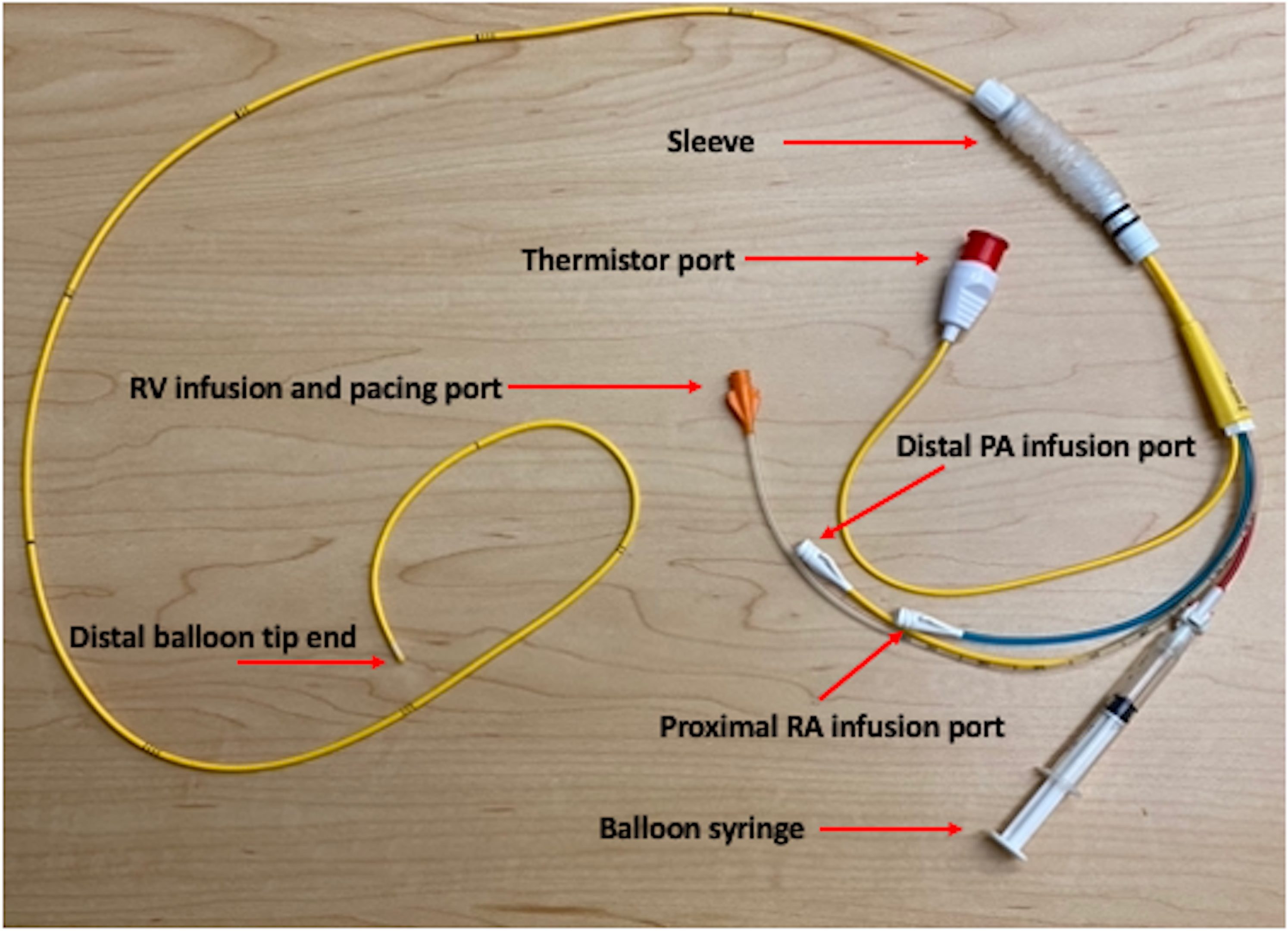
Thermodilution Paceport pulmonary artery catheter.
Bedside Assessment of Right Ventricular-Pulmonary Arterial Interaction
The efficiency of ventriculoarterial coupling, or the coupling between the RV and the pulmonary circulation is integral to understanding the RV's ability to adapt to various loading states. While echocardiography and PAC measurements provide valuable metrics of RV function, they are subject to the influence of different loading conditions.
Assessing RV contractility and afterload as independent entities and using their ratio to describe the mechanical interaction between the RV and pulmonary circulation, or ventricular-vascular (VV) coupling is potentially useful in the critical care setting both for understanding the nature of RV dysfunction and evaluating the response to an intervention (eg, volume loading, inotrope or vasopressor titration, acute pulmonary vasodilator therapy). The reference standard for characterizing VV coupling involves plotting the continuous relationship between pressure and volume (PV loops) over a range of preload to define RV contractility as ventricular Ees and total afterload as end-systolic PA Ea 72 (Figure 4, panel A). While this is a powerful tool that has been applied clinically, use of RV PV analysis in critical care environments remains largely impractical due to the need for: (a) cost-effective and readily calibrated beat-to-beat measurements of RV volume; (b) application of controlled maneuvers that acutely alter the RV preload (Valsalva, external abdominal compression, balloon occlusion of the inferior vena cava); and (c) a user-friendly platform for rapidly analyzing multi-beat PV loops. As an alternative, a single beat method has been described that combines analysis of the RV pressure waveform with SV to derive Ees and Ea without the need for preload variation or measurement of RV volume.72,75 This approach involves prediction of “Pmax,” the maximum RV pressure that would have been achieved if no ejection occurred and estimation of RV end-systolic pressure (ESP). With these data, Ees is calculated as (Pmax-ESP)/SV, and Ea calculated as ESP/SV (Figure 4, panels B and C). This method has been widely used clinically for characterizing VV coupling in patients with PH and commercially available software has simplified the process. However, Pmax prediction is affected by shape and fidelity of the RV pressure signal, and derived values for both Ees and Ea are dependent upon how end-systole is defined 75 complicating comparison of data between studies. Despite these limitations, Pmax and ESP can also be used to estimate RV ejection fraction (RVEF) without any measurement of RV volume as RVEF = 1−(ESP/Pmax) 73 (Figure 5). Recent data demonstrate that using real-time algorithmic analysis of RV pressure waveforms to derive Pmax and ESP, RVEF can be calculated on a beat-to-beat basis with reasonable accuracy 76 (Figure 6). Ultimately, the potential for estimation of VV coupling and RVEF at the bedside is attractive for rapidly gauging the dynamic RV response to diagnostic or therapeutic interventions encountered in various critical care clinical scenarios.
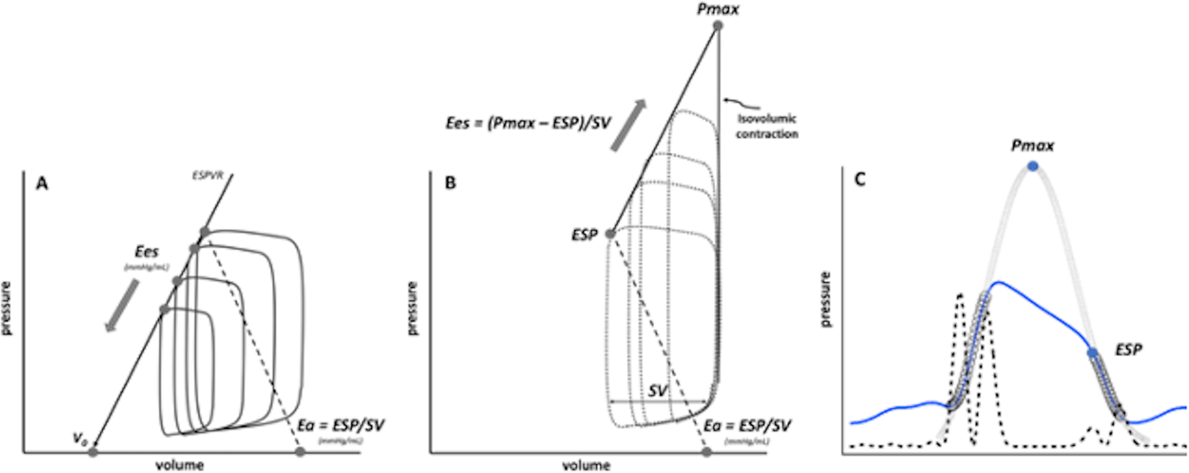
Comparison of multi-beat and single beat derivation of ventricular end-systolic elastance (Ees) as a load-independent measure of contractility, and end-systolic arterial elastance (Ea) as a measure of total afterload. The multi-beat analysis example (Panel A) shows pressure volume loops created during decreasing preload. Regression along the declining end-systolic pressure/volume points defines the end-systolic pressure volume relationship (ESPVR). The ESPVR slope defines Ees with the volume intercept at a pressure of 0 mm Hg designated as Vo. Afterload summarized as Ea is typically calculated from the end-systolic pressure (ESP) and stroke volume (SV) for the first beat. In contrast, the single beat model (Panel B) is based upon the theoretical construct of a fixed preload with increasing afterload to the point of no ejection (ie, clamping the aorta or pulmonary artery). Within this model, idealized pressure volume loops (dotted lines) show increasing pressure with decreasing SV until the contraction becomes isovolumic with pressure at this point designated as Pmax. Ees is then slope of the theoretical ESPVR defined by increasing end-systolic pressure/volume points and calculated from the estimated Pmax and measured ESP and SV. Importantly, the single beat model does not create actual pressure volume loops since actual volume is unknown and does not define Vo. Panel C shows an example of Pmax prediction from the rise and fall of ventricular pressure (blue line) using segments (open circles) with an event marker (hatched line) derived from the second derivative of pressure as previously described.75,76
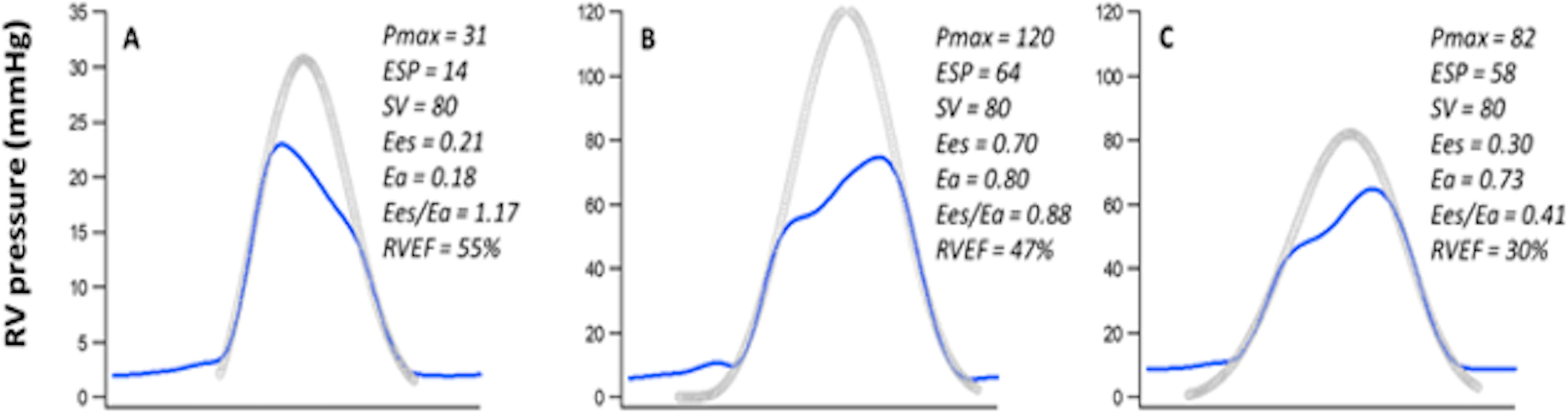
Examples of single beat analysis of right ventricular pressure (RVP) waveforms to define the maximal isovolumic and end-systolic pressures (Pmax and ESP, respectively) and derive ventricular end-systolic elastance (Ees), end-systolic pulmonary arterial elastance (Ea), and ejection fraction (RVEF) assuming a constant stroke volume (SV). From these data, Ees represents contractility and Ea total afterload with ventricular: vascular coupling summarized as Ees/Ea. Panel A shows a normal RV pressure waveform with peak of 23 mm Hg occurring early in systole, Ees/Ea > 1, and RVEF > 50%. Panel B shows a pulmonary hypertensive waveform with peak of 68 mm Hg occurring late in systole. A compensatory increase in Ees is evident with reasonably preserved coupling and RVEF relative to normal. Panel C shows a pulmonary hypertensive waveform with peak of 62 mm Hg occurring late in systole but only a modest compensatory increase in Ees thus impaired coupling and decreased RVEF relative to normal.
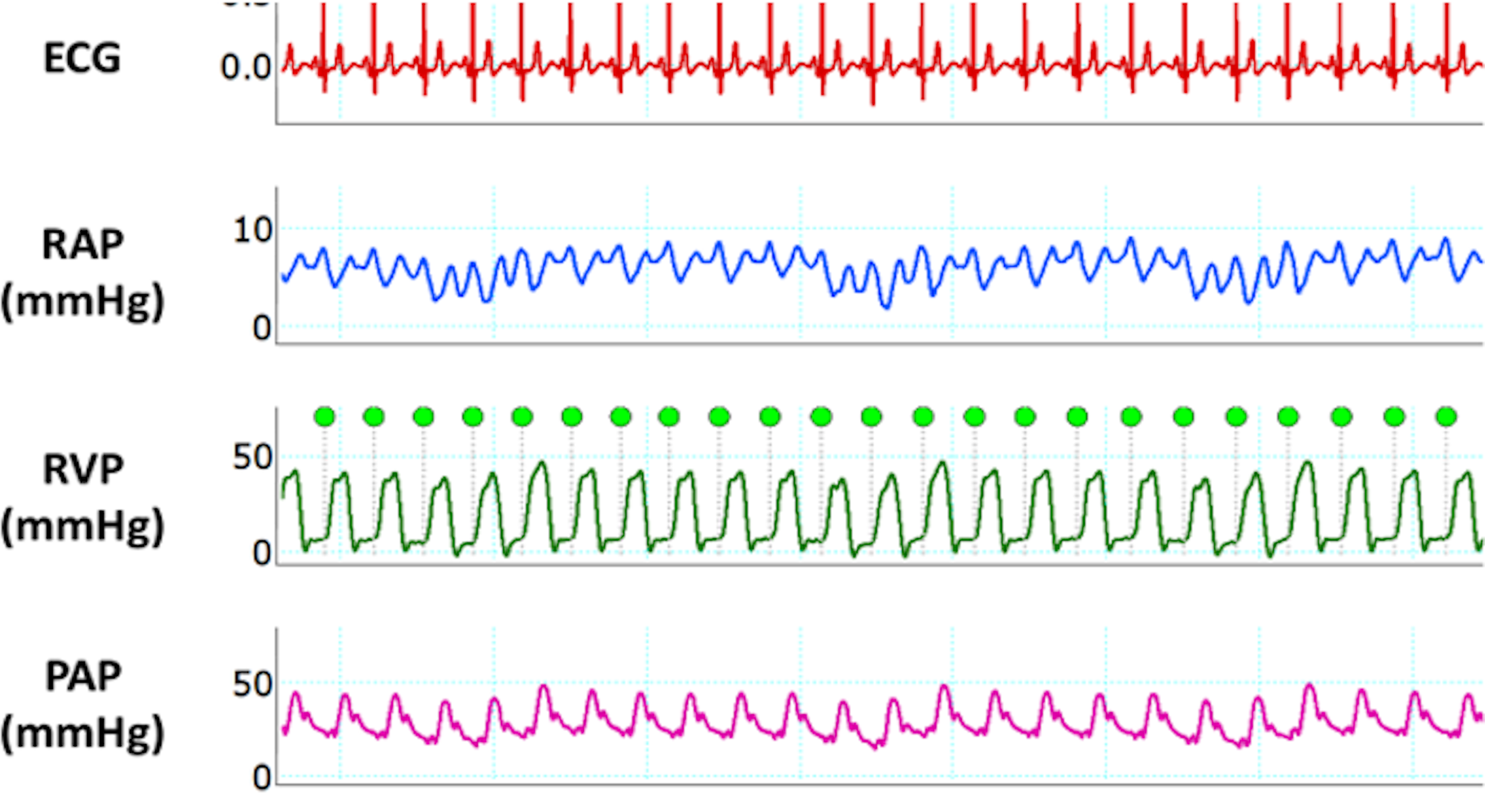
Data recorded using a Paceport pulmonary artery (PA) catheter to yield right atrial (RAP), right ventricular (RVP), and PA pressure (PAP). From these data right ventricular ejection fraction (RVEF) was calculated on a beat-to-beat basis at the bedside as previously described. 78 .
RH Failure Management
RH failure is a complex disorder that can lead to multiorgan failure and circulatory collapse. Extracardiac manifestations include bowel ischemia leading to bacterial translocation, renal failure requiring renal replacement therapy, and liver dysfunction leading to life-threatening coagulopathy and encephalopathy. 25 Initial steps in management should be focused on correcting any underlying illness (ie, treating sepsis with antibiotics or RV ischemia with coronary reperfusion therapy). Second, underlying pathology felt to be contributing to overall RV dysfunction should be addressed; this involves managing processes such as alveolar hypoxia, metabolic acidosis, and optimizing the mechanical ventilator to ensure RV protection. 77 In particular, it is critical to ensure patients have peripheral oxygen saturations greater than 90% to minimize the afterload effects of hypoxic pulmonary vasoconstriction. 23 In parallel, management must be directed at optimizing RH function through careful adjustments in preload, afterload, and contractility.17,25 Given the potential need for advanced therapies including mechanical circulatory support (MCS) and pulmonary vasodilators, patients with RH failure should be considered for transfer to a tertiary or quaternary center that has experience in the management of RH failure.
Preload Management of the RH
Careful volume management is a critical component of caring for patients with acute RH failure, and initial clinical evaluation should include assessment of volume status; this should involve a clinical exam evaluating peripheral edema, jugular venous distention, and pulmonary congestion as well as potentially more invasive hemodynamic measurements such as CVP/RA and RV pressures using a PAC as described above. 2 Optimal preload should aim to keep RV transmural pressure moderately elevated with CVP goal in the upper limit of normal range (ie, 8-12 mm Hg) with real-time adjustments made to support RV function and CO (which can be approximated by the central or mixed venous oxygen saturation). 17 It is worth mentioning that it is often said that acute RH failure is a “preload dependent state,” as adequate right-sided filling pressure maintains CO in this patient population. 17 Indeed there is a subset of patients with RH failure who may be preload dependent, including those who present with RV MI. However, if volume loading is attempted it should be guided by ongoing hemodynamic measurements and generally only in the setting of low arterial systolic pressures in conjunction with decreased filling pressures. 30 Volume loading especially in the setting of increased RV afterload can lead to superimposed pressure overload and/or RV dilatation, at which point there may be simultaneous LV dysfunction due to underfilling from septal displacement and reorientation of myocardial fibers. 3 More commonly, particularly in the setting for chronic PH, it is necessary to optimize volume status by decongesting patients to restore optimal interventricular interactions.2,23 This can be achieved with the use of intravenous loop diuretics with or without the addition of thiazide diuretics with titration based on response to therapy recognizing that because the RV has a flatter Starling curve than the LV a greater change in filling pressure may be needed to improve SV. 17 On a practical note, during diuresis in this patient population clinicians will often observe worsening renal function (WRF). There has been increasing interest in diuretic associated WRF with recent literature suggesting that this response is not associated with worse clinical outcomes. 78 Additionally, it has been shown that diuresis-associated declines in GFR may represent benign changes in renal filtration instead of tubular injury. 79 As such, in patients with established RH dysfunction who have evidence of elevated right-sided filling pressures and are clinically volume overloaded, an initial increase in creatinine with diuresis should not deter further efforts of decongestion. Further, if hypotension is encountered in the setting of diuresis, it is reasonable to support systemic blood pressure with vasopressors. In the event of persistent congestion despite escalating diuretic therapy, it may be necessary to implement continuous veno-venous hemofiltration or ultrafiltration with renal replacement therapy. 2
Afterload Management of the RH
In acute RH failure, decreasing afterload is often the most effective strategy to improve RH function. The usefulness of this strategy is clearly demonstrated by studies that show that there is immediate relief from PH following pulmonary thromboendarterectomy or lung transplantation, and that the RV can recover to normal architecture over time regardless of the severity of initial disease following these procedures. 80 Initially, it is critical to correct reversible causes of increased RV afterload that are common in the intensive care unit. One example is alveolar hypoxia with resulting hypoxic vasoconstriction which leads to increased PVR and thus increased RV afterload, a response that is notably compounded by acidemia.17,81 Thus, one of the initial intervention strategies in the afterload management of RH failure should be aimed at correcting alveolar hypoxia and acidemia while ensuring optimal ventilator management. 17
Afterload Management of the RH: Pulmonary Vasodilator Use
The initiation of pulmonary vasodilators is an important intervention to consider in the setting of RH failure as a means of reducing RV afterload, and often patients with RH failure and longstanding PH will arrive at the ICU on established pulmonary arterial vasodilator therapy. Over the past 2 decades, several drugs have been identified targeting 3 main signaling pathways: (1) prostacyclin, (2) nitric oxide (NO), and (3) endothelin-1 pathways.82,83 Trials for these medications have supported their use in Group 1 PH (PAH), Group 3 PH (PH due to interstitial lung disease), and Group 4 PH (PH due to chronic pulmonary thromboembolic disease), though none have validated their use in the setting of acute RH failure with randomized controlled clinical trials.82,83 It is important to note that in addition to pulmonary vasodilation, many of these systemically delivered therapies can cause systemic vasodilation and thus care must be taken with their use in hemodynamically unstable patients. Additionally, pulmonary vasodilators have been shown to worsen arterial oxygenation in patients with underlying parenchymal lung disease (due to diversion of blood flow to poorly ventilated alveolar units, ie, ventilation/perfusion mismatch).84–86
Inhaled administration of these medications enables targeted delivery to the lungs, which augments pulmonary selectivity and reduces systemic side effects, including hypotension.86,87 Furthermore, inhaled versions of these medications may offer more rapid onset of action, short half-lives, and may lead to improvements in ventilation-perfusion matching.86,87 Accordingly, for the purpose of this review, we will focus on pulmonary vasodilators delivered via the inhaled route since these are of greatest interest in caring for critically ill ICU patients.
Inhaled NO (iNO) works by stimulating soluble guanylate cyclase to synthesize cGMP activating cGMP-dependent PKG ultimately causing vascular relaxation. 88 In its inhaled form, iNO has been shown to selectively induce pulmonary vasodilation, reverse hypoxic vasoconstriction, and improve oxygenation without causing systemic vasodilation 89 in patients with ARDS90,91 and following heart and lung transplant. 92 In patients with RV dysfunction with shock and respiratory failure, iNO has also been shown to improve CO, SV, and mixed-venous oxygen saturation. 93 While in patients with PH undergoing cardiac surgery, iNO use is associated with improved RV performance. 94 These features in conjunction with its rapid onset of action and short half-life makes iNO an attractive agent for RV-afterload reduction in the ICU, especially in intubated patients. 17
Agents that target the prostacyclin pathway—including epoprostenol, treprostinil, and iloprost—bind prostaglandin receptors and exert vasodilatory effects via cAMP signaling. 83 All 3 medications can be delivered through the inhaled root, though only inhaled epoprostenol can be given as a continuous medication. In cardiothoracic operating rooms and the ICU, inhaled epoprostenol has been shown to selectively vasodilate the PA in patients with PH, improve oxygenation in patients with refractory hypoxemia, and maintain systemic mean arterial pressure (MAP). 95 In PAH patients, it has been shown to lead to a significant reduction in mean PA pressure and a rise in CO with no change in systemic arterial pressure. 96
Afterload Management of the RH: Vasopressor Use
Maintaining systemic blood pressure is paramount to optimizing end organ perfusion. In the setting of RH failure, it is critical to maintain systemic systolic and diastolic blood pressure above the RV systolic and diastolic blood pressure, respectively, to ensure adequate RV perfusion since the RV is particularly sensitive to ischemic injury. 97 Theoretically, the ideal vasopressors of choice for this patient population should act to increase systemic arterial pressure and RV contractility without significantly increasing PVR or RV afterload. 17 Norepinephrine is an alpha-adrenergic agonist with additional beta-adrenergic agonist effects 98 that works both to increase systemic MAP via systemic vasoconstriction and to increase RV CO and SV, respectively. 99 Norepinephrine may improve RV myocardial oxygen delivery via its effects on systemic vascular resistance. 100 It has also been described to improve RV contractility and augment RV-PA coupling in animal models.101–103 These favorable effects of norepinephrine on the RV myocardium may help to explain why RVEF can be maintained with norepinephrine in the face of increasing PVR. 100 Vasopressin, or antidiuretic hormone, exerts its hemodynamic effects mostly through action on V1a and V2 receptors, which facilitates constriction of the systemic vascular smooth muscle and stimulation of water reabsorption in the renal collecting duct, respectively. 103 In animal models, vasopressin has been shown to cause PA vasodilation. Evora and colleagues demonstrated that in dogs, vasopressin caused endothelium-dependent vasodilation of the PA from vasopressin V1-mediated production of nitric oxide. 104 Additionally, Russ and colleagues showed that in rat lungs preconstructed with a synthetic thromboxane analogue (ie, a potent PA vasoconstrictor), the administration of arginine vasopressin produced PA vasodilation. 105 This effect of vasopressin, however, has not been consistently demonstrated in humans 106 and therefore, its clinical relevance remains unknown. Epinephrine is a mixed alpha and beta agonist that acts both on myocytes and vascular smooth muscle to cause an increase in blood pressure via positive inotropic and chronotropic effects in addition to systemic vasoconstriction. It can also cause systemic vasodilation through B2 stimulation. 17 In a small study, epinephrine was shown to improve not only RV contractility in patients with septic shock, 107 but also increase systemic arterial and venous pulmonary pressures. 98 Phenylephrine is a pure alpha-1 receptor agonist. While it augments right coronary artery perfusion by increasing the systemic vascular to RV perfusion gradient, 17 use of phenylephrine can cause a baroreceptor-mediated reflex bradycardia after rapid increases in systemic MAP. 98 In our practice, norepinephrine is generally the first-line agent in all-comers to the ICU with RH failure and systemic hypotension with vasopressin being the preferred adjunctive therapy. Table 1 provides a summary of the different vasopressor options available for RH failure.
Vasopressor and Inotropic Agents Used in the Management of Right Heart Failure.
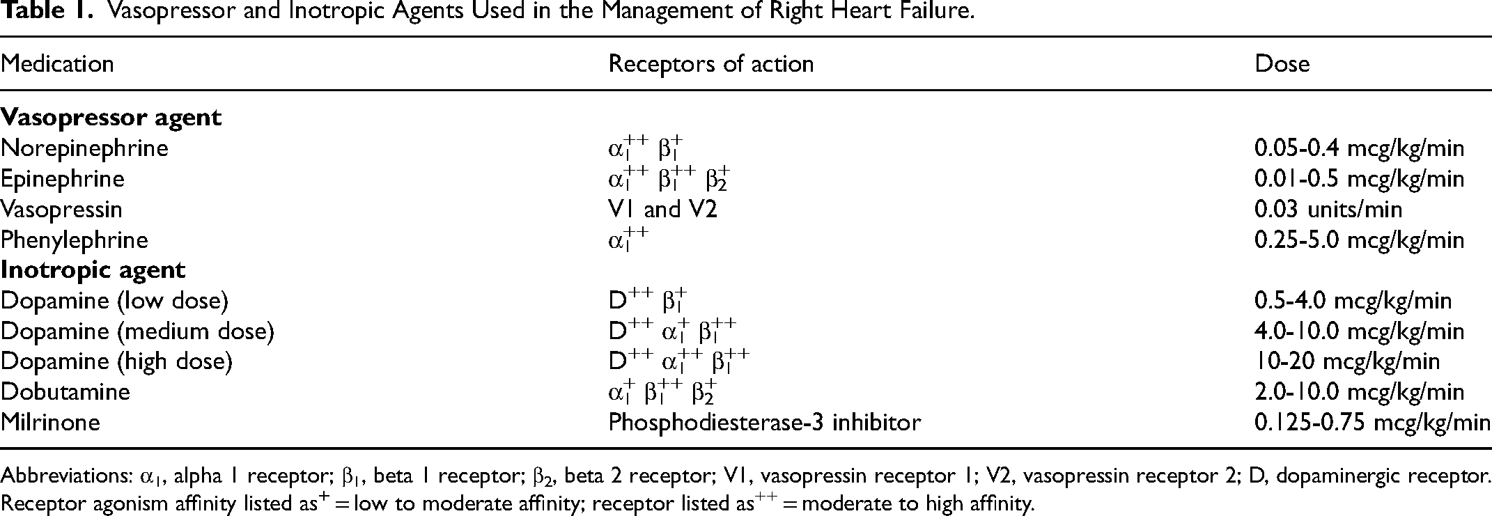
Abbreviations: α1, alpha 1 receptor; β1, beta 1 receptor; β2, beta 2 receptor; V1, vasopressin receptor 1; V2, vasopressin receptor 2; D, dopaminergic receptor. Receptor agonism affinity listed as
Right Ventricular Inotropic Support
Dopamine acts on both dopaminergic and adrenergic receptors; it theoretically has dose-dependent effects, but its effects may be unpredictable in critically ill patients. 99 At low doses, dopamine causes a decrease in systemic vascular resistance which in turn can reduce the systemic to RV perfusion gradient particularly in a hypertensive RV. At intermediate doses it binds to B1 adrenergic receptors causing increased inotropy and chronotropy with mild increases in systemic vascular resistance. At high doses, the a1-adrenergic receptor binding property of dopamine predominates, causing systemic vasoconstriction.98,108 Dobutamine has affinity for both B1 and B2 receptors, but its predominant effect is inotropic and chronotropic. 99 At higher doses, Dobutamine can precipitate systemic hypotension through B2 stimulation and affect the systemic to RV perfusion gradient. 2 Milrinone is a phosphodiesterase-3 inhibitor that increases levels of cyclic adenosine monophosphate (cAMP) to cause increase in myocardial contractility and lusitropy while also causing systemic vasodilation when given through the parenteral route. 98 In addition to these systemic vasodilatory effects, milrinone is a potent pulmonary vasodilator 108 and causes significant reduction in RV end-diastolic pressures. 2 Milrinone is almost exclusively cleared by the kidney and is preferably avoided in patients with CrCl <10 ml/min, as its accumulation can lead to arrythmias and hypotension. 109 Inhaled aerosolized milrinone presents an alternative to the intravenous form and has lower reported association with systemic hypotension when studied in patients undergoing cardiac surgery. 110
Inotropic medications should be used judiciously with the understanding that they can be pro-arrhythmogenic. 2 This is of particular significance in the hypertensive RV (as in patients with established PH), which is reliant on active atrial contraction or “kick” rather than passive filling of the RV during its diastolic phase. As such, loss of the active atrial contraction in the setting of an atrial arrythmia can contribute to worsening hemodynamics. Recent clinical guidelines have recommended against routine use of these agents in hospitalized patients with acute HF noting lack of evidence supporting improved survival in patients with HF 109 and thus clinicians should proceed cautiously and in consultation with a multidisciplinary management team. They should generally only be used in patients who have continued evidence of end organ hypoperfusion (eg, increasing serum lactate, increasing CVP or RVEDP, worsening mixed or central venous oxygen, and/or WRF) despite optimization of RV preload and afterload. Table 1 provides a summary of the different inotropic options available for RH failure.
Advanced Therapies for RH Failure
Durable MCS or acute mechanical circulatory support (AMCS) devices may be necessary for patients who have refractory RH failure that is not responsive to therapies aimed at optimizing preload, contractility, and afterload with a goal of enhancing CO and augmenting end-organ perfusion. 111 In general, the decision to use MCS/AMCS should be a short-term intervention used as a bridge to recovery or a bridge to decision/bridge to bridge. The latter refers to the implementation of these support devices to clinically stabilize patients allowing for evaluation of more durable support or intervention (eg, placing a patient with high-risk PE with RH failure on veno-arterial extracorporeal membrane oxygenation (VA-ECMO) as a bridge to mechanical thrombectomy).111,112
Current options for durable MCS include surgical RV assist devices (RVADs) with extracorporeal centrifugal-flow pulps, isolated pulsatile RVADs, rotary-flow RVADs, and biventricular support with pulsatile ventricular assist devices (VADs). 113 Percutaneous AMCS options include central or peripheral VA-ECMO, the TandemHeart centrifugal-flow pump, and the axial flow Impella RP catheter. 2 The percutaneous AMCS devices differ in their configuration, estimated flow rates and effect on hemodynamics as outlined in Table 2. VA-ECMO is considered to indirectly bypass the RV as the return cannula supplies blood in a retrograde fashion via the femoral artery. In contrast, the Impella RV, Tandem RVAD, and Protek Duo devices directly bypass the RV as they displace blood from the RA directly to the PA. 113 Oxygenators can be spliced into the circuit of the Protek Duo and Tandem RVAD devices providing additional oxygen support, however, the addition of an oxygenator is not available for the Impella RV device. Differential hypoxia may be encountered in VA-ECMO and the additional venous cannula may be required to assist with oxygenation leading to a veno-arterial-venous (VAV) ECMO configuration. Each MCS device has its own unique mechanism of action, advantages, and potential complications. While emerging guidelines support the careful use of AMCS/MCS in patients with acute cardiogenic shock, 111 high quality evidence regarding positive outcomes data and data supporting its use in isolated RH failure are limited. 114 Table 3 provides an overview of the different percutaneous AMCS/MCS devices for isolated RH failure.
Examples of Devices for Continuous Cardiac Output Monitoring Based Upon Arterial Blood Pressure Pulse Contour.
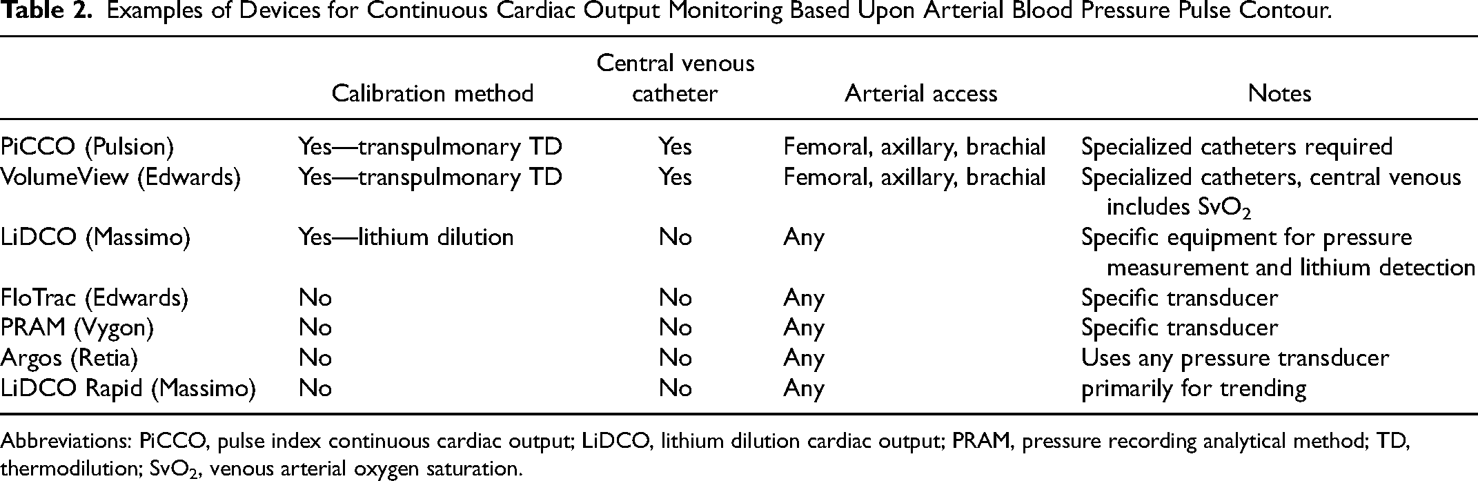
Abbreviations: PiCCO, pulse index continuous cardiac output; LiDCO, lithium dilution cardiac output; PRAM, pressure recording analytical method; TD, thermodilution; SvO2, venous arterial oxygen saturation.
Overview of Percutaneous Acute Mechanical Circulatory Support Devices for Isolated Right Heart Failure.
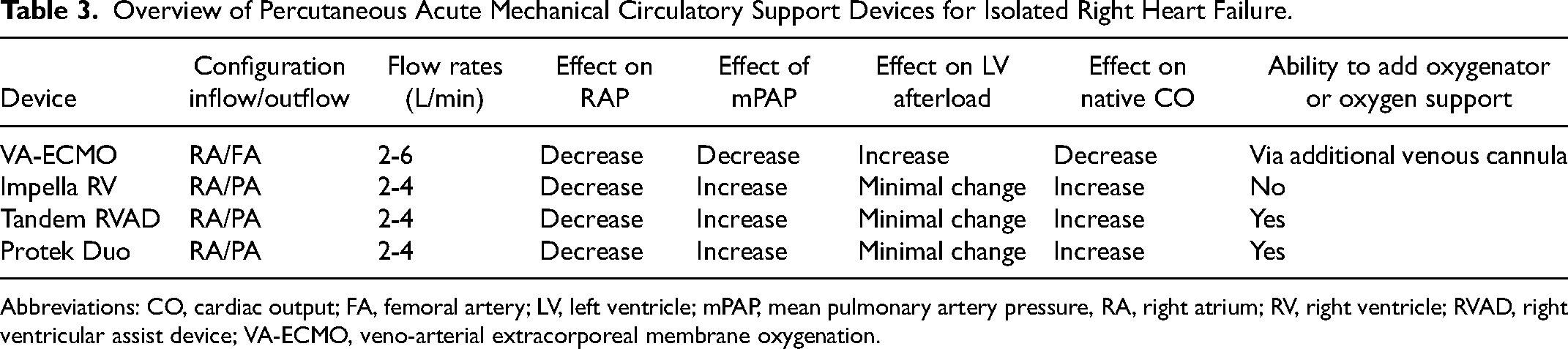
Abbreviations: CO, cardiac output; FA, femoral artery; LV, left ventricle; mPAP, mean pulmonary artery pressure, RA, right atrium; RV, right ventricle; RVAD, right ventricular assist device; VA-ECMO, veno-arterial extracorporeal membrane oxygenation.
For patients with PAH, non-mechanical percutaneous and surgical approaches can be also considered in those who have progressive clinical deterioration despite maximal medical therapy. Options include percutaneous balloon atrial septostomy, where balloon dilation of the atrial septum is performed to create an atrial communication that allows for right-to-left atrial shunting and subsequent decompression of the RH. 115 While this is a reasonable therapeutic approach for highly select group of end-stage PAH patients, 115 it should be avoided in patients with acute RH failure due to the high likelihood of post-procedural mortality from insufficient pulmonary blood flow and severe hypoxemia. A Potts shunt, performed either via a surgical or catheter-based approach to create an anastomosis between the left PA and the descending thoracic aorta, is another palliative option that attempts to approximate the hemodynamic milieu of Eisenmenger disease associated with a patent ductus arteriosus.116–118 A Potts shunt carries relative advantages over atrial septostomy including sparing the brain and myocardium from exposure to desaturated blood and paradoxical emboli, and the use of a pressure-restrictive shunt device prevents critical hypoxemia. 117 Emerging data on longer term outcomes of pediatric patients who have undergone these procedures have produced mixed results. 118 More recently, a unidirectional-valved shunt was developed to permit flow from the main PA to the descending aorta only during periods of supra-systemic PA pressure, although more data is needed to assess its long-term use and efficacy. 119 Ultimately, the goals of the above interventions are to extend life in the absence of heart and/or lung transplant options, defer time to transplant, or to provide palliation at end of life.
Respiratory Support in RH Failure
The process of intubation presents substantial risk due to the amalgam of hemodynamic effects of sedation, paralysis, hypoxemia, hypocapnia, sympathetic stimulation, and alterations in intrathoracic pressure that can lead to systemic hypotension and circulatory collapse in the setting of acute RH failure.120,121 The European Society of Cardiology/European Respiratory Society recommends that intubation be avoided, if possible, in patients with advanced RH failure for these reasons. 82
High-flow nasal cannula should be considered for patients with hypoxia given its minimal contribution to positive pressure. In patients with ongoing hypoxia or combined hypoxemic and hypercapnic respiratory failure, a trial of non-invasive positive pressure ventilation (NIV) can be considered as a temporizing measure. Intubation should not be delayed if medically necessary, but it may be helpful to consult with a cardiac anesthesia team. Invasive arterial blood pressure monitoring should be strongly considered to assess beat-to-beat coronary perfusion and central venous line to monitor the CVP/RA pressure can be utilized. 77 Central venous access or dedicated adequate peripheral venous access should be obtained for the potential use of vasopressor agents in the peri-intubation period. Anesthesia induction has a myriad of potentially detrimental hemodynamic effects including decreased heart rate via reduction of sympathetic drive, decreasing RV contractility, and increasing PVR through inhibition of spontaneous ventilation leading to atelectasis and subsequent alveolar hypoxia. Preload and systemic MAP should first be optimized; small fluid boluses or the administration of vasopressors prior to anesthesia can be considered to maintain the systemic MAP above the mean PA pressure or systemic systolic and diastolic BP over the RV end-systolic and end-diastolic pressures, respectively (if RV pressure waveform is available).25,122 Inhaled pulmonary vasodilators can be started prior to induction to improve RV function and oxygenation and continued via endotracheal tube once mechanical ventilation is established. For patients with evidence of shock prior to intubation or have severely reduced RV function, pre-intubation ECMO should be considered to assist in preserving cardiopulmonary function during the peri-intubation period. For induction, our preferred medications are hemodynamically neutral agents such as etomidate or ketamine. Acknowledging that all induction agents may cause some hypotension, even the hemodynamically neutral agents should be titrated to effect, or considered dose reduced rather than being administered as a large bolus. 123 Propofol should be avoided given its effect on reducing systemic blood pressure and therefore the systemic to RV perfusion gradient.23,25,77 The most experienced operator should be performing the procedure to minimize time to intubation and maximize first pass success without complications.
To ameliorate the substantial risk of anesthesia induction and intubation in this population, an experienced operator can consider using awake fiberoptic intubation in a spontaneously breathing patient supported by peri-intubation oxygenation with high flow nasal canula or nasal NIV. In addition to oxygenation, this procedure involves keeping the patient in the upright position while providing topical anesthesia to the airway (eg, topical, or nebulized lidocaine) with low dose sedation (eg, ketamine, low dose fentanyl or midazolam) prior to oral intubation with bronchoscopic guidance (or via a nasal route in the appropriate clinical setting, eg, non-coagulopathic and non-pregnant patients).124,125 Johannes and colleagues evaluated this technique in 9 patients with acute RH failure from PH with severe hypoxemic respiratory failure and demonstrated 100% first pass intubation success of the technique in the immediate peri-intubation period, making it an appealing alternative to conventional methods. 126 Following intubation, regardless of approach, maintenance of anesthesia on mechanical ventilation is generally maintained with low dose opioids or ketamine together with benzodiazepines or propofol. 25 Systemic vasopressors may be used to ensure maintenance of coronary perfusion pressure in the setting of decreased systemic blood pressure following induction and maintenance of anesthesia. 77
Once intubated, every effort should be aimed at achieving an “RV protective strategy,” which may seem at odds with well accepted approaches of managing mechanical ventilation in critically ill patients founded in “lung protective ventilation.”17,77 This involves ensuring adequate oxygenation, using low tidal volumes while achieving plateau pressures <30 cmH2O while avoiding hypercapnic acidosis and minimizing PEEP to mitigate increases in PVR. 122 Peripheral oxygen saturation should generally be kept above 90%.17,25 Early extubation is preferred with the caveat that weaning and subsequent withdrawal of positive pressure ventilation can cause an increase in right-sided venous return to an already volume and pressure overloaded RV and also increase LV afterload which can lead to worsening RH failure and flash pulmonary edema, respectively. 127
It is also important to recognize how positive pressure ventilation impacts RH hemodynamics. During positive pressure ventilation, the ventilator generates positive airway pressure (transmitted into the alveolar pressure and pleural pressure) and thus there is a positive transpulmonary pressure. 122 This can work to impede RV preload by increasing intrathoracic pressure and reducing RV transmural filling pressures. 17 Additionally, PEEP has a U-shaped effect on PVR; initially PEEP can sustain open alveoli, increase lung volume, and positively tether blood vessels which works to decrease PVR and RV afterload, but later it can cause alveolar distension thereby compressing vasculature which increases PVR and RV afterload and decreases RV preload. 122 In critically ill patients with underlying circulatory compromise, increases in pleural pressure (from either high PEEP or high airway pressures) can compromise RV perfusion if pleural pressure or RV pressure is allowed to increase beyond aortic pressure. 122 In poorly compliant lungs, changes in intrathoracic pressure as above have more pronounced effects on hemodynamics than in patients with no underlying pulmonary pathology.
Positive pressure ventilation has distinct effects on the LV afterload. During positive pressure ventilation, LV wall tension (which is made up by the difference between LV systolic pressure and mean intrathoracic pressure) 128 remains constant, since the pleural pressure, aortic pressure, and LV pressure increase equally. 122 The net effect creates a flow gradient between the thorax and peripheral organs. 122 Additionally, during positive pressure ventilation, mean intrathoracic pressure is increased and transmitted to the intrathoracic arterial system, leading to aortic pressure autoregulation secondary to baroreceptor stimulation.128,129 This works to lower systemic vascular resistance and LV afterload. 122 This desirable effect is important in managing patients with PH due to left heart disease as they may benefit from positive pressure ventilation mediated reduction is systemic afterload. Conversely, following endotracheal extubation or withdrawal of positive pressure support, acute LV decompensation from increased systemic afterload may occur. 129
End of Life Care of the RH Failure Patient
For all patients in the ICU, mortality is high ranging from 6.4% to as high as 40%.130,131 In this context, despite the bold efforts of the myriad teams caring for critically ill patients with RH failure, there are patients who will ultimately die because of their illness and those who will decide that the functional limitations imposed by their illness and treatments are incompatible with their care goals. In fact, it has been shown that 44% of patients with PAH die of progressive RH failure or sudden death, and most of these patients die in the ICU, 132 despite the fact that most patients with advanced lung disease generally prefer to die at home. 133 Recognizing the morbidity and mortality associated with this particular syndrome, timely goals of care discussions should be had with patients and their families specifically addressing the circumstances in which a transition away from life-prolonging measures would be preferred. 134
Recommendations exist to assist in guiding a transition to end-of-life care in the ICU, which are applicable to patients with RH failure. The American College of Critical Care Medicine highlights several principles aimed at improving the care of patients throughout the dying process. These include ensuring patient and family-centered care and decision making; guaranteeing adequate symptom management in end-of-life care including those related to pain, dyspnea, and delirium; ethical and compassionate decisions around withdrawing life-sustaining treatment; and ensuring the provision of bereavement and support services. 135 Throughout this process, a dedicated palliative care team should be involved early to provide a comprehensive and person-centered approach to address physical, psychological, social, and spiritual well-being of patients and family members.136 Palliative care teams can be particularly helpful in navigating several end-of-life considerations applicable to patients with RH failure including management of severe symptoms of respiratory failure, decisions surrounding whether to offer MCS or transplant, questions around the withdrawal of MCS or mechanical ventilation, and timing around transitioning to hospice care. Additionally, for PAH patients receiving continuous pulmonary vasodilator therapy, the palliative care team alongside PH physicians can assist with developing a weaning protocol to prevent the hemodynamic and symptomatic decompensation associated with abrupt discontinuation of these medications as patients transition to comfort-based care.137
Conclusion
Acute RH failure in the ICU is an increasingly recognized disease process with multiple distinct etiologies associated with significant morbidity and mortality. Understanding the unique anatomy and physiology of the RH circulatory system and its response to distinct hemodynamic states is critical to rapidly identifying and successfully managing this condition by intensivists. Future studies are needed to optimize pharmacologic and mechanical treatment of RH failure and to understand the molecular and genetic underpinnings of this syndrome to help understand individual risk, tailor management, and improve patient outcomes.
Footnotes
Declaration of Conflicting Interests
The author(s) declared no potential conflicts of interest with respect to the research, authorship, and/or publication of this article.
Funding
The author(s) received no financial support for the research, authorship, and/or publication of this article.