





Abstract
Background
Venous thromboembolism (VTE), whether pulmonary embolism (PE) or deep vein thrombosis (DVT), is common in patients with COVID-19. Recommendations on systematic screening in the intensive care unit (ICU) are lacking.
Research question
Is there any clinical benefit of systematic screening for DVT in critically ill patients with severe COVID-19?
Study design and methods
Single-center randomized clinical trial (RCT) of COVID-19 cases admitted to the ICU. Patients were randomized into two groups: a study group that underwent ultrasound (US) screening for DVT Mondays and Thursdays, and a control group that was treated according to the unit protocol. The primary outcome was the presence of DVT. Secondary outcomes were ICU total stay, death within 21-day follow-up and bleeding complications (minor or major). A composite outcome of poor prognosis variables was analyzed. We tested a superiority hypothesis with a confidence level of 95% and an equivalence limit of 20%.
Results
163 patients (84 screening group, 79 control group) were enrolled between April and July 2021. There were 90 men (55.2%) with a mean ± SD age of 49.8 ± 13.58 years. In screening group 16.7% developed DVT versus 3.8% in control group (p = .007), and 3.6% versus 5.1% developed PE, respectively (p = 0.7). Poor outcome variables were male sex, age, COVID-19 vaccination status, Fibrinogen, Urea, Creatinine and Interleukin 6 (IL6) levels; Acute Physiology and Chronic Health Evaluation II (APACHE II) and Sequential Organ Failure Assessment (SOFA) scales. The superiority comparison, with a power of 95%, showed no statistically significant differences for a composite endpoint (p = .123). After adjusting by group, the OR for poor outcome is 1.966 (0.761-5.081) p = 0.163
Interpretation
Among these patients, a strategy of systematic US screening for DVT was not associated with any significant improvements to clinical outcomes compared with usual care.
Clinical Trial Registration
Clinicaltrials.org registration number: NCT05028244.
Introduction
COVID-19 is associated with a high incidence of venous thromboembolism (VTE) in all patient settings.1–3 This numbers are particularly high on patients admitted to the intensive care unit (ICU)3,4 with rates of 24.1% versus 7.7% in non-ICU settings. 1 High rates have been observed in studies with systematic screening,5,6 with figures as high as 30% for patients in the ICU compared to non-ICU settings. 7 This is relevant, as thrombosis is associated with an increased risk of mortality.8–10
COVID-19 is recognized as a pro-thrombotic state, which is a hallmark of severe cases. Endothelial damage caused by viral infection, along with cytokine storm-mediated inflammation, triggers excessive tissue factor expression and thrombin generation. Elevated levels of D-dimer and fibrin degradation products observed in COVID-19 patients mirror those seen in disseminated intravascular coagulation (DIC), supporting the connection between these pathologies.11–13
While early diagnosis and treatment should favorably impact patient outcomes, this has not been reliably demonstrated. Patients receiving anticoagulation in this setting have a decreased risk of VTE but may also experience potentially serious bleeding.14,15
Although systematic ultrasound screening results in a higher detection of deep vein thrombosis (DVT) in other studies,4,5 these studies do not prospectively or repeatedly evaluate patients throughout the disease's evolution as was done in the present work and all of them are limited to a single screening exploration.
Regarding recommendations only one non-systematic review has advocated routine screening for thrombotic complications in this setting (COVID-19 in the ICU), but the association between early detection and favorable clinical outcomes is not well established. 16
Available guidelines recommend extended compression US from the common femoral vein to the distal calf veins as the best test to diagnose lower extremity DVT when clinical signs are present and there is an absence of other more probable diagnostic (American Society of Hematology, 17 American College of Chest Physicians, 18 Society of Radiologists in Ultrasound. 19 None of these guidelines address this diagnoses approach in the context of the COVID-19 pandemic. We were unable to find any more screening protocol recommendations in the available evidence to date. Most studies, when present, only perform one single screening study. Moreover, guidelines do not provide detailed recommendations regarding the optimal bedside procedure to be performed in the critical care setting.
We hypothesized that twice weekly ultrasound screening for DVT would not significantly modify mortality, ICU stay, or the presence of symptomatic VTE. The aim of this study was to assess the impact of twice-weekly ultrasound screening for DVT in patients with COVID-19 in the ICU.
Methods
This was a single-center, non-blind, randomized, superiority clinical trial with an intent-to-treat analysis. The primary objective of this study was to assess the impact of twice-weekly ultrasound screening for DVT in patients with COVID-19 in the ICU. Secondary objectives included measuring the incidence of venous thromboembolic events in both study groups and their type of presentation; comparing demographic variables and outcomes between both groups; and defining the variables of poor prognosis and compare them between the two study groups using a composite outcome.
The inclusion criteria were patients aged 18 years or older admitted to the ICU at Hospital Universitari Vall d’Hebron in Barcelona with severe acute respiratory syndrome due to SARS-CoV-2 confirmed by polymerase chain reaction, between April and July of 2021. Exclusion criteria included patients with newly detected DVT, known DVT or pulmonary embolism (PE) in the previous 3 months, chronic anticoagulation, patients on extracorporeal membrane oxygenation therapy and pregnant women.
The intervention in the study was bilateral venous Doppler ultrasound of the lower limbs. This was performed within 72 h of admission to the ICU in all cases. All examinations were performed bedside by two vascular surgeons with extensive experience in venous US. All safety measures were taken. Patients with positive Doppler ultrasound findings for DVT were excluded.
Patients included in the study were randomly assigned to a screening group or a usual care (control) group using a computer-generated random sequence. Patients in the screening group received usual care plus systematic twice-weekly screenings (Mondays and Thursdays) consisting of venous Doppler ultrasound of the lower extremities (femoral, popliteal, and gastrocnemius veins). Patients in the control group received usual care. Both groups underwent Doppler ultrasound examination for clinically suspected DVT.
Suggestive signs of DVT were entire leg swelling, an increase of >3 cm in calf circumference compared with the contralateral calf, major pitting edema in one leg, or recently developed collateral superficial veins and based on the Well's probability score for DVT. 20 Femoral veins were omitted in patients in the prone position. Ultrasound was considered positive when the vein was incompressible, when there was no evidence of flow on manual distal compression, and when intraluminal hyperechogenic foci were observed partially or completely occluding a given vein segment. Iliac veins were indirectly assessed by interpreting the Doppler flow in the common femoral vein at the arch of the great saphenous vein during respiration.
Similarly, testing for suspected PE was left to the discretion of the medical team, and when performed, characteristics, date, and results were recorded. Suggestive signs of PE included sudden worsening of respiratory condition without an apparent cause combined with a significant increase in D-dimer levels.
Symptomatic VTE was diagnosed based on clinical manifestations of DVT or PE confirmed by imaging techniques. Asymptomatic VTE was diagnosed solely by positive imaging results. Jugular and femoral venous catheter–associated thrombosis was also assessed by ultrasound when suspected by the ICU team.
The number of asymptomatic and symptomatic VTE (PE, and DVT) events that occurred over a follow-up period of 21 days was recorded, including events of death or discharge from the ICU during this period. After 21 days, the only variable recorded were all-cause mortality and length of ICU stay.
Other variables analyzed included age (years), sex (male or female), weight (kg), height (m), and history of hypertension, diabetes mellitus, dyslipidemia, chronic kidney disease, renal replacement therapy, atrial fibrillation, heart disease, stroke, peripheral artery disease, DVT, and PE, as well as occurrence of minor and major bleeding events according to the definitions of the International Society on Thrombosis and Hemostasis. 21 This information was obtained from clinical records, as it was impossible to collect a full history from most patients.
Additional variables included long-term antiplatelet or anticoagulation therapy, need for intubation, prone positioning, venous catheter–related events, and mechanical or pharmacological thromboprophylaxis (drugs and doses).
The laboratory parameters recorded on admission to the ICU included platelet count (×109/L), prothrombin time (s), fibrinogen (g/L), lymphocyte count (x109/L), glomerular filtration rate (mL/min/1.73m2), lactate dehydrogenase (IU/L), C-reactive protein (mg/dL), ferritin (ng/dL), and interleukin-6 (pg/mL). D-dimer levels (ng/mL) were measured daily when available.
The final variables analyzed were disease severity according to the Acute Physiology and Chronic Health Evaluation II (APACHE II) 22 score and to the Sequential Organ Failure Assessment (SOFA) 23 score, when available. These assessments were made on the day of VTE diagnosis, when applicable.
The primary outcome was the presence of symptomatic DVT in the two groups of patients. Secondary outcomes included ICU length of stay, hospital discharge, death within the 21-day follow-up period and bleeding complications (minor or major bleeding).
To achieve greater power in the study, three independent poor prognostic factors were grouped into a composite variable labeled “poor prognosis”. This variable included time to death, symptomatic VTE, and major bleeding during follow-up compared between patients in the screening and control groups.
The study was approved by the Drug Research Ethics Committee at Hospital Universitari Vall d’Hebron (code PR(AD) 176/2021) and was registered at clinicaltrials.org (NCT05028244). Informed consent was obtained for every participant.
The sample size was calculated to achieve a power of 90% to detect differences when testing the null hypothesis (H0) using a normal one-sided, asymptotic test for the two independent groups. A confidence level of 95% was assumed, along with a VTE prevalence rate of 30% in the screening group and 10% in the control group based on rates observed in a previous study of our group [26.5%] 24 and other large series1–3,8–10 . The calculations were performed using Ene 3.0, a sample size calculation program developed by the Servei d'Estadística Aplicada and distributed by the GlaxoSmithKline laboratory. A sample size of 134 patients (67 in each group) was determined to be necessary. Statistical analyses were performed using IBM SPSS Statistics Version 26.0 (International Business Machines Corporation, Armonk, New York, USA). For the descriptive analysis, frequency tables were created for categorical and nominal variables. Measures of central tendency (mean, median, standard deviation, and interquartile range) and dispersion were calculated for continuous variables. The normality of the distribution of numerical variables was checked using Q-Q plots and the Shapiro-Wilk test.
To ensure robust power in the analysis we constructed a composite outcome incorporating known poor outcome factors. Patients were classified as having a poor outcome if they presented any of the components of the composite outcome, which included symptomatic VTE, major bleeding, an ICU stay >21 days, or all-cause mortality.
Baseline characteristics were compared between patients in the screening and control groups as well as between patients with good and poor outcomes. Significant between-group differences were analyzed using the Pearson chi-squared test or Fisher exact test for categorical variables, the chi-squared test for linear trends for ordinal variables, and the t test or Mann-Whitney U test for continuous variables. Variables with a p-value <0.1 in the univariate analysis were included as independent variables into a stepwise multiple logistic regression model to identify independent predictors of poor outcomes. An additional logistic regression model with the same variables was constructed to adjust for the influence of group (screening vs usual care). For the composite outcome, statistical significance was assessed using the log-rank test. Significance was set as p < .05.
Results
Of the 229 consecutive patients admitted to the ICU between April 1sts, 2021, and July 31st, 2021, 163 were enrolled. 84 in the screening group and 79 in the control group. The patient inclusion flowchart and exclusions are shown in Figure 1.
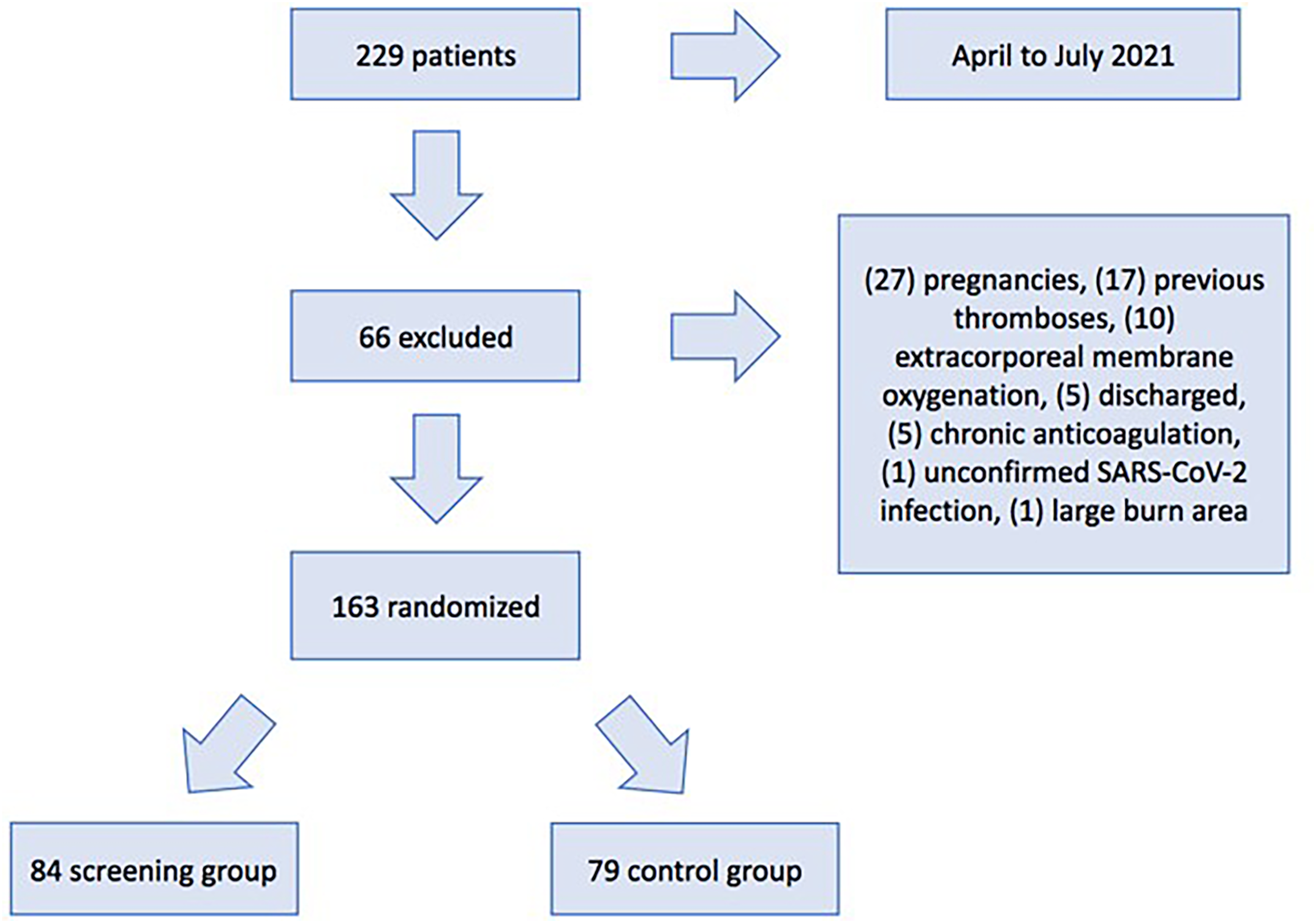
CONSORT diagram.
The main characteristics of patients following randomization are summarized by group in Table 1. The vast majority (84.7%) had never smoked and 15.3% were current or former smokers. Only 12.9% of patients were fully vaccinated; the remaining 87.1% were partially vaccinated or unvaccinated.
Clinical Characteristics of Patients with Severe COVID-19 on Randomization to Systematic Screening and Usual-Care Groups.
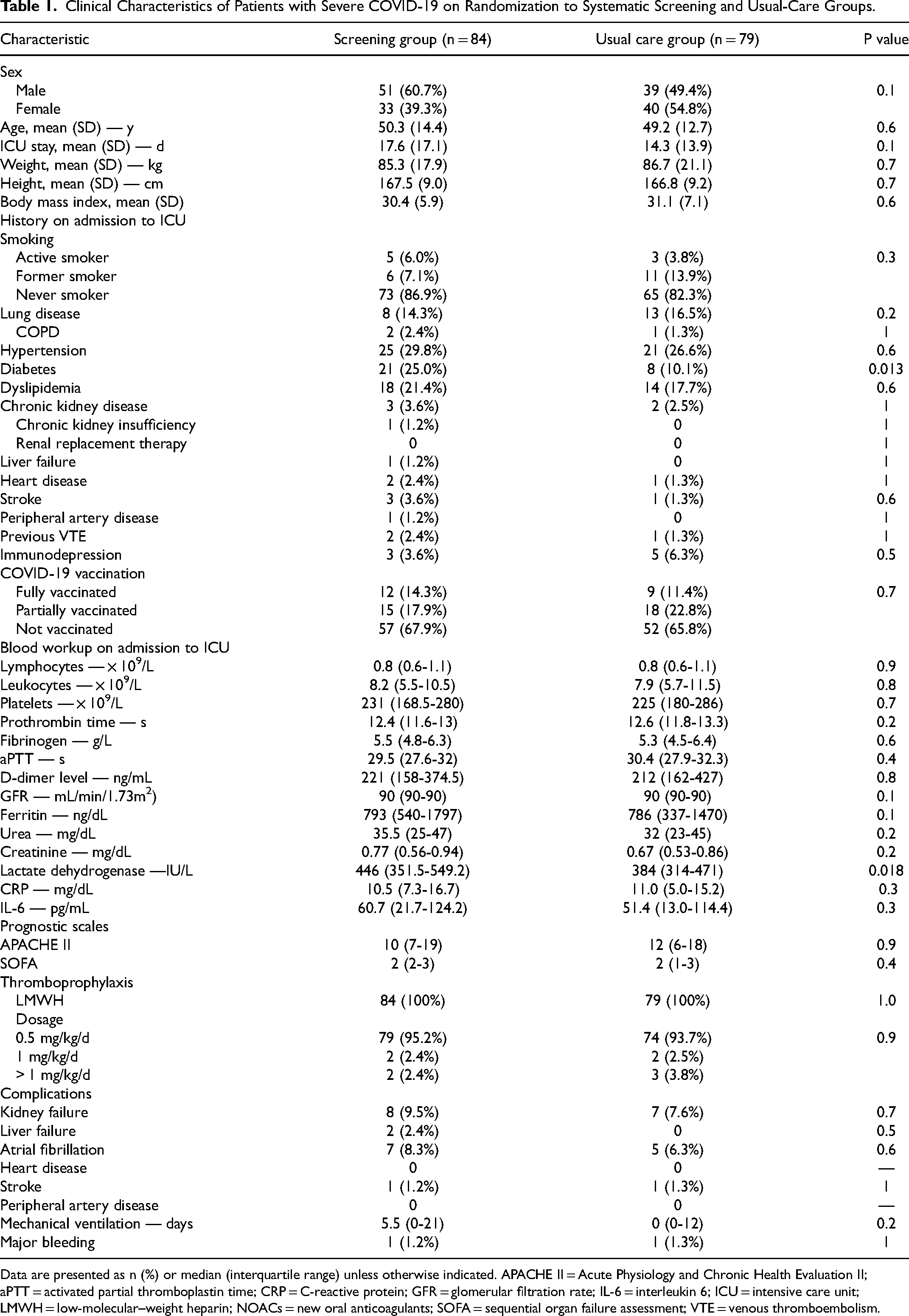
Data are presented as n (%) or median (interquartile range) unless otherwise indicated. APACHE II = Acute Physiology and Chronic Health Evaluation II; aPTT = activated partial thromboplastin time; CRP = C-reactive protein; GFR = glomerular filtration rate; IL-6 = interleukin 6; ICU = intensive care unit; LMWH = low-molecular–weight heparin; NOACs = new oral anticoagulants; SOFA = sequential organ failure assessment; VTE = venous thromboembolism.
Enoxaparin thromboprophylaxis was initiated at a dosage of 0.5 mg/kg/day on admission to the ICU in 154 patients (94.5%) following standard protocol of the unit. The other patients received enoxaparin as anticoagulation therapy at higher dosages: 1 mg/kg/day in four patients (2.4%) and 1.5 mg/kg/day in seven (3.1%).
Primary Outcome
Symptomatic and asymptomatic VTE events were significantly more frequent in the screening group (18 [21.4%] vs 8 (10.1%) in the control group, p = .049). DVT accounted for most of the difference, with 14 cases (16.7%) in the screening group and 3 (3.8%) in the control group (p = .007) (Tables 2 and 3). All positive cases were treated with full dose anticoagulation (1.5 mg/kg/day of enoxaparin) on the day of diagnosis.
Type of Thromboembolic Event by Study Group and Main Coexisting Factors.
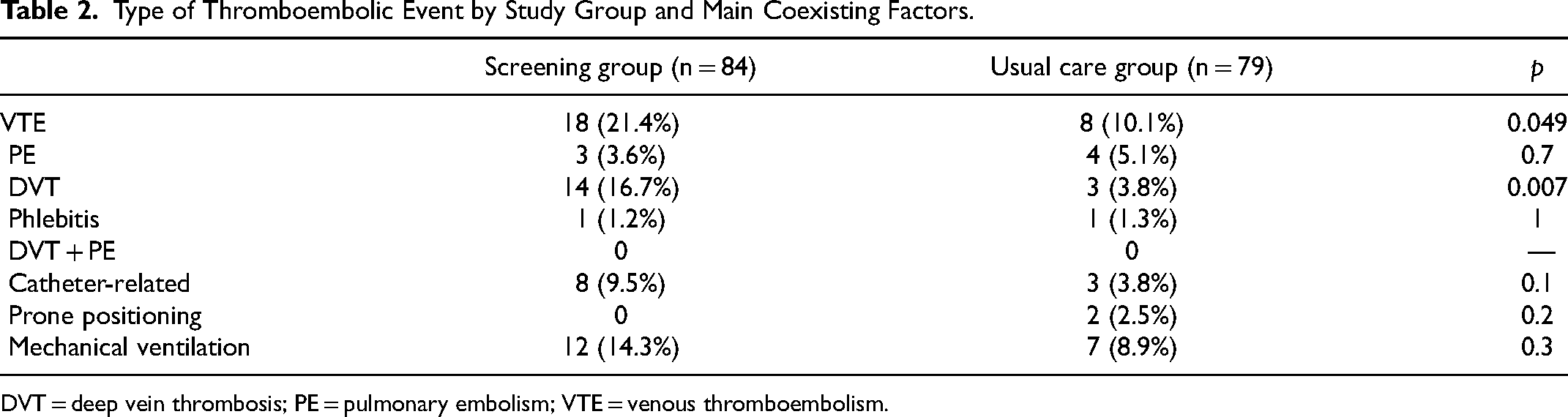
DVT = deep vein thrombosis; PE = pulmonary embolism; VTE = venous thromboembolism.
CTPA Procedures and Results per Study Group (p = 0.7).
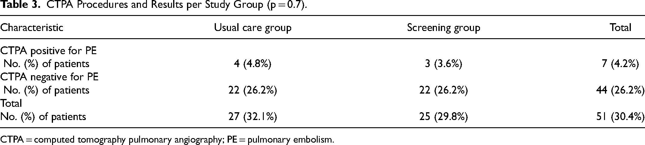
CTPA = computed tomography pulmonary angiography; PE = pulmonary embolism.
Secondary Outcomes
No significant differences were observed in ICU length of stay (mean 17.6 days in the screening group [SD: 17.1] vs 14.3 days in the control group [SD: 13.9], p = .1). There were three deaths (3.6%) in the screening group and two (2.5%) in the control group during the 21-day follow-up period. Hospital discharge rates were 60 (71.4%) and 59 (74.7%). After 21 days, 21 patients in the screening group (25%) and 18 (22.8%) in the control group were still in the ICU after 21 days. There were no statistically significant differences between the two groups for this variable (p = .9). Three patients in the screening group (3.6%) and two in the control group (2.5%) developed bleeding complications (p = .7). Only one in each group was categorized as major bleeding (p = 1).
Composite Outcome
A total of 22 patients in the control group (27.8%) and 33 in the screening group (39.3%) experienced a poor outcome based on the composite endpoint, which included all-cause mortality, major bleeding, all VTE, and an ICU stay >21 days. This difference was not statistically significant (p = .123). Table 4 lists the variables significantly associated with a poor outcome based on the composite outcome. Independent predictors of a poor outcome in the multiple regression analysis included male sex (odds ratio [OR] 3.01, 95% CI 1.25-7.27, p = .014]), APACHE II score (OR 1.08, 95% CI 1.02-1.14, p = .011]), and SOFA score (OR 1.59 [1.17-2.15, p = .003]) (Table 5). After adjusting for screening versus non-screening and the independent predictors of a poor outcome, differences in clinical outcomes remained non-significant (OR: 1.966 (0.761-5.081) p = 0.163) (Table 6).
Variables Associated with a Poor Outcome (Composite Endpoint Integrating Death, Symptomatic VTE, ICU Stay >21 Days, and major Bleeding).
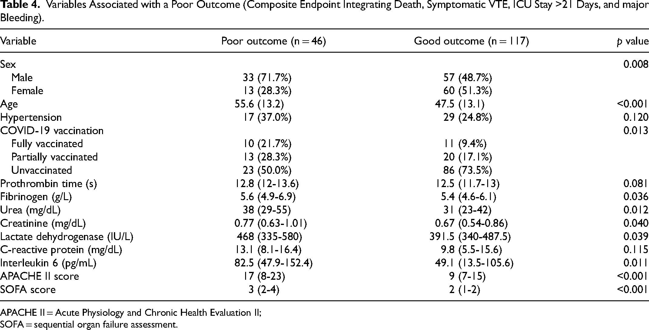
APACHE II = Acute Physiology and Chronic Health Evaluation II; SOFA = sequential organ failure assessment.
Independent Predictors of a Poor Outcome (Logistic Regression Analysis).

Logistic Regression Model Adjusting for the Relationship Screening Versus No Screening by Predictors of Poor Outcome.

Interpretation
This study is the first to prospectively evaluate the impact of lower limb ultrasound screening in patients admitted to the ICU for COVID-19, providing valuable insights to enhance routine clinical practice and protocols. Although the incidence of DVT remains high, it was lower than reported in previous global series. A statistically significant increase in detection rates was observed in the group undergoing systematic screening, although this finding did not translate into significant differences in the clinical outcomes studied.
I. High prevalence of VTE and association with poor outcomes
In this study, systematic ultrasound screening for DVT did not improve clinical outcomes in ICU patients with COVID-19 compared to usual care. As anticipated, screening identified more cases of DVT (14 vs 3 in the control group, p = .005), consistent with previous findings that detection rates are significantly higher with routine screening than with selective screening based on clinical suspicion, for both DVT and PE.4–7,12,25–27 Most detected cases were asymptomatic and involved distal pulmonary and lower limb vessels. To date, early detection in this period has not been associated with clinical benefit in systematic.12,27
Both DVT and PE are associated with significantly worse outcomes in patients with COVID-19. Xiao et al, 8 in one of the largest meta-analyses investigating the association between COVID-19 and VTE (332,915 patients), showed that thrombosis significantly increased ICU admissions (OR 2.9, 95% CI 1.6-5.24, p < .05) and the risk of COVID-19-related mortality (OR 2.61, 95% CI 1.91-3.55, p < .05). Although the meta-analysis (which included 25 studies) primarily focused on VTE events, it also encompassed arterial events such as stroke, myocardial ischemia, and mesenteric ischemia, which may have a greater impact on prognosis. Similar conclusions were reached in smaller meta-analyses, that also included arterial events.10,28 In a prospective study of 184 patients with a mean follow-up of 14 days, Klok et al 29 identified an increased risk of mortality in patients with venous or arterial thrombotic complications (hazard ratio 5.4, 95% CI 2.4-12).
In a previous study by our group, involving 230 patients with COVID-19, we observed no significant differences in mortality after a 7-day follow-up between patients with and without VTE (6.6% vs 5.3%, p = .70).
24
In that study, we developed a predictive model comprising three variables: an age >66 years, D-dimer levels >1500 ng/mL on admission, and a lymphocyte count <0.45 × 10⁹/L. The three variables combined predicted a mortality rate of 100% (area under the receiver operating curve 0.81, 95% CI, 0.73-0.89, p < .001).
30
In the current trial, we analyzed mortality rates in ICU patients who developed VTE during the 21-day initial follow-up and subsequent periods for patients with longer ICU stays. Mortality was higher in patients with VTE than in those without (19.2% vs 4.2%, p = .015).
II. Anticoagulation and thromboprophylaxis therapy
Recommendations for antithrombotic prophylaxis in this patient population vary. In the ATTACC, REMAP-CAP, and ACTIV-4ª RCTs, full-dose anticoagulation was discontinued prematurely due to futility. Randomization of 1098 patients revealed no improvement in organ failure–free survival, while patients receiving therapeutic doses experienced an increase in bleeding.
14
In a systematic review comparing the safety and efficacy of intermediate- and therapeutic-dose anticoagulation in hospitalized patients with COVID-19, Reis et al 15 analyzed eight RCTs with more than 5000 patients. Intermediate doses did not increase the relative risk (RR) of bleeding in patients with moderate to severe COVID-19 (RR 1.03 (95% CI 0.86-1.24). Intermediate-dose anticoagulation had minimal or no effect on thrombotic events or death (RR 1.03, 95% CI 0.86-1.24), but may increase major bleedings (RR 1.48, 95% CI 0.53-4.15) in this population. Therapeutic doses, on the other hand, did not reduce thrombotic events and death in patients with moderate COVID-19 (RR 0.64, 95% CI 0.38-1.07). These results are also present on those with severe disease (RR 0.98, 95% CI 0.86-1.12). The authors also reported that therapeutic-dose anticoagulation was consistently associated with an increased risk of major bleeding, regardless of COVID-19 severity or treatment arm (RR 1.78, 95% CI 1.15-2.74).
93.5% of patients received enoxaparin thromboprophylaxis. This was the standard treatment for confirmed VTE in our center (1 mg/kg/12 h subcutaneous). No differences were observed for bleeding events between patients who underwent systematic screening and those who received usual care (three compared two events, with one major episode in each group). Bleeding occurred in 2 of the 26 patients with VTE (7.7%) compared with 3 of the 142 patients without (2.1%). The difference was not significant, but it reflected a trend (p = .071), possibly due to the use of higher enoxaparin doses after detection of VTE.
III. Role of ultrasound screening
Clear recommendations for the use of ultrasound screening for DVT in patients with COVID-19, particularly in critically ill patients, are limited and primarily based on narrative reviews and expert opinions. A systematic review of 2928 patients found a higher incidence of VTE in critically ill patients with COVID-19 despite standard thromboprophylaxis and recommended systematic screening for VTE in the ICU.
5
However, this study did not detect a significant association between these events and mortality. Conversely, Sebuhyan et al,
16
recommended against routine ultrasound in asymptomatic patients with COVID-19, particularly those with clinically or radiologically diagnosed PE. They argued that routine screening in asymptomatic patients in non-ICU settings could increase unnecessary exposure to SARS-CoV-2 infection among medical staff and monopolize limited resources.
It is evident that systematic screening results in higher detection rates than selective screening based on clinical suspicion, and our results are consistent with this observation. Still, our findings also contribute to the growing body of evidence indicating that absence of systematic ultrasound screening in this setting does not negatively affect patient outcomes.
Study Limitations
This study has several limitations. First, there is a potential risk of measurement bias, as ultrasonography is operator-dependent. However, all the procedures were performed by two vascular surgeons with expertise in venous ultrasound blinded to the other's results, nevertheless no other blinded examinator reviewed the scans. In addition, inclusion of calf veins in the study may lead to overdiagnosis. On the other hand, the exclusion of femoral veins when the patient was in prone position may lead to underdiagnosis. Second, this trial was a single-center study, and the findings may not be generalizable to other centers. Multicenter studies and larger samples could show different results. Third, the follow-up period in this study was limited to 21 days. A longer follow-up might have provided more comprehensive insights. We plan to conduct a long-term follow-up with these patients in a future study to better assess the impact of COVID-19 and its relationship with thrombosis, incorporating adjustments for quality-of-life measurements. Fourth, the study itself could have induced more inquiries about the possibility of deep vein thrombosis, however, during the study period only two “extra” inquiries were received for the non-screening group, of which only one case was positive, and it was associated with central venous catheter. For the rest of the patients, the screening scheme of each group and the usual clinical practice of intensive care were followed. Fifth, the study population was calculated based on the incidence of DVP obtained by Bellmunt-Montoya et al (26.5%). 24 However, in this study the incidence was lower (18%). Therefore, we cannot exclude the possibility of obtaining different results with a larger study population. Furthermore, the assessment of mortality was conducted in a population calculated using the DVT incidence data, and this observation should be considered when interpreting the results. Sixth, the open-label design of the study could have included biases from routine clinical practice.
Our group is considering conducting future studies on this topic to provide further evidence regarding this disease. Based on the prospectively collected patient sample, we plan to carry out a 5-year follow-up study to assess the impact of the disease using quality-of-life scales. This study will involve comparisons with healthy controls as well as patients who have experienced severe pneumonia in the ICU.
Conclusions
Among patients with severe acute respiratory syndrome due to SARS-CoV-2 admitted to the ICU, systematic ultrasound screening for DVT did not result in significant improvements in clinical outcomes compared to usual care.
Footnotes
Abbreviations
Acknowledgements
This study was conducted as part of the “Doctorat en Cirurgia i Ciències Morfològiques” doctoral program at Universitat Autònoma de Barcelona (UAB). The authors thank Doctor Manuel Armengol for his mentorship in this program. The authors thank Manuel Quintana for providing statistical and technical support and to the librarians at Hospital Universitari Vall d’Hebron for their bibliographic assistance.
Authors Contributions
Carlos Ernesto Marrero Eligio De La Puente, David Flota Ruiz, Lluis Sánchez Besalduch, Xavier Faner Capó and Daniel Gil Sala: Contributed to the design of the study.
Carlos Ernesto Marrero Eligio De La Puente and David Flota Ruiz: Performed all ultrasounds examinations.
Clara Palmada Ibars, Ivan Bajaña Mindiolaza, Luis Silvestre Chiscano Camon and Adolfo Ruiz Sanmartin: Contributed to data collection and analysis.
Carlos Ernesto Marrero Eligio De La Puente, Juan Carlos Ruiz-Rodríguez, Ricard Ferrer and Sergi Bellmunt Montoya: Contributed to the writing and revision of the manuscript.
Consent to Participate
Verbal consent was obtained from all patients prior to the procedure. In cases where patients were intubated and unable to provide consent themselves, oral consent was sought from the family members present. If no family was available in person, consent was requested by phone.
Consent for Publication
Not applicable
Data Availability
The datasets used and analyzed during the current study are available from the corresponding author upon reasonable request.
Declaration of Conflicting Interests
The authors declared no potential conflicts of interest with respect to the research, authorship, and/or publication of this article.
Ethical Considerations
The study was approved by the drug research ethics committee at Hospital Universitari Vall d’Hebrón (code PR(AD) 176/2021) and is registered at clinicaltrials.org (NCT05028244). Informed consent was obtained for every subject.
Funding
The authors received no financial support for the research, authorship, and/or publication of this article.
