
Abstract
Recently, biomaterials have been widely used in a variety of medical applications. We previously reported that a poly-
Introduction
Ultra-thin films have attracted a great deal of attention because of their unique mechanical properties such as huge aspect ratio or significant flexibility, which endows them with high levels of adhesiveness and mechanical stability in both water and air. 1 – 3 These films include a variety of materials such as cross-linked Langmuir-Blodgett membranes, 4 layer-by-layer (LbL) membranes composed of polycations and polyanions, 5 hybrid type of organic and inorganic compounds, 6 and organic/inorganic interpenetrating networks. 2 A fabrication method using a spin-coater has been developed that simplifies the construction of LbL membranes, facilitating greater throughput and thereby reducing the overall cost.7,8 We have developed biocompatible and biodegradable PLLA with a film thickness of several tens of nanometer (PLLA nanosheet) as well as an LbL film (LbL nanosheet), composed of chitosan/sodium alginate, using spin-coating techniques.9,10 These materials are suitable for applications in wound dressing or tissue regeneration. When murine fibroblast NIH3T3 cells were seeded on the PLLA nanosheet, there was no cell attachment observed on the nanosheet. PLLA is known for its nontoxicity, ease of fabrication and biodegradability. Nonetheless, surface modification of PLLA is still required to provide cytocompatibility.11,12 This observation explains the absence of tissue adhesion in vivo. On the other hand, collagen is a fundamental biomacromolecule that has many domains recognized by integrin on the surface of the cell membrane. Collagen, like laminin, fibronectin, vitronectin, and elastin, is a major component of the extracellular matrix (ECM) 13 and is widely used for fabricating ECM-like scaffolds5,14 or coating materials for cell adhesion and proliferation.15,16 Here, we propose a biomaterial where one side is made up of a PLLA nanosheet, which behaves as an adhesion barrier, and the other side contains immobilized collagen, which is predicted to enhance wound healing. Typically, several methods such as covalent bonding, LbL assembly and graft-coating are used for constructing collagen-coated materials. 11 In this report, two immobilization methods were tested: (i) collagen cast on the surface of a PLLA nanosheet (Col-Cast-PLLA) and (ii) collagen spin-coated on the nanosheet (Col-Spin-PLLA). We anticipated that both kinds of nanosheet would improve the cell adhesion properties. However, it was difficult to generate a homogeneous nanosheet for Col-Cast-PLLA and it took considerably longer to prepare because of the dry-in-air procedure. By contrast, Col-Spin-PLLA gave a homogeneous collagen surface with a short preparation time. Finally, we compared the morphology, wettability, and cell behavior on the surface of three types of nanosheet: PLLA, Col-Cast-PLLA, and Col-Spin-PLLA.
Materials and methods
Freestanding poly-L-lactic acid nanosheet
Poly-
Collagen spin-coated PLLA nanosheet (Col-Spin-PLLA)
A collagen solution: Atelo Cell 1AC-30 (native collagen bovine dermis) at pH 3.0, was purchased from KOKEN Corp. (Tokyo, Japan) and diluted 6-fold (f.c. 0.5 mg/mL) with 70% ethanol. A PLLA nanosheet was spin-coated with the solution for 20 s at 4000 rpm. After casting a PVA supporting film on the resulting Col-Spin-PLLA nanosheet, the complex film was peeled from the substrate.
PLLA nanosheet attached to a dish
The nanosheet with the supporting film was attached to a dish. Water was then added to the dish to allow the PVA to dissolve overnight. The dish was washed twice with water to completely remove the PVA and air-dried for at least 3 h.
Collagen-coated PLLA (Col-Cast-PLLA)
A PLLA nanosheet prepared with a spin-coater as described above was transferred onto a polystyrene dish (PS) and 50 µL of diluted solution was coated on the 24-well dish of PLLA. Alternatively, an equivalent amount of collagen was coated onto PLLA on a silicon substrate.
Immunostaining reagent
Alexa Fluor®594 phalloidin was purchased from Invitrogen Corp. (Carlsbad, CA), and 4′,6-diamidino-2-phenylindole (DAPI) was purchased from Wako Chemical Co. (Tokyo, Japan).
Atomic force microscopy measurement
Atomic force microscopy (AFM) was used to observe the surface morphology of the nanosheets at 0, 1.0, and 4.5 h incubation in 10% FBS-containing medium at 37°C. The thicknesses of the nanosheet samples prepared on the silicon wafer at 0 h were also determined.
Assessment of the surface profile
The thickness of each nanosheet was determined using a surface profiler α-step (KLA-Tencor Corp., San Jose, CA).
Infrared measurement
The PLLA, Col-Cast-PLLA and Col-Spin-PLLA nanosheets were measured using FT-IR (FT/IR-410 Fourier Transform Infrared Spectrometer, JASCO, JAPAN). The nanosheets were scooped onto a NaCl plate and air-dried for 1 day. Infrared transmittance was performed with 32 times resolution in the range of 500–4000 cm−1.
Cell culture
Murine fibroblast cell line NIH3T3 was purchased from DS Pharma (Osaka, Japan). NIH3T3 cells were seeded on a nanosheet sample and cultured under a DMEM-containing 10% fetal bovine serum and 1% penicillin streptomycin solution (Wako Chemical Co.) in a humidified atmosphere of 5% CO2 at 37°C. After reaching confluence, the cells were dissociated with a 0.05 w/v% trypsin–0.53 mmol/L EDTA/4Na solution supplemented with phenol red (Wako Chemical Co.). The nanosheet samples of PLLA, Col-Spin-PLLA and Col-Cast-PLLA with the PVA supporting film were attached to a 24-well PS dish (IWAKI Corp, Japan). PVA was dissolved in distilled water and then dried. The dish was filled with medium that contained the same number of NIH3T3 cells and cultured for an appropriate time, followed by observation with an optical microscope (Olympus, Co., Tokyo, Japan).
Cell attachment test
The attachment assay was carried out using the 24-well dish prepared with PLLA, Col-Cast-PLLA, Col-Spin-PLLA or untreated 24-well dish (PS) as a control. We initially seeded 1.5 × 10⁵ cells in 500 µL of medium and incubated the cells for 60 min. The cells were gently washed twice in PBS and then fresh medium (450 µL) was added, which was supplemented with MTT (3-(4,5-dimethyl-2-thiazolyl)-2,5-phenyl-2H tetrazolium bromide, 5 mg/mL, 60 µL; Sigma-Aldrich, St Louis, MO). After the cells were incubated for 5 h in this medium, the supernatant was removed, and then 450 µL of DMSO was added to solubilize the formazan precipitates. The 24-well dish was shaken for 10 min and the absorbance at 550 nm measured with a Benchmark Plus microplate spectrophotometer (BIO-RAD Corp., Hercules, CA).
Cell spreading test
The cell spreading assay was performed on a 24-well-dish prepared with PLLA, Col-Spin-PLLA or Col-Cast-PLLA. Initially, 1.5 × 10⁵ NIH3T3 cells were plated on the 24-well dish and incubated for 60 min at 37°C. The cells were washed and then stained with 6 µM Fluo3-AM (Molecular Probes, Dojin Chemical, Tokyo) in a fresh medium for 35 min at 37°C and then for 15 min at room temperature. Cell spreading was estimated as described previously. 17 After washing the cells, the dish was mounted on a fluorescence microscope BIOREVO BZ-9000 (KEYENCE, Tokyo, Japan). Cell spreading area on the substrate or nanosheet was then quantified using Image J software (JAVA).
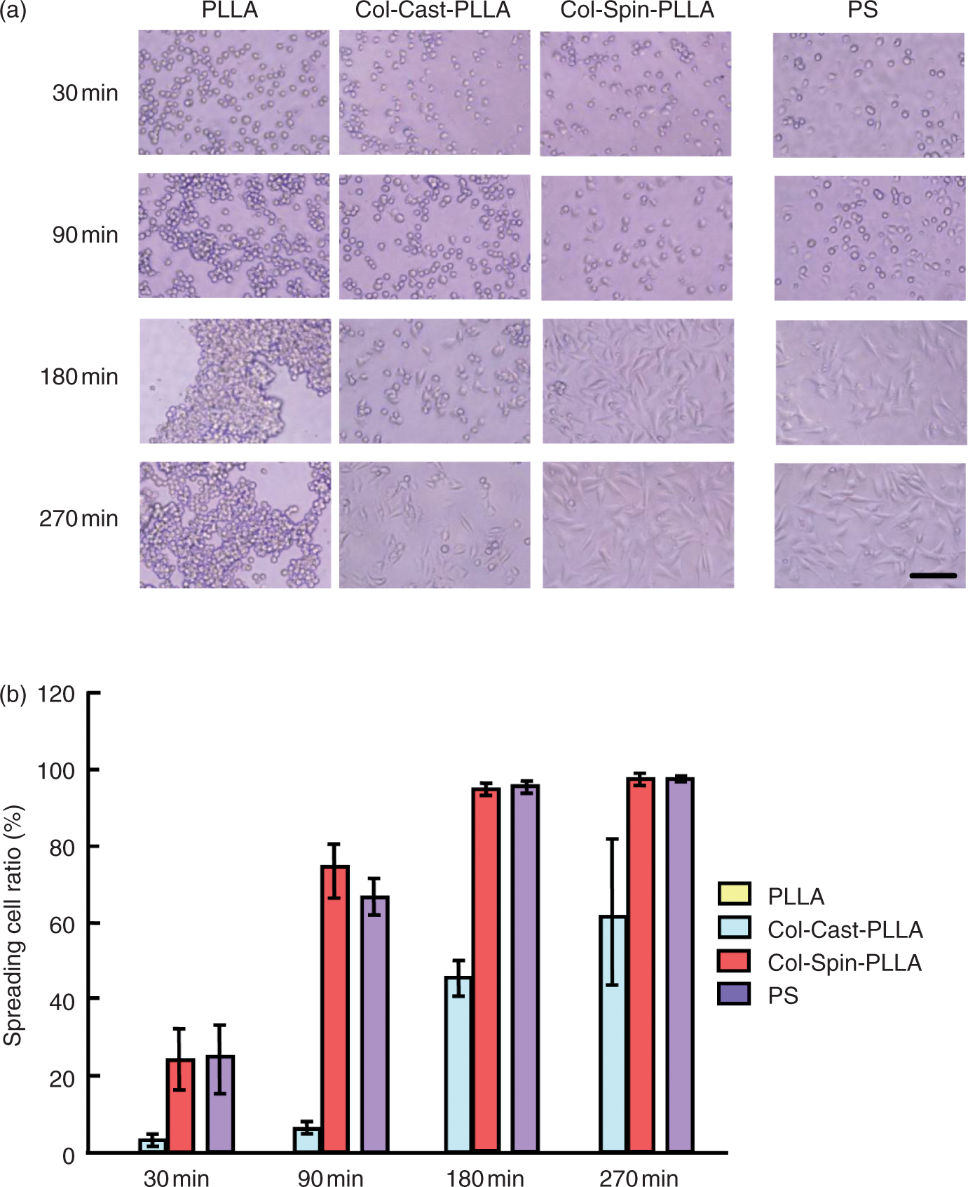
Analysis of cell spreading ratio with time
The attachment assay was carried out using the 24-well dish prepared with PLLA, Col-Cast-PLLA, Col-Spin-PLLA or untreated 24-well dish (PS) as described above. 1.5 × 10⁵ NIH3T3 cells in 500 µL of DMEM medium were re-plated and incubated under a humidified atmosphere containing 5% CO2. Cells were viewed under a phase-contrast microscope, and random fields were photographed after 30, 90, 180, and 270 min at a total magnification of 200×. We defined cells that had failed to spread as phase-bright and rounded. 18 By contrast, spread cells were not phase-bright, exhibited extensive membranous protrusions, and lacked a rounded morphology. 18 The numbers of spread and rounded cells were counted in three fields in triplicate. The spreading cell ratio was calculated and presented graphically.
Immunofluorescence of the cells on actin filaments
The NIH3T3 cell line was incubated in DMEM containing 10% fetal bovine serum and 1% trypsin-streptomycin for 4.5 h after the cells were seeded on the PS, PLLA, Col-Cast-PLLA, or Col-Spin-PLLA. The medium was removed from the dish and the cells were washed with PBS. Cells were then fixed with 4% p-formaldehyde (Wako Chemical Co.) for 1 or 2 h at room temperature. After quenching in p-formaldehyde with 50 mM NH4Cl for 20 min, the dish was washed with PBS three times for 10 min each. The cells were incubated in a 1% BSA PBS solution containing 1/200 Alexa Fluor®594 phalloidin for 45 min. After three washings with PBS, the cells were stained with 1/1000 of the DAPI PBS solution (1 mg DAPI was dissolved in a 1 mL methanol solution 2 µL was put into the 2 mL of PBS solution) and the morphology of the actin filaments was observed.
Heterofunctionality of the Col-Spin-PLLA nanosheet
We used a PVA sacrificial film method for evaluating the heterofunctionality of the Col-Spin-PLLA nanosheet. Initially, we spin-coated 2.0 wt% PVA at 4000 rpm for 20 s and dried on a hotplate at 80°C and the Col-Spin-PLLA nanosheet was prepared on a PVA sacrificed film. Then, the PVA layer was dissolved in distilled water and the floating Col-Spin-PLLA membrane adhered to the Ecoli petri dish and dried in air for 3 h. To evaluate the heterofunctionality of the nanosheet, the edge of the nanosheet was folded. Confluent NIH3T3 cells in 10-cm PS dish were detached and 25% of the confluent cells were seeded on the nanosheet and cultured for 16 h.
Results and discussion
The nanosheets were prepared with a spin-coater (Figure 1). The thickness of the nanosheet was adjusted by the rotating speed and/or the concentration of the PLLA solution. It was initially difficult to coat the hydrophilic collagen homogeneously on the hydrophobic PLLA nanosheet because the collagen aqueous solution is repelled. Then, we diluted the collagen solution (Atelo Cell 1AC-30 Native collagen Bovine dermis, KOKEN, Tokyo) to 0.5 mg/mL with 70% ethanol in order to increase the affinity of the collagen solution for the PLLA surface. These nanosheets are suitable for decal transfer to other substrates via a polyvinyl alcohol (PVA) cast membrane as a supporting film (Figure 2). Using the ethanol solution, we coated collagen homogeneously onto the PLLA surface and prepared three kinds of nanosheet samples: PLLA, Col-Cast-PLLA (where collagen was cast on the PLLA), and Col-Spin-PLLA (where collagen was spin-coated on the PLLA). For evaluating the correlation between morphological change of collagen and cell adhesiveness, we determined the thickness, surface morphology, and contact angle, which is thought to be closely related to collagen morphology.
Preparative scheme of nanosheets: (a) PLLA nanosheet and (b) Col-Spin-PLLA nanosheet. Transference of a PLLA nanosheet to a PS dish.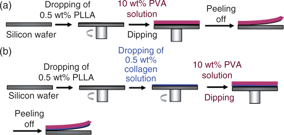
We used atomic force microscopy (AFM) and ellipsometry to analyze the surface morphology and thickness of each nanosheet. The AFM results are shown in Figure 3(a). Both the PLLA and Col-Spin-PLLA samples formed a smooth surface, whereas Col-Cast-PLLA showed some roughness. This roughness is likely to be caused by deposition of collagen from the 70% ethanol solution due to the water enriched condition in the late stage of the dry-in-air process. The ellipsometric data also supported the proposition that a homogeneous collagen layer was generated by Col-Spin-PLLA. The thickness of the PLLA nanosheet was 59.5 ± 9.5 nm, whereas Col-Spin-PLLA and Col-Cast-PLLA had a thickness of 67.1 ± 5.2 nm and 180.2 ± 62.5 nm, respectively (Figure 3(b)). The difference in thickness between PLLA and Col-Spin-PLLA suggested a 5–10 nm thick collagen layer was formed on the PLLA nanosheet. The spin-coater is thought to instantaneously generate an ultra-thin homogeneous layer of hydrophilic collagen on the hydrophobic PLLA surface. Unevenness in the collagen layer is derived from differences in the distribution of collagen density during the dry-air-process on Col-Cast-PLLA. Surprisingly, there was no collagen fibril on both Col-Cast-PLLA and Col-Spin-PLLA, which was generally seen on the type-I-collagen substrates. This result suggests that the collagen is denatured in both Col-Cast-PLLA and Col-Spin-PLLA (Figure 3(a)). Taken together, the AFM and ellipsometry results indicate that the preparation of a homogeneous nanosheet covered with an ultra-thin collagen layer is feasible using a spin-coating rather than casting methodology.
(a) AFM images of nanosheets prepared on silicon wafers: A; top view (left; silicon wafer, right; nanosheet, scale bar: 10 µm), B; cross-sectional images. (b) Thickness of nanosheets prepared on silicon wafers measured by ellipsometry. Mean values ± SD of three measurements are shown.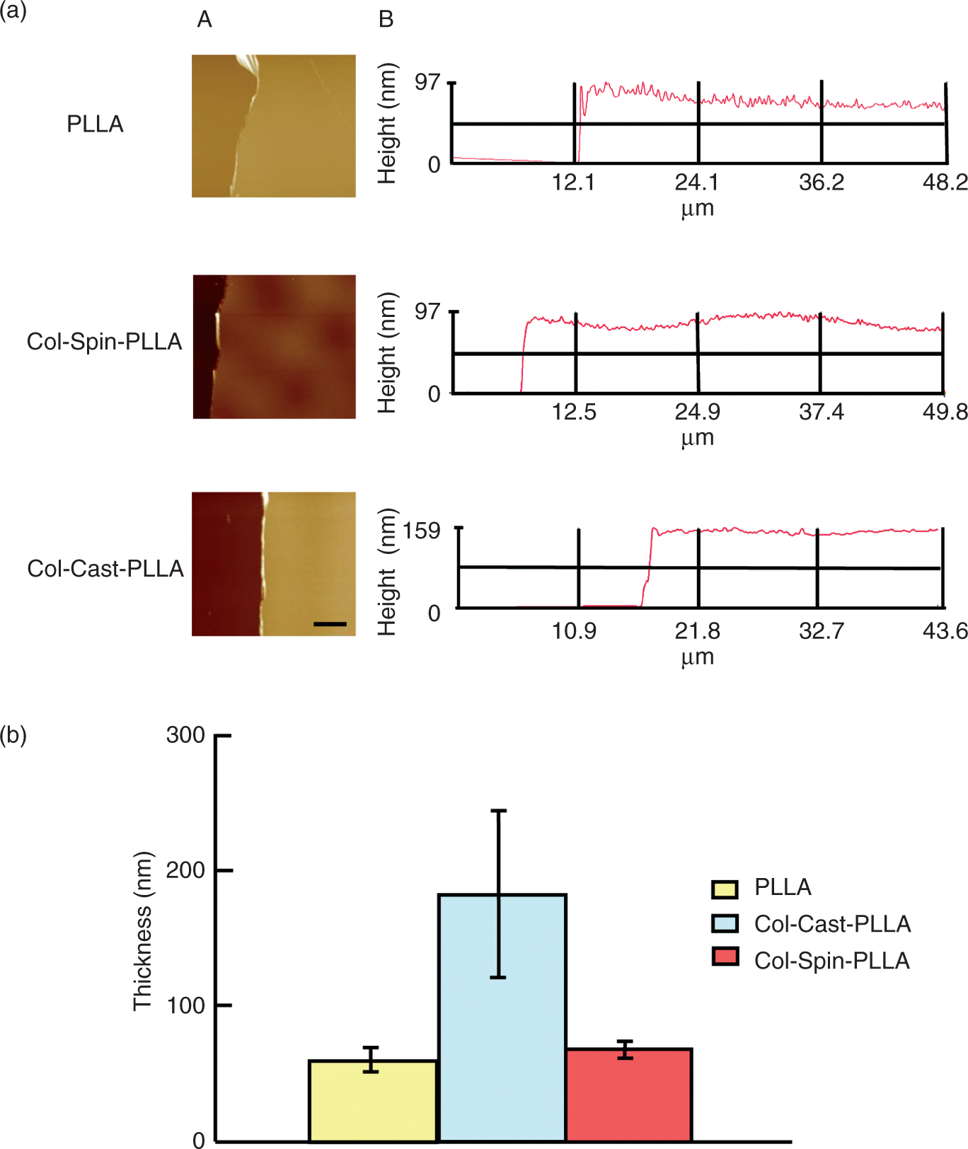
The wettability of each nanosheet was evaluated by measuring the contact angle as outlined in ‘Materials and methods’ section. The Col-Spin-PLLA (53.1 ± 1.06°) showed the highest hydrophilicity, whereas Col-Cast-PLLA (72.3 ± 1.39°) showed almost the same hydrophobicity as PLLA (71.3 ± 0.22°) (Figure 4). As a reference, PS showed a higher hydrophilicity (36.5 ± 1.46°) than Col-Spin-PLLA. The high hydrophilicity of Col-Spin-PLLA compared with PLLA indicates the coating effect of collagen on the PLLA surface is completely different from that of Col-Cast-PLLA.
The contact angles of water on various substrates. Data are expressed as means ± S.E. mean of values obtained from three different fields.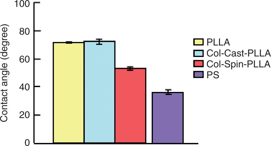
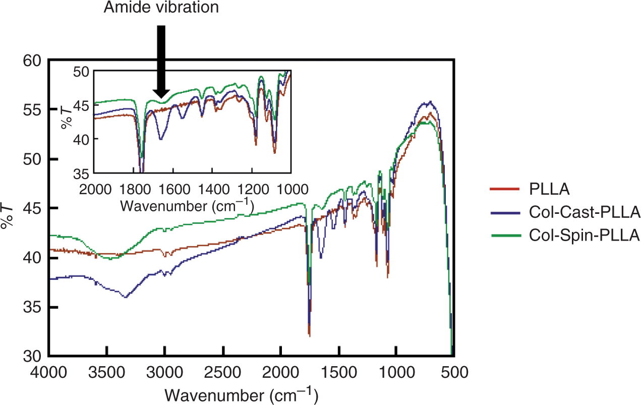
AFM measurements were also conducted on the nanosheet samples at 0, 1.0, and 4.5 h after incubation under the same medium conditions as used for cell cultivation. Noteworthy was the appearance of many collagen fibrils on the Col-Cast-PLLA after 1.0 h incubation, which was not apparent for the PLLA and Col-Spin-PLLA preparations. Moreover, it was also clarified that the fibrils grew during incubation for 4.5 h (Figure 5). The unevenness of Col-Cast-PLLA is due to the formation of collagen fibrils as mentioned above. However, there was no fibril formation on Col-Spin-PLLA even after 4.5 h incubation, although the amide vibration mode of collagen was detected at around 1640 cm−1 derived from the amide I peak of collagen in IR spectrum (Figure 6), suggesting the presence of procollagen as a precursor of collagen fibrils. Liang et al. reported that collagen morphology was induced by dewetting and self-assembly.
19
The collagen layer was less rough after fast drying than after slow drying. The spin-coating method is considered to be a fast drying process, which leads to the formation of smaller and more filamentous collagen fibrils. Furthermore, we believe that the hydrophobic interaction between collagen and the PLLA nanosheet is too strong to allow formation of collagen fibrils. Alternatively, the amount of collagen on the Col-Spin-PLLA is too small to initiate the development of collagen fibrils as evident from the AFM measurements. The hydrophilic RGD moiety of the nonfibril collagen is present on the molecular surface of Col-Spin-PLLA (Figure 5). Indeed, Col-Spin-PLLA is more hydrophilic than Col-Cast-PLLA.
20
Changes in collagen morphology with time. Samples were incubated at 37°C in medium containing 10% FBS (Left; low magnification, Right; high magnification, scale bar: 5 µm). IR spectrum of PLLA, Col-Cast-PLLA and Col-spin-PLLA nanosheet.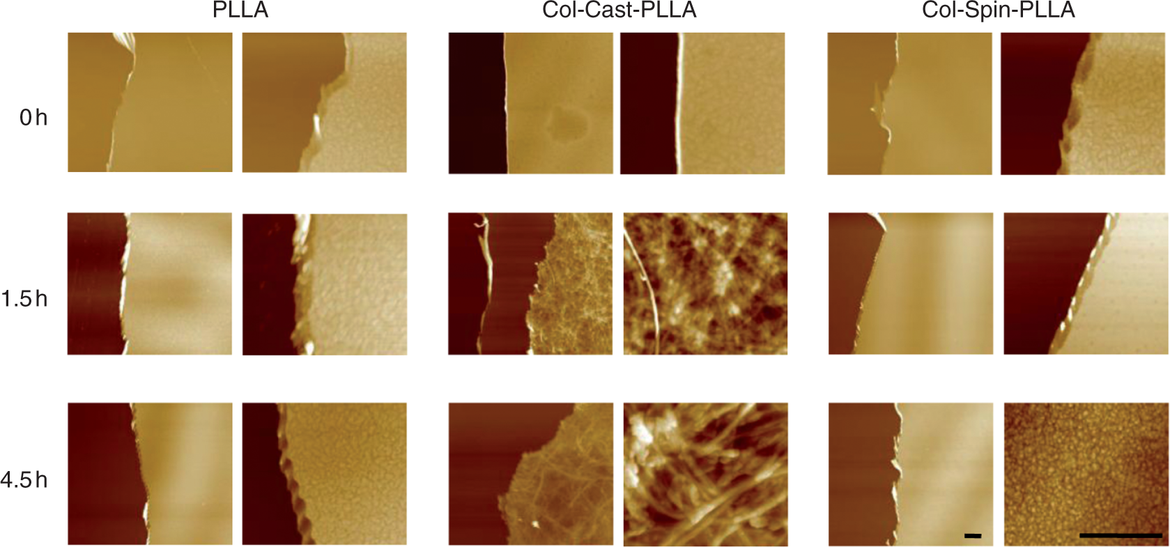
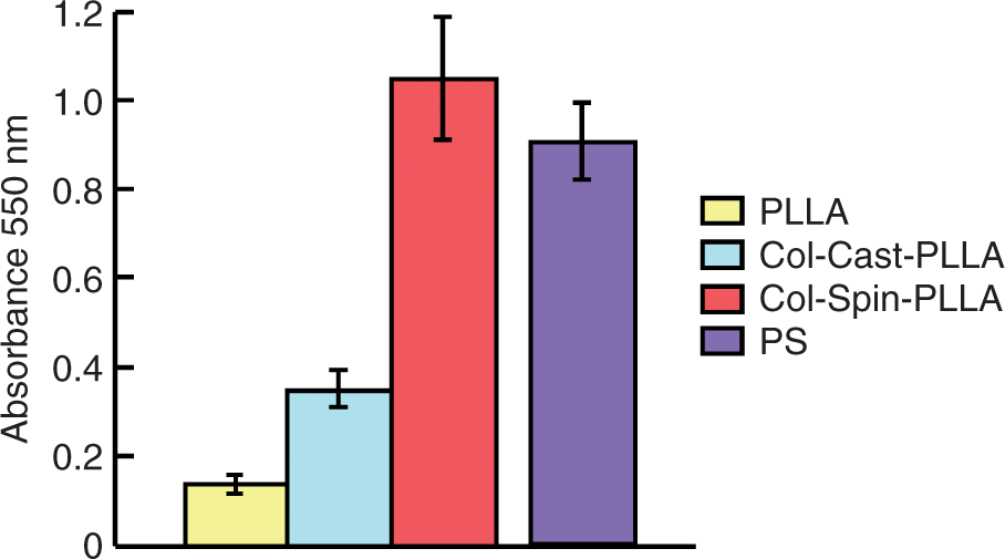
We studied the cell behavior on the nanosheet samples using a NIH3T3 cell line. The appearance of NIH3T3 cells on each sample was evaluated by using Fluo3-AM (Figure 7(a)). The cells on both PLLA and Col-Cast-PLLA did not spread, but instead maintained a rounded morphology (tethering). The majority of cells were confirmed to be strongly attached to Col-Spin-PLLA, where they adopted an elongated shape. The commercially available PS culture dish gave almost the same number of attached cells in comparison with Col-Spin-PLLA. Quantification of the data using Image J software (Figure 7(b)) indicated that the order of the contact area of cells on the nanosheet sample was Col-Spin-PLLA≧PS>Col-Cast-PLLA =PLLA. This result was confirmed using the MTT assay (compare Figure 8 to Figure 7(a)). Specifically, the Col-Spin-PLLA nanosheet showed the greatest amount of cell attachment, with Col-Cast-PLLA having less than half that of Col-Spin-PLLA, and PLLA the lowest. As a reference, the commercial PS surface showed a slightly lower level of cell attachment than that of Col-Spin-PLLA. Furthermore, we studied the early stage of cell attachment at 30, 90, 180, 270 min after seeding in order to analyze the rate of cell adhesion (Figure 9). Cells were photographed at the appropriate time, and the numbers of spread and rounded cells were counted. After 30 min, almost all cells showed a rounded shape on Col-Cast-PLLA, whereas as many as 25% of cells had begun to spread on the PS and Col-Spin-PLLA. After 90 min, >90% of cells still showed rounded morphology on Col-Cast-PLLA, whereas > 60–70% of cells had begun to spread on Col-Spin-PLLA and PS. By 180 min most of the cells had spread and formed membranous protrusions on Col-Spin-PLLA and PS, whereas approximately half of the cells still showed rounded morphology on Col-Cast-PLLA. Indeed, even after 270 min approximately 40% of cells on Col-Cast-PLLA had failed to spread. For PLLA, the cells neither attached nor spread throughout the observation period and had aggregated with each other by 270 min. In the early stage of cell attachment, the number of spreading cells showed the following order: Col-Spin-PLLA≧PS>>Col-Cast-PLLA>PLLA. From the above data, the amount of spreading cells is closely related to the collagen morphology. Specifically, the number of spreading cells was considerably greater for Col-Spin-PLLA in comparison with that for Col-Cast-PLLA. Thus, the spin-coating method generates a better cell adhesive surface compared with PLLA or Col-Cast-PLLA.
(a) Image of fluo3-AM stained NIH3T3 cells deposited on the various samples (scale bar: 50 µm). (b) Cell spreading area on the various samples. The amount of cell attachment at 1-h incubation after washing the various substrates with PBS. NIH3T3 cells were detached from the culture dish and re-suspended in fresh medium containing 10% FBS. After 1 h incubation, the substrates were washed with PBS and cells were incubated for 5 h in fresh medium containing MTT (3-(4,5-dimethyl-2-thiazonyl)-2,5-diphenyl-2H tetrazolium bromide. 5 mg/mL). Then the supernatants were decanted, and the formazan precipitates were solubilized by the addition of 450 µL of 100% DMSO and placed on a plate shaker for 10 min. Absorbance at 550 nm was determined on a microplate reader. Living cell number was proportional to the absorbance of MTT at 550 nm. (a) Spreading cell ratio on the various substrates. NIH3T3 cells were detached from their culture dishes and re-plated in fresh medium containing 10% FBS. The cells were then allowed to adhere and spread at 37°C for 30 min, 90 min, 180 min or 270 min, at which times they were photographed with the use of a phase-contrast microscope and the numbers of spread and rounded cells were counted. The results are representative of three separate experiments (scale bar: 100 µm). (b) The spreading cell ratio with time was calculated and is shown graphically.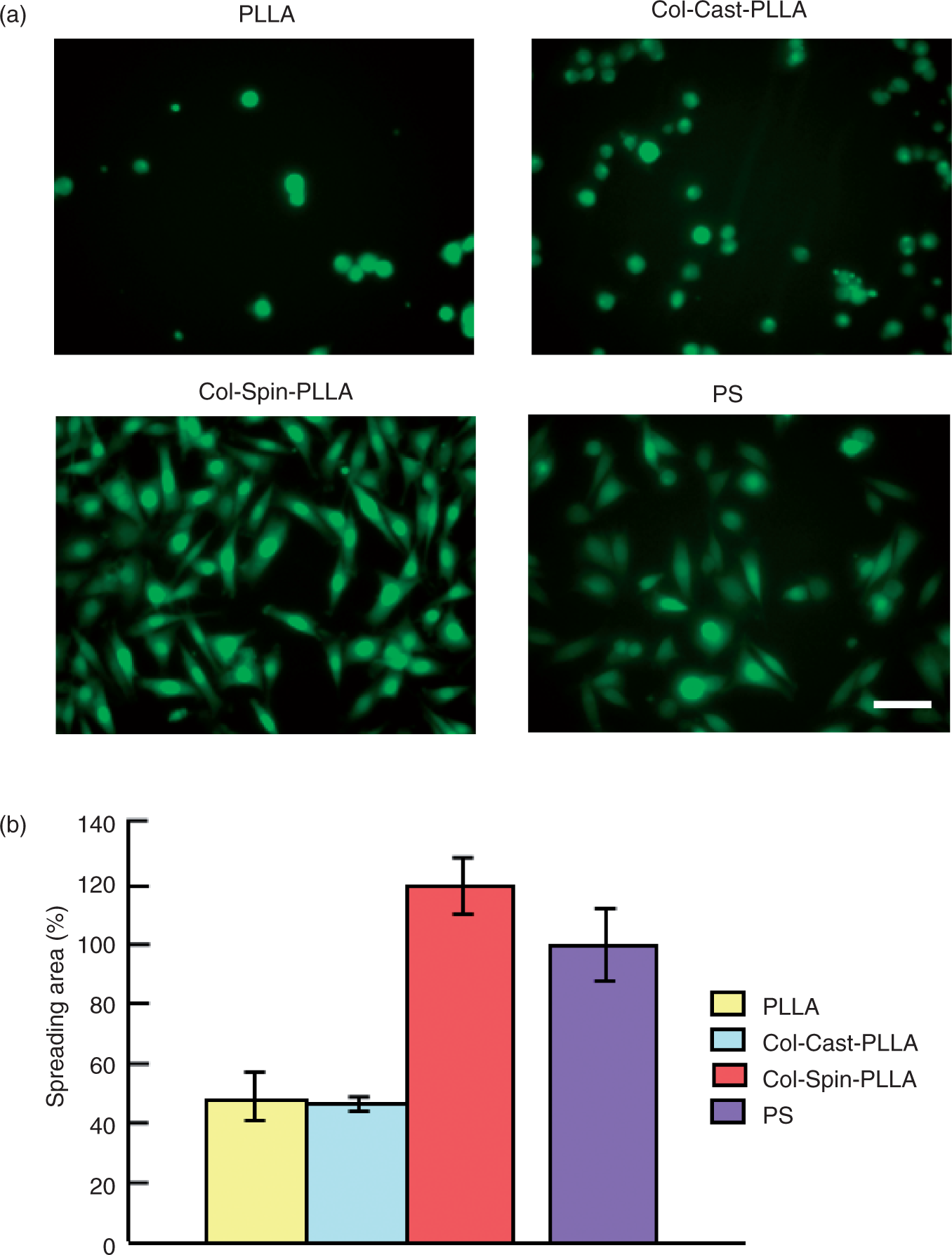
In order to obtain information concerning focal adhesion, we observed fluorescent-labeled actin filaments inside the cells to evaluate cell adhesiveness with each surface (Figure 10). There was no detectable fluorescence intensity in the case of PLLA due to the lack of cell adhesion after the extensive washing step in the immunostaining procedure. The elongation of actin filaments was barely observed for the Col-Cast-PLLA sample. However, quite good elongation was observed on Col-Spin-PLLA, indicating a reasonable biological affinity with the cells. Intriguingly, PS as a control seemed to show poorer actin filament elongation than Col-Spin-PLLA.
Actin filaments of the NIH3T3 cell line were stained using phalloidin 4.5 h after seeding (40× image). The cell nucleus was stained with DAPI (scale bar: 50 µm).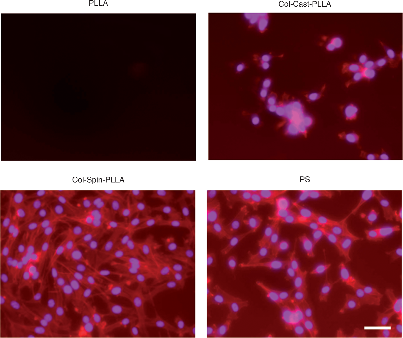
Taken together, our results show that cell adhesive properties tend to be closely related to collagen morphology. Indeed, our results are in good agreement with previous reports, which showed that the cells expand on the films of native collagen with a lower density of large fibrils.21,22 These earlier papers suggest that collagen fibrils control excessive proliferation of cells. 21 – 25 The differences in cell behavior between Col-Cast-PLLA and Col-Spin-PLLA were reproduced in our findings described here. Daniel et al. reported that the thermal or proteolytic denaturation of collagen I unwinded the triple-helical structure to expose RGD-motifs. 26 – 29 RGD-motifs trigger the binding of α5β1- and αv-integrins, which signals the initiation of cellular processes such as adhesion, spreading, motility, and differentiation. Our results from collagen morphology and contact angle measurements show that nonfibril Col-Spin-PLLA is more hydrophilic than fibril Col-Cast-PLLA. Our contact angle result of Col-Spin-PLLA showed almost the same value as RGD-immobilized PLLA film. 30 Hence, the RGD moiety might be exposed in Col-Spin-PLLA. Therefore, the Col-Spin-PLLA nanosheet appears to mimic the situation during wound repair. 26
Finally, we prepared a heterofunctional free-standing PLLA nanosheet with a spin-coated collagen side and an uncoated side. The sacrificial film was used for the convenient collection and manipulation of the nanosheets.
3
Next, we attached cells to the nanosheet with the collagen side on top and the uncoated PLLA side exposed by folding the edge of the nanosheet. There was no cell attachment on the folded area, while cells were confluent on the collagen side after 16 h incubation (Figure 11). The extensive formation of actin filaments was confirmed inside the cells on the collagen side of the nanosheet. Therefore, we were able to prepare a heterofunctional nanosheet that controls cell adhesive properties. Moreover, cell attachment was confined specifically to one side of the nanosheet.
Heterofunctionality of the Col-spin-PLLA nanosheet. The PLLA side (a), and Col side (b) of the Col-Spin-PLLA, Upper side of (c); PLLA side of the folded Col-spin-PLLA nanosheet, down side of (c); Col side of Col-spin-PLLA nanosheet on the E-coli culture dish.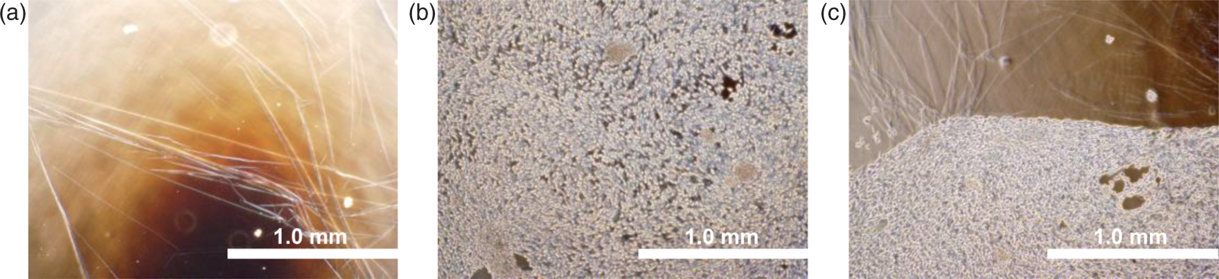
Conclusion
In conclusion, we have established a technology, using a modified spin-coating process, to homogeneously coat collagen onto a noncell adhesive PLLA nanosheet, thereby furnishing the surface with cell adhesive properties. The development of a heterofunctional nanosheet will be extremely valuable for the generation of novel biomaterials, such as post-surgical wound dressings. Specifically, these nanosheets have cell adhesive properties on one side, for attachment to the wound, and a nonadhesive surface on the other side, which acts as an adhesion barrier. Additional in vivo experiments are currently being conducted to further evaluate these heterofunctional nanosheets.
Footnotes
Acknowledgement
This work was partially supported by GCOE ‘Practical Chemical Wisdom’ (D.N.), ‘Scientific Research (B) 21300181’ (S.T., N.G.), ‘High-Tech Research Center’ Project for Waseda University (S.T., N.G.): matching fund subsidy from Mext, Japan.