
Abstract
The present research focused on determining the effect of hydroxyapatite-20 wt% mullite (H20M) particle eluates on apoptosis and differentiation of human fetal osteoblast (hFOB) cells. The H20M particles (257 ± 37 nm) were prepared, starting with the production of a nanocomposite using a unique route of spark plasma sintering, followed by a repeated grinding-cryo treatment and elution process. Tetrazolium based cytotoxicity assay results showed a time- and dose-dependent effect of H20M particle eluates on hFOB cytotoxicity. In particular, the results revealed statistically reduced cell viability after hFOB were exposed to the above 10% H20M (257 ± 37 nm) eluates for 48 h. The apoptotic cell death triggered by H20M treatment was proven by the analysis of molecular markers of apoptosis, that is, the Bcl-2 family of genes. hFOB expression of Bcl-xL and Bcl-xS significantly increased 25.6- and 25.2-fold for 50% of H20M concentrations, respectively. The ratio of Bcl-xL/Bax (4.01) decreased 2-fold for hFOB exposed to 100% of H20M eluates than that for 10% H20M eluate (7.94) treated hFOB cells. On the other hand, the Bcl-xS/Bax ratio for the 10% H20M eluate was 4.15-fold, whereas for 100% H20M eluates, it was 11.55-fold. Specifically, the anti-apoptotic effect of the H20M particle eluates was corroborated by the up-regulation of bone cell differentiation marker genes such as, collagen type I, cbfa, and osteocalcin. In summary, the present work clearly demonstrated that H20M submicron to nanometer composite particle eluates have a minimal effect on hFOB apoptosis and can even up-regulate the expression of bone cell markers at the molecular level.
Keywords
Introduction
There are numerous applications for nanostructured biomaterials in tissue engineering and in human health care pertaining to their unique properties.1–3 In particular, nanostructured metals, ceramics, polymers and their composites have been studied for various orthopedic applications to repair and regenerate human tissues. For orthopedic applications, hydroxyapatite (HA) based nanomaterials have been some of the most preferred materials due to their unique biocompatibility and bioactivity properties. The development of nanomaterials (or materials with constituent dimensions less than 100 nm in at least one direction) for orthopedic applications is driven by the following factors: (a) increased cellular functionality that can be expected due to the presence of a large volume fraction of grain boundary, 4 (b) the enhanced physical properties (hardness, strength) of nanomaterials, and (c) the ability of nanomaterials to mimic constituent components of natural bone, which also contains nanosized HA particles. Suffice to say, it has been broadly reported that materials with coarse or micron sized grain sizes have different properties than nanometer sized particles of the same chemical composition, which has led to the exploitation of biomaterials in numerous orthopedic applications. 5 Moreover, the size differences have a significant effect on cellular behavior and gene expression. Here, we are interested in studying the changes at the cellular and genetic level, when osteoblasts are exposed to wear debris particles generated inside the body during wear at the implant site.
In our previous study, bulk hydroxyapatite-mullite (HM) composite materials exhibited enhanced in vitro cell adhesion, cell viability (fibroblast) and osteogenic differentiation with osteoblasts (bone forming cells), as compared to monolithic HA. 6 In another study, the size and chemical composition of nano/submicron HA-20% mullite (H20M) eluates governed the cytotoxicity and genotoxicity properties for L929 fibroblasts. 7 It was concluded that above a 25% concentration of submicron to nanometer H20M in biomaterial eluates, toxicity via DNA damage resulted, as measured using a comet assay. It, however, remained to be investigated whether signaling mechanisms in genotoxicity was only due to the fact that these were nanoparticles or was due to the HM chemical composition. It was also not clear how the fraction of live, apoptotic and necrotic cells in H20M-treated osteoblast cells evolved with respect to time and dose. This requires cell sorting in combination with a fluorescent activated cell sorter (FACS). Although a number of biomaterials are being developed with a goal to obtain a desirable combination of physical and biocompatibility properties,8–11 cytotoxicity analysis involving apoptosis/necrosis of such biomaterials are limited.12,13 Also limited is our understanding of how specific genes are expressed during the differentiation process of bone cells, when treated with biomaterials. 14
Among only a few studies, Setzer et al. investigated the role of surface roughness of Ti/ZrO2 on the gene expression of hFOB 1.19 osteoblast cells. Their study revealed a greater influence of surface roughness than material substrate composition on gene expression during the initial phase of cell attachment and proliferation. 11 In a different study, Ye et al. reported a time- and dose-dependent expression of p53 and Bax genes in nano-SiO2-treated cells, while the expression of the Bcl-2 gene did not significantly increase after exposure to nano-SiO2. 15 None of these studies reported the effect of biodegradable products of biomaterials (such as HA) on cellular apoptosis/necrosis as well as specific gene expression.
In the present work, an attempt was made to study the concentration and time dependent effects of submicron to nanometer HM composite exposure on cytotoxicity, apoptosis, genetic expression of apoptosis, and bone cell differentiation markers by quantitative real time gene expression in a human osteoblast cell line (hFOB). In particular, as a continuation of our previous study on the genotoxicity and cytotoxicity of H20M nano/submicron eluates on L929 cells, the current work focused on the molecular dynamics of the Bcl-2 family of genes and bone cell specific markers cbfa, OCN (osteocalcin) and COL1 (collagen type I). Together, such studies have a broad relevance for the potential application of H20M materials in load-bearing orthopedic implants.16–18 Therefore, a design criteria of these experiments was to study the response of H20M nano/submicron eluates on an osteoblast cell line, as clearly such bulk materials may degrade during extended use. Most importantly, the results of this study demonstrated that submicron to nanometer H20M-treated osteoblasts could exhibit a concentration dependent up regulation of cbfa, COL1 and OCN, suggesting a range of suitable concentrations with no toxicity, and thus promise for the further study of H20M for orthopedic applications. The results of the present work is highly relevant as the use of HM composites as bone replacement materials would lead to the release of fine particles in vivo due to friction and wear and the effects of such particles on biological system biological are therefore of immediate concern.
Materials and methods
Powder synthesis
For the present work, HA was synthesized by the widely reported solution-precipitation method.
19
HA preparation followed the previously published work of Basu and co-workers.20,21 Briefly, the precursor material for the HA was calcium oxide (CaO) and phosphoric acid (H3PO4):

Commercially available phase pure mullite (KCM, Japan) was used as second phase in the production of the composites and the H20M composition was ball milled for 16 h in a wet medium (acetone).
An appropriate amount of dried powder mixtures was placed in a graphite die having a ∼15.4 mm internal diameter and proper dressing with a graphite sheet inside the die to avoid contamination. This entire die assembly was placed inside the Spark Plasma Sintering chamber (SPS Syntex INC, model: SPS-515S, Kanagawa, Japan). A vacuum of 50 torr and a pressure of 30 MPa were maintained throughout the experiment. A DC current of 0.5–1.5 kA and a DC voltage of 5–10 V were applied during the SPS experiments. The powder mixture was sintered at 1100°C with a heating rate of 100°C/min and a soaking time of 5 min. The temperature during the entire SPS processing was monitored by focusing an optical pyrometer on the graphite die. The final thickness of the spark plasma sintered HM (20 wt%) composite was around 2–3 mm. The removal of the graphite sheet around the sample was ensured before further characterization.
The density, measured using Archimedes’s principle, revealed the attainment of near full density (>99.5%
Preparation of H20M particles
In a new approach, the spark plasma sintered H20M biocomposite was initially ground using a mortar and pestle and the obtained powders were ball milled for 20 h at 300 rpm using agate balls (3 mm size) with the jar one-third filled with toluene to serve as a milling medium. At each ball milling cycle, 3 g of powder were taken and the ball to powder weight ratio was kept at 25:1. Subsequently, the repeated cycles of freezing and thawing using liquid nitrogen (−196°C) was adopted to induce brittleness in the powder sample between two consecutive ball mill cycles. After tailoring the above mentioned process sequence, particle size stocks were prepared (Figure 1), which were further used for the cytotoxicity, apoptosis, and gene profiling assays.
Schematic of the preparation of biomaterial submicron to nanometer eluates.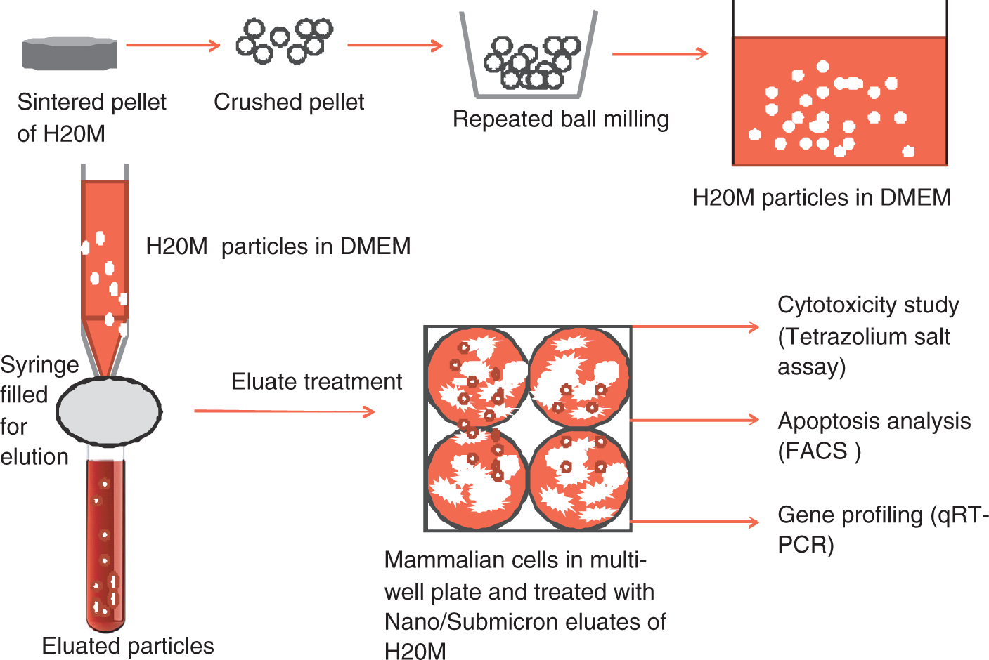
Preparation of biomaterial eluates
The as-prepared H20M powders were sterilized by autoclaving at 121°C for 15 min. A 100 mg of sterilized submicron to nanometer H20M powder were added into Dulbecco’s modified Eagles medium (DMEM) supplemented with a 1% antibiotic (penicillin, streptomycin, and amphotericin) cocktail. The prepared H20M solution was rocked on a gel rocker (continuous/reciprocal) for 5 days. After 5 days of the elution process, the culture medium was filtered with 0.45 µm Millipore filters. 100% of the filtered biomaterial eluate was stored at −20°C and the obtained eluate was considered as 100% after filtration for the following studies.
Particle size analysis
The particle size of the H20M eluates were measured using dynamic light scattering (DLS, Zetasizer Nano-ZS model equipped with 4.0 mW, 633 nm laser, Model ZEN3600, Malvern Instruments Ltd., Malvern, UK). The parameters used in the measurements were viscosity of 0.34 poise, reflective index of 1.054 and temperature of 25°C. The particle distribution size was further confirmed by a transmission electron microscope (TEM, JEOL 2010FS, Japan). For TEM observations, the samples were prepared by drop-coating on copper grids and the samples were dried overnight at 100°C. TEM analysis was carried out at an accelerating voltage of 200 kV.
Osteoblast cell line
In this study, a human osteoblast cell line (hFOB 1.19; ATCC No. CRL-11372) was used for cytotoxicity, apoptosis and gene profiling studies. The as-received cryo-vial was rapidly thawed and cultured in DMEM, supplemented with 10% fetal bovine serum (Sigma Aldrich) and a 1% antibiotic cocktail (10,000 IU penicillin, 10 mg streptomycin and 25 µg amphotericin /mL) (Sigma Aldrich) at 37°C in 5% CO2 humidified atmosphere. The exponentially growing cells were used for in vitro experiments. For this, a 90% confluent monolayer was trypsinized using a 0.25% trypsin EDTA solution (Sigma Aldrich).
Treatment preparation
The 100% H20M eluate was used for cytotoxicity, apoptotic cell death and gene profiling studies. 100% pure sterilized H20M eluates were diluted in cell culture medium to obtain eluates at doses of 10%, 25%, 50%, 75%, and 100%. As further described below, MTT, FACS, and real time gene experiments were performed at the above mentioned concentrations. The hFOB cells without treatment were used as a negative control for MTT, FACS and qRTPCR. 1 mM Camptothecin (Sigma Aldrich) was used as a positive control for the FACS assay for apoptosis.
Mitochondrial activity (cytotoxicity assay)
The first indication of material toxicity is generally observed by a cytotoxicity study. 22 It is known that a cytotoxicity study confirms the live/dead status by the formation of formazan crystals by the living cells that correlate with the colorimetric assessment of cell–material cytotoxicity. In view of this, cell viability activity was assessed by the MTT assay. hFOB cells were seeded into a 96-well plate at approximately 1 × 10⁴ and incubated overnight in a CO2 incubator. Following this, hFOB cells were exposed to H20M for eluates of different doses for 12, 24, and 48 h. 100 µL of each dilution of H20M eluates (10%, 25%, 50%, 75%, and 100%) dilution was added into each well. After 12, 24 and 48 h treatment, solution was removed and subsequently, 10 µL of MTT (5 mg/mL) / 100 µL of 1 x PBS was added into each well between image acquisition in a phase contrast microscope to visualize blue formazan crystals. Thereafter, the reactant MTT solution was carefully removed and gently washed with 1 x PBS. Following this, formazan crystals were solubilized by adding 200 µL of DMSO and mitochondrial activity of the viable cells were quantified by measuring absorbance at 540 nm by a spectrophotometer (Molecular Devices).
Fluorescent activated cell sorter by FITC-AnnexinV/PI assay (apoptosis assessment)
To determine apoptosis, hFOB cells were plated in 24-well plates and treated with H20M eluate of various dilutions. FITC AnnexinV/PI double staining was used to differentiate the apoptotic and necrotic cell populations. Standard protocols were followed according to the Annexin V- FITC Apoptosis detection Kit protocol from Sigma (APOAF). Briefly, after seeding, hFOB cells were treated with H20M eluates for 12, 24, and 48 h. Subsequently, cells were washed with ice cold PBS and harvested with 0.25% trypsin and resuspended in 200 µL of a 1X binding buffer. Cell suspensions were incubated at room temperature (RT) for 5 min and subsequently, 10 µL of Annexin V-FITC was added to the cell suspensions. Thereafter, the tube was incubated for 15 min at RT. After incubation, 5 µL of propidium iodide (PI) was added into the cell suspention and apoptosis was conducted by a FACS BD Biosciences Aria uploaded with BD Biosciences DiVa 5.0 software with two different channel filters for PI and FITC. In the FACS unit, the PI was in the channel LP Mirror 655 and BP Filter 695/40 and FITC was in the channel LP Mirror 502 and BP Filter 530/30. While a linear scale was used for the forward and side scatter, the log scale was used for the fluorescence readings. A blue laser at 488 nm was used to read the fluorescent of both the colors.
Total mRNA isolation
hFOB cells were seeded at an approximate density of 7 × 10⁴ cells/well of a 6-well plate and treated with eluates of H20M for 6 h. After the eluate treatment, the cells were washed with 1X PBS (ice cold), then 1 mL of TRIzol (TRIzol® LS Reagent Cat. No. 10296-028) and 0.2 mL of chloroform were added before the cells were scrapped for total RNA isolation. Subsequently, the solution was vortexed, incubated, and centrifuged at 12,000 g for 15 min at 8°C. After the isolation, 0.5 mL of isopropyl alcohol was added for the RNA precipitation and incubated at room temperature for 10 min. Subsequently the samples were recentrifuged at 12,000 g for 15 min at 4°C. An invisible pure RNA pellet was formed on the bottom of the centrifuge tube. Following, precipitated, RNA was washed with 75% ethanol twice. Finally, RNA was air dried and the RNA pellet was re-dissolved in the DEPC (nuclease free water) treated water. RNA purity and concentration was measured at A260/A280.
cDNA synthesis and real time quantitative reverse transcription-polymerase chain reaction
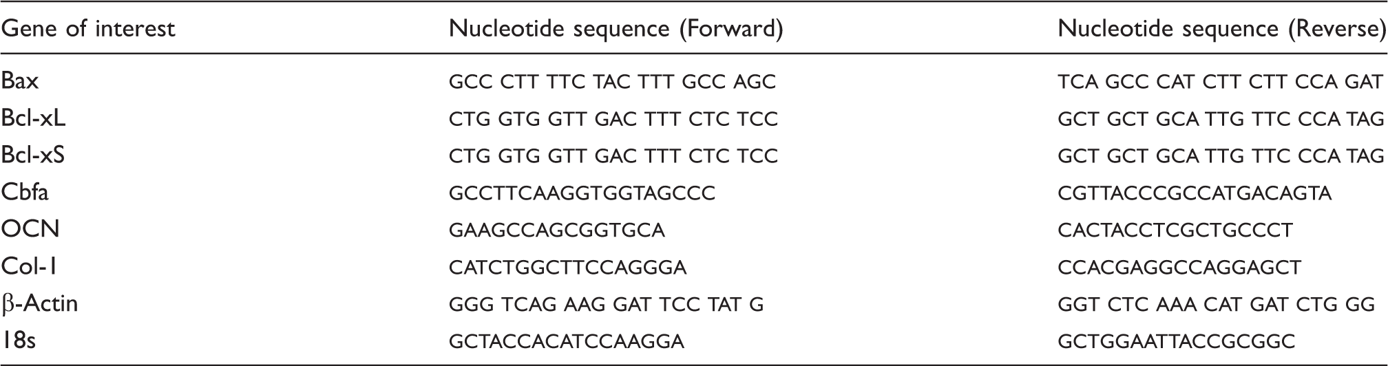
Primer sequence of bone cell, apoptosis, and housekeeping genes used in the present investigation.
Statistical analysis
Statistical analysis was carried out by one-way analysis of variance (ANOVA) using commercial SPSS-13 software. All experiments were run in triplicate (n = 3). Post hoc comparisons were performed using Dunnet’s test and Tukey tests at a statistically significant value of p < 0.053.
Results
Biomaterial eluate characterization
The average hydrodynamic particle size of the H20M eluates, as obtained using dynamic light scattering (DLS) was 257 ± 37 nm (Figure 2(b)). The size distribution revealed that 49% of the particles were in the range of 257 nm, 24% were in the range of 220 nm and 26% were in the range of 295 nm. A representative bright field TEM image illustrates that particles were in a micron to nanometer regime (Figure 2(a)), which corroborated with the particle sizes obtained by DLS. TEM analysis also confirmed that most of the particles were spherical in shape. Some of the particles agglomerated, which possibly increased the size of the H20M eluate particles.
Representative bright field TEM micrograph of the H20M particles prepared in DMEM medium (a) and particle size distribution as measured using dynamic light scattering (b).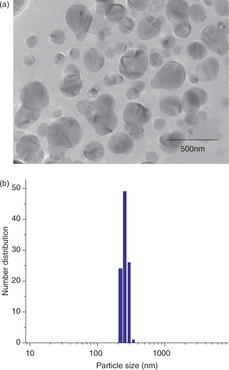
As reported in our recent paper, 26 the XRD analysis of H20M eluates and the as-synthesized H20M powder revealed the presence of HA and mullite. The presence of TCP, CaO, Al2O3, or SiO2 was not detected within the resolution limit of XRD.
Cellular toxicity
The cytotoxicity effects of H20M (257 ± 37 nm) eluates on osteoblasts (hFOB 1.19) were analyzed by determining the mitochondrial activity of living cells. The percent viability of the live cells was calculated with respect to controls (cells without eluate treatment). Figure 3 shows the concentration as well as the time dependent nature of the viability of the H20M treated hFOB cells. Cell viability was above 100% after treatment for 24 h with all the eluate doses, except for the 100% concentration eluate. After 24 h of treatment, the lowest percent viability was 81%, even at 100% H20M eluate concentrations. Moreover, for up to 75% concentration of the eluates, cellular viability was 100% or more with respect to the negative control. After 48 h of treatment, cellular viability reduced, up to 65.14% at 100% of eluate treatment. A stastistically significant (p < 0.05) difference was noticed only at 100% of the eluate treatment for 48 h with respect to the control material, whereas after 12 h of treatment, the percent viability of hFOB cells were statistically (p < 0.05) different at 10%, 25%, and 75% doses of H20M eluates. The absence of any systematic trend with respect to either eluate concentration or treatment time can be attributed to the fact that the cell populations are invariably at different stages of cell cycle/cell growth stage.
MTT analysis revealing the percent viability of hFOB cells, after treatment with HA-20% mullite biomaterial eluates. Cytotoxicity (MTT) of H20M treated hFOB for 12, 24, and 48 h. Note: x-axis: The concentration of H20M eluates. y-axis: Percent viability of cells with respect to the control (untreated cells). *Shows statistical significant differences with respect to control between different concentrations at 12, 24 and 48 h of treatment at p < 0.05. **Shows statistical significant differences with respect to control between 12, 24, and 48 h of treatment at p < 0.05.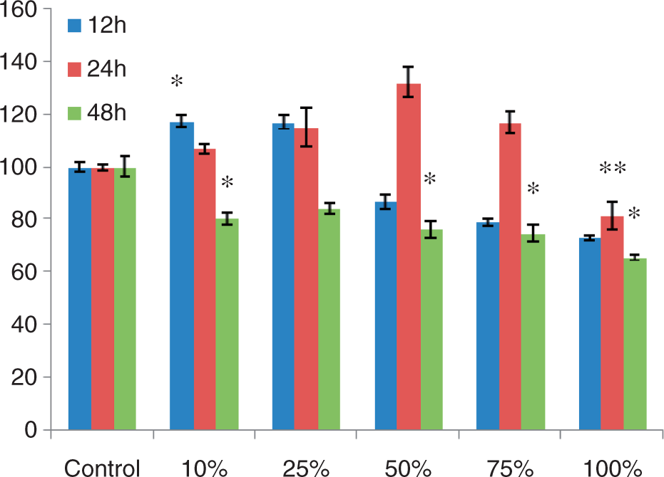
Apoptotic cell death analysis
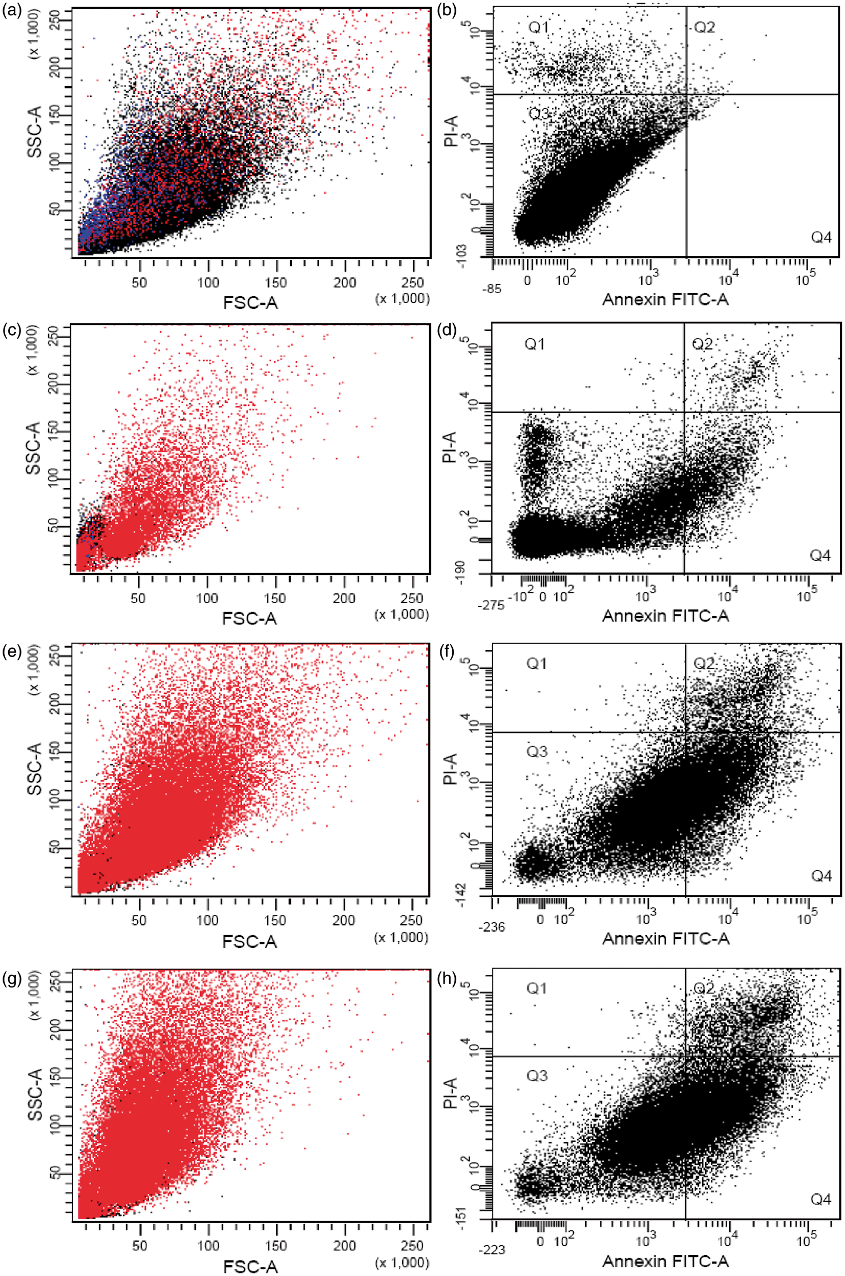
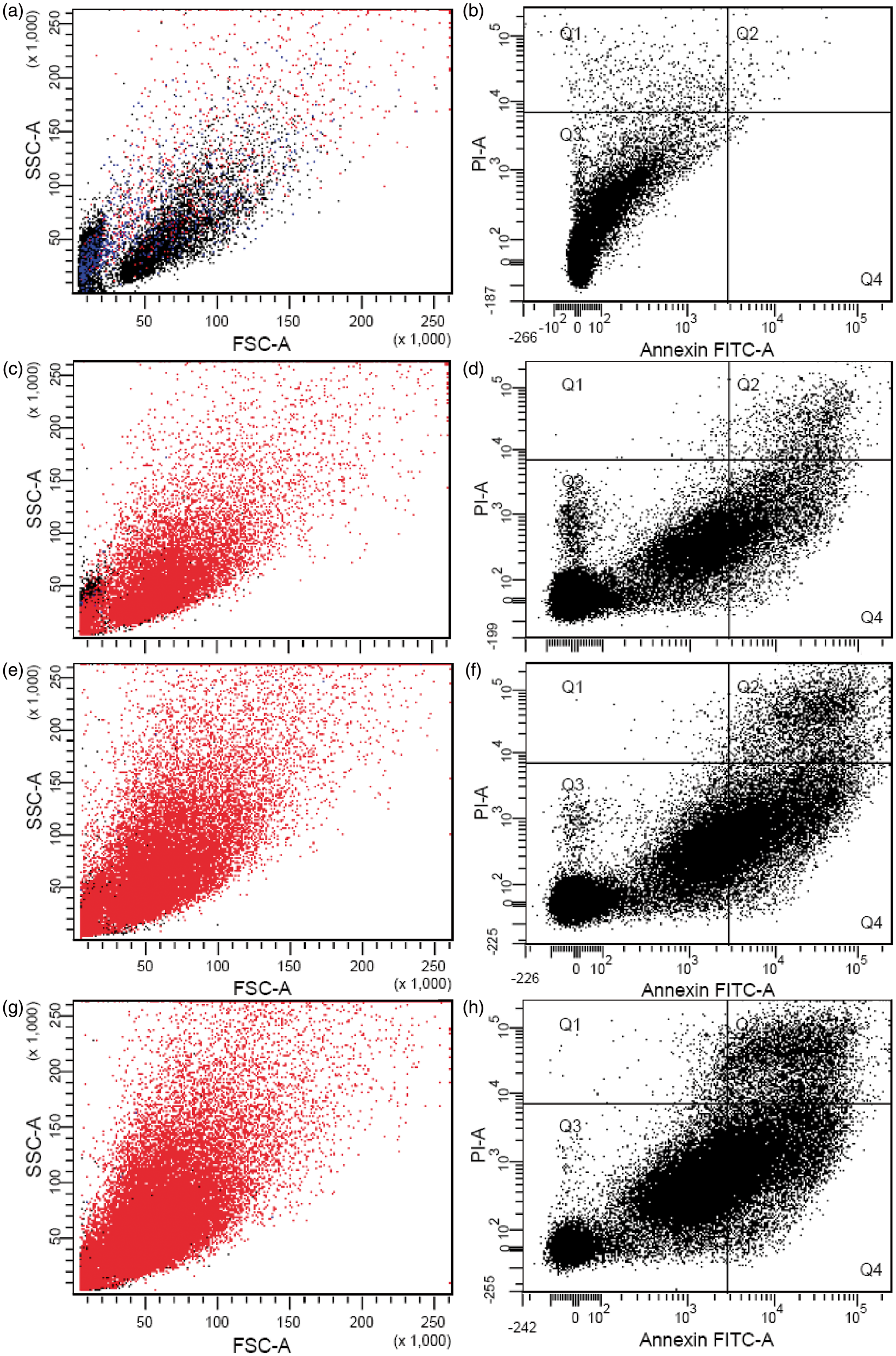
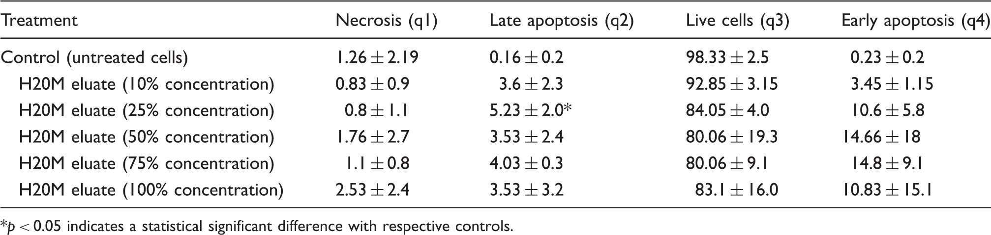
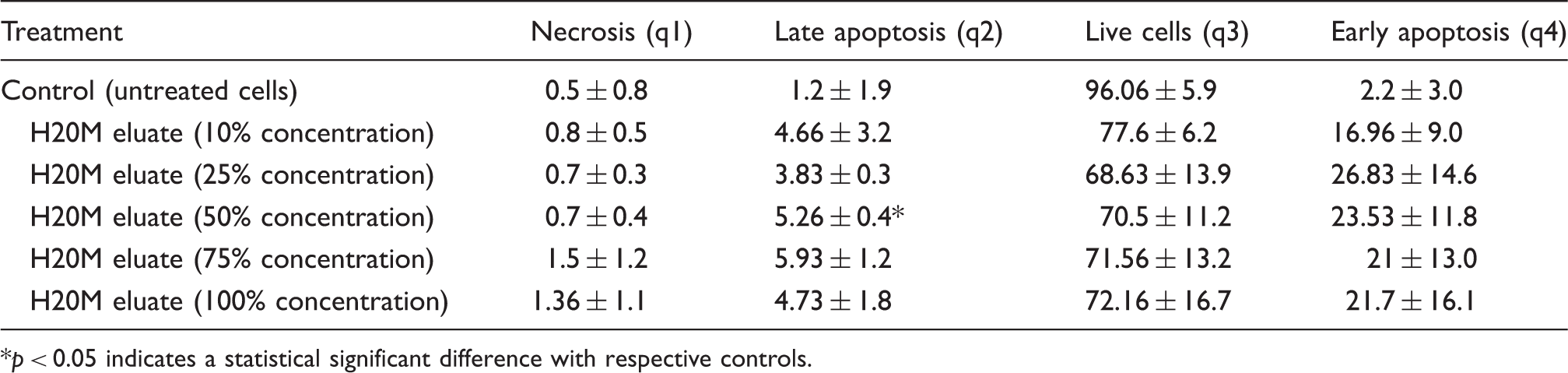
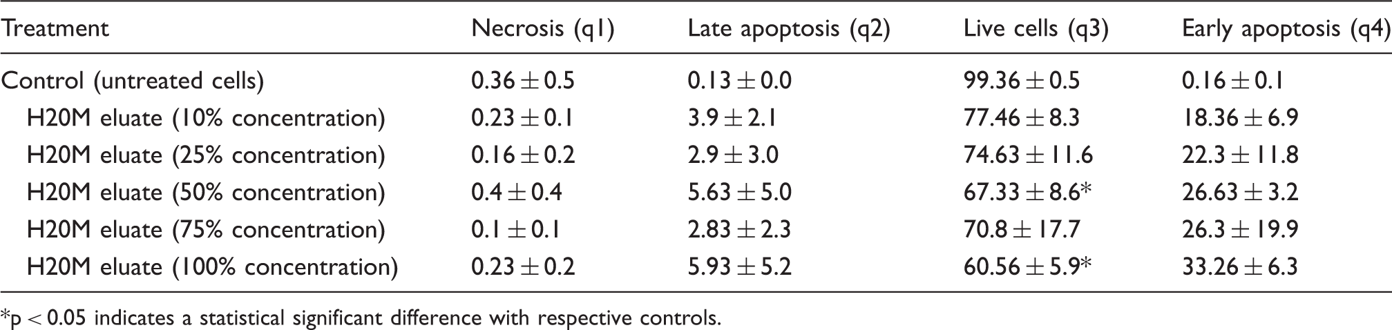
In order to determine how the necrotic, apoptotic cell population changes in a time and concentration dependent manner, the treated hFOB cells were sorted using FACS. In particular, the apoptotic cell death caused by H20M eluates was examined by flow cytometry after 12, 24, and 48 h of treatment. Figures 4, 5, and 6 plot the results of divariate FITC annexinV-PI hFOB treated cell population with varying dilution of H20M eluates for 12, 24, and 48 h, respectively. In analyzing FACS data, it can be reiterated that the lower left quadrant, q3 (both FITC Annexin V and PI negative) represents live cell populations, while the upper right quadrant, q2 (both FITC annexin V and PI positive) represents late apoptotic stage. In the present study, we considered that sorted cells in the lower right quadrant, q4 (FITC annexinV positive and PI negative), are in the early apoptotic stage, while the upper left quadrant, q1 (FITC annexin V negative and PI positive), signifies dead/necrotic/nonviable cells. Broadly, it was observed that apoptosis and necrotic cell death was dependent on H20M concentration as well as treatment time. It was noticed that the apoptotic data corroborated with the cytotoxicity results (Figures 6–8). From FACS analysis, the percentage of live (q3), apoptotic (q2 and q4), and necrotic (q1) cells at various concentrations/times were determined and are provided in Tables 2–4. From Table 2, it was clear that after 12 h of treatment, cells did not show a concentration-dependent apoptotic death. At less than or equal to 50% H20M concentrations, the q3 quadrant (live cells) was 80% or more of the total cell population, whereas at greater than or equal to 50% H20M concentration, the early apoptotic cell population was 10% or less of the total population. Moreover, after 24 h of H20M treatment, 65–80% of the entire cell population was in the live state (Table 3). Interestingly, after 24 h of treatment, apoptotic and necrotic cell populations showed a concentration-dependent effect. At 25% or more of H20M concentrations, 15–30% of the cell population showed early apoptotic cell death (Table 3). The apoptotic cell population decreased in a concentration-dependent manner above H20M concentrations of 25% or more (Table 3). Also, necrotic cell populations increased with increasing H20M concentrations of 50% or more (Table 3). On the other hand, the live cell populations were 60% of the entire cell populations after 48 h of treatment with the highest H20M concentration 100% (Table 4). Also, it was clearly revealed from Table 4 that after 48 h of H20M eluate exposure to hFOB cells, early apoptotic cell death at 100% H20M treatment increased to more than 30% of the total cell population, which was significantly higher than that in the case of the lower treatment time of 12/24 h. Figures 3–6 and Tables 1–3 (MTT and FACS) collectively indicated a similar trend of percent viability and apoptotic cell population. The overall conclusion from the FACS data was that at less than 50% dose after 24 h treatment, apoptosis is low. Whereas longer exposure times of up to 48 h, a greater than or equal to 50% and less than or equal to 100% H20M eluate dilution showed time and dilution dependent apoptosis.
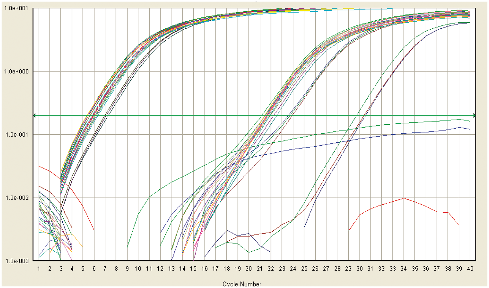
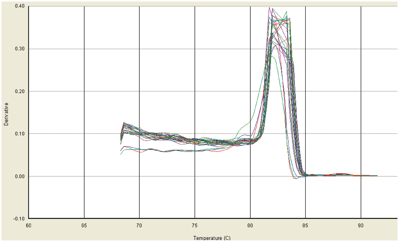
FACS graph showing hFOB populations after 12 h of treatment with HA-20% mullite nanoparticle eluates. A-B (Control), C-D (10%), E-F (50%), and G-H (100%). Right panel (A, C, E, and G) shows total population; left panel (B, D, F, and H) shows distribution of population. Q3 – live populations, Q4 – apoptotic populations, Q1 – necrotic populations and Q2 – late apoptotic populations. FACS graph showing hFOB populations after 24 h of treatment with H20M nanoparticle eluates. A-B (Control), C-D (10%), E-F (50%), and G-H (100%). Right panel (A, C, E and G) shows total population; left panel (B, D, F, and H) shows distribution of population. Q3 – live populations, Q4 – apoptotic populations, Q1 – necrotic populations, and Q2 – late apoptotic populations. FACS graph showing hFOB populations after 48 h of treatment with H20M nanoparticle eluates. A-B (Control), C-D (10%), E-F (50%) and G-H (100%). Right panel (A, C, E and G) shows total population; left panel (B, D, F, and H) shows distribution of populations. Q3 – live populations, Q4 – apoptotic populations, Q1 – necrotic populations and Q2 – late apoptotic populations. Representative amplification curve for realtime gene expression of hFOB cells after treatment for 6 h with H20M eluates. Representative dissociation curve for realtime gene expression of hFOB (osteoblast) cells after treatment for 6 h with H20M. The single peak shows the specific binding of the primers. Summary of FACS data obtained with H20M eluates of treated osteoblasts after 12 h of treatment. p < 0.05 indicates a statistical significant difference with respective controls. Summary of FACS data obtained with H20M eluates of treated osteoblasts after 24 h of treatment. p < 0.05 indicates a statistical significant difference with respective controls. Summary of FACS data obtained with H20M eluates of treated osteoblasts after 48 h of treatment. p < 0.05 indicates a statistical significant difference with respective controls.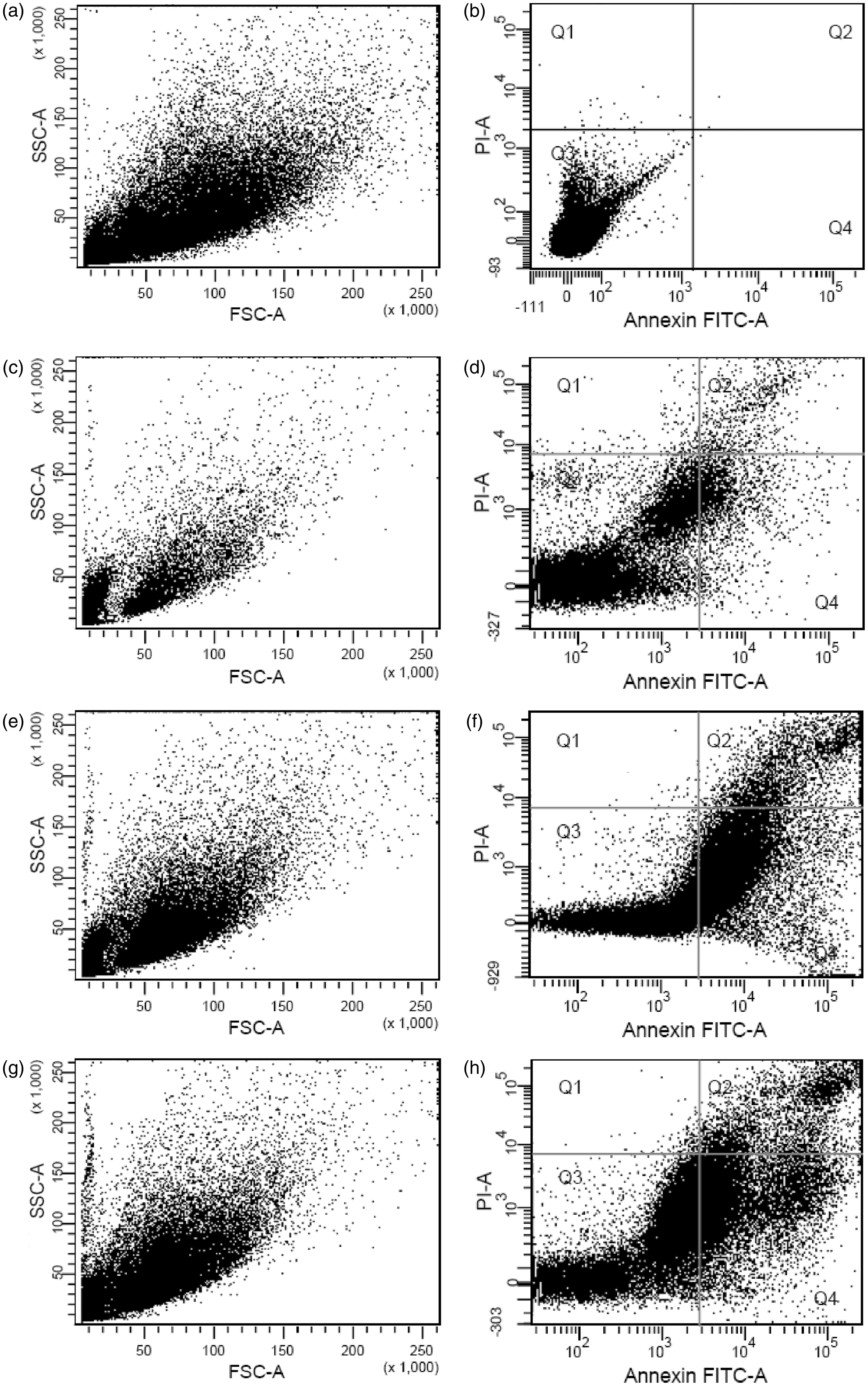
Gene expression analysis of apoptotic cell markers
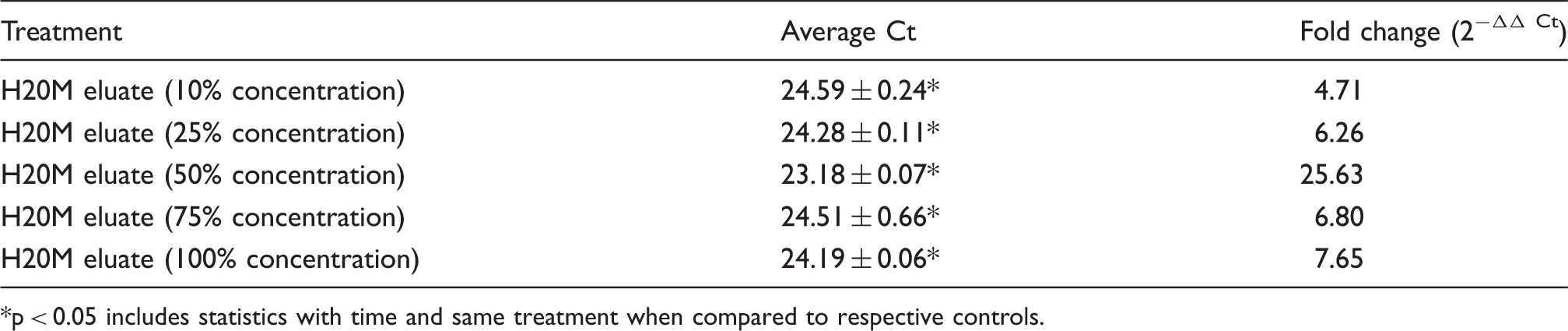
Quantitative real time gene expression of the apoptosis marker

p < 0.05 includes statistics with time and same treatment when compared to respective controls.
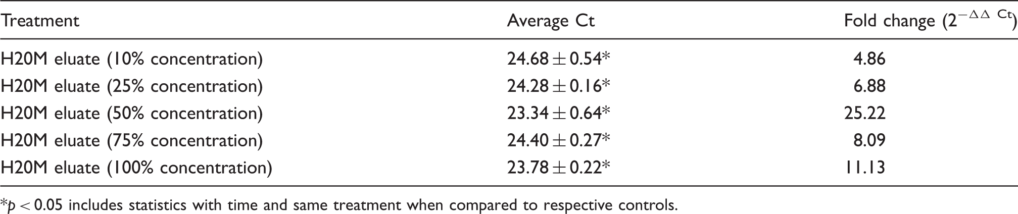
Quantitative real time gene expression of the apoptosis marker,
p < 0.05 includes statistics with time and same treatment when compared to respective controls.
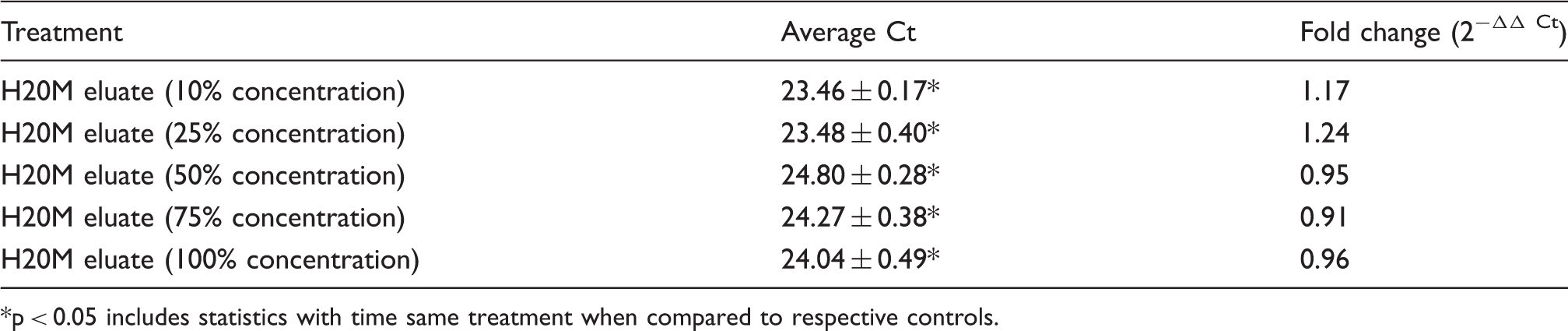
Quantitative real time gene expression of the apoptosis marker,
p < 0.05 includes statistics with time and same treatment when compared to respective controls.
Quantitative real time gene expression of the apoptosis markers
p < 0.05 includes statistics with time same treatment when compared to respective controls.
Gene expression analysis of bone cell markers
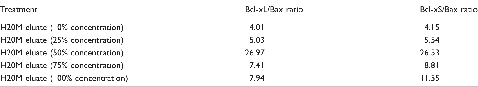
Ratio of the Bcl-xL/Bax and Bcl-xS/Bax determined with hFOB cells after treatment for 6 hrs with varying concentrations of submicron particle eluates of H20M.
Quantitative real time gene expression in terms of fold change for bone cell marker
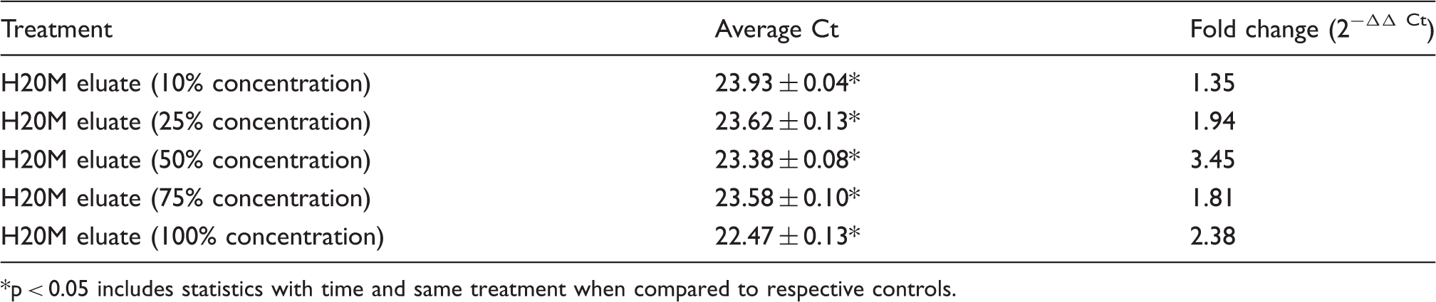
p < 0.05 includes statistics with time and same treatment when compared to respective controls.
Quantitative real time gene expression in terms of fold change for bone cell marker
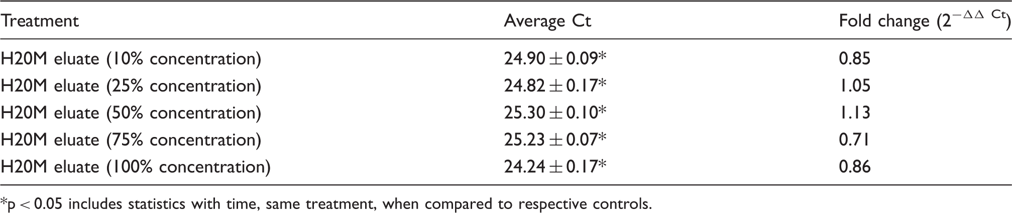
p < 0.05 includes statistics with time, same treatment, when compared to respective controls.
Quantitative real time gene expression in terms of fold change for bone cell marker

p < 0.05 includes statistics with time and same treatment when compared to respective controls.
Discussion
This study is a continuation of a recent study 26 and in this work, H20M-treated osteoblasts were investigated using FACS to sort apoptotic and necrotic cell populations. 27 Apoptotic cell death at the molecular level is identified by flip-flop translocation mechanism of phosphatidyleserin (PS) from the inner to outer side of cell membranes that indicates programmed cell death. Moreover, PS is also associated with signaling mechanisms through protein kinase activation as well as bone tissue remodeling.28–30 Therefore, apoptotic cell death in H20M-treated osteoblasts was further confirmed by gene expression of the Bcl-2 family at the transcription level. 31 It has been widely reported that the expression of apoptotic genes is commonly performed at the mRNA level rather than the protein level. 32 The Bcl-2 family of proteins is known to contribute to apoptosis (pro- or anti-apoptotic regulators) and also regulate cellular activities.33,34 In particular, Bax and Bcl-xS are responsible for apoptotic cell death, whereas Bcl-xL promotes cell survival. The ratio of Bax/Bcl is therefore considered as a factor that determines the live/dead status of the cell, when exposed to any physiological and chemical stimulus.35–37 In addition to apoptosis, uncontrolled regulation of Bcl-2 causes autoimmunity, neurodegenerative, and cancer.38,39
Our present findings indicated that HM particles play a significant role in the up and down regulation of the Bcl-2 family of genes, in time- and concentration-dependent manner. Apoptotic cell death was studied here in osteoblasts (hFOB) by a fluorescent activated cell sorter and Bcl-xL (anti-apoptotic) and Bax, Bcl-xS (pro-apoptotic) expression at different concentrations in H20M-treated hFOB cells. Also, the bone cell molecular markers Cbfa, COLI (collagen type I) and OCN (osteocalcin) were studied to further confirm the effect of H20M particles on bone cell proliferation, differentiation and extracellular matrix synthesis in vitro by quantitative real time gene expression.
In this section, an attempt was made to understand the cellular and molecular response to novel nanostructured orthopedic biomaterial particulates via cellular and genomic profiling of H20M-treated human osteoblasts (hFOB). Based on MTT and FACS data analysis, various aspects concerning cellular viability and cellular apoptosis of human osteoblasts (hFOB) treated with H20M particulate eluates will be discussed in this section. Gene expression was also studied to understand the underlying molecular dynamic aspects of cellular apoptosis caused by the biomaterial eluate treatments. It can be reiterated here that the HM system has been well characterized concerning in vitro dissolution, in vitro biocompatibility, and physical properties In addition to this, in vivo implantation tests with H20M composites in a rabbit model demonstrated biocompatibility in the bulk form. 11
Although H20M bulk composites support comparable or even better cell adhesion as well as osteoblast differentiation than monolithic HA, the toxicity of ultrafine particles (micron/nanoparticles) of H20M remains an issue, should they become dissolved during long term use in vivo. Therefore, an attempt was made to study the cellular as well as molecular basis of apoptotic cell death of hFOB cells treated with H20M eluates. In addition, the molecular markers of bone cell proliferation and differentiation were also studied for a better understanding of cell–material responses by quantitative real time gene expression.
From the analysis of the cytotoxicity data, the results revealed that H20M caused cell death after 24 h of exposure to hFOB with respect to the untreated hFOB cells (control). Importantly, after 12 h of treatment, the results showed more than 100% of cell viability up to a 25% concentration of H20M eluates. Here, it is argued that above 25% concentration of H20M, H20M doses appear to have important consequences as cell viability reduced with an increase in the concentration of H20M eluates. Likewise, with increasing exposure time of hFOB to H20M submicron to nanometer particles, it was hypothesized that the mitochondrial dehydrogenase enzyme of the hFOB-treated cells were more active to overcome the effect of H20M exposure on cellular viability.
The apoptotic cell death studied by FACS was consistent with the cytotoxicity data with respect to time and concentration of H20M exposure. From Tables 2–4 and Figures 4–6, it was evident that apoptotic cell death increased with increase in H20M concentration and treatment time. Apoptotic cell death involves biochemical and genetic regulation in response to physical and chemical stimuli to the cells. FACS results showed that the number of apoptotic cell populations increased with increases in the H20M concentration. It is also known that molecular mechanisms of apoptotic cell death involves physical and chemical stimuli that change the membrane integrity by translocation of surface molecules phosphatidyleserin (PS) from the inner to outer surface of the membrane. 40 From Tables 2–4 and Figures 4–6, it is clear that the percent of apoptotic cells was more than that of necrotic cells. It is, thus, hypothesized that H20M eluates trigger a cascade of signaling that induces the translocation of the PS molecules on the outer surface of the plasma membrane, which thereby increases the availability of PS molecules at the outer leaflet of the cell membrane.
H20M is a composite material of two different phases, mullite (3Al2O32SiO2) and HA. It is possible that mullite stimulates apoptotic cell death by up and down regulation of the Bcl-2 family of genes. In contrast, the major phase of the eluate contains Ca-P, which stimulates bone cell differentiation. It can be reiterated here that the expression of various GOI was quantified after 6 h of H20M eluate treatment in an attempt to achieve higher mRNA expression (transcriptional study). The Bcl-2 family of genes encodes many proteins through splicing and Bcl-2 shows similarity with Bcl-xL in terms of possessing four homology domains. 41 In contrast, the up-regulation of the ratio of Bcl-xL/Bax reduces cell death and the maximum fold change was 26.97 at a 50% dose of the H20M. The Bcl-xL, a homolog of Bcl-2, promotes cell survival by increasing the rate of transcription than the Bax, a pro-apoptotic transcript. From Table 9, it is apparent that the ratio of Bcl-xL/Bax and Bcl-xS/Bax was up regulated. Unlike Bax, Bcl-xS showed similarity with the BH3 domain of anti-apoptotic proteins that are responsible for the anti-apoptotic nature of the Bcl-2 family gene. The germane discussion of the anti-apoptotic properties of H20M is corroborated with the presence of 80% of the HA.
Most importantly, the anti-apoptotic property of H20M was further supported by the up regulation of osteoblast differentiation markers. Apoptotic cell death through the Bcl-2 family of genes is controlled by different signaling pathways through up and down regulation of mitochondrial enzyme cytochrome c and p53 gene. The cytochrome c, a key apoptogenic factor, controls cell death by releasing the apoptotic factor from mitochondria and this is an extrinsic pathway of programmed cell death. The up regulation of the Bcl-2 family of genes, by the H20M eluates, provided evidence of the extrinsic signaling pathway of apoptosis. From Tables 2–4 and Figures 3–6 (MTT and FACS data), it can be stated that cell surface receptors and mitochondrial enzymes are indispensible factors that control cell death or cytotoxicity caused by the H20M eluate treatment. A number of studies on the mechanism of apoptosis induced by physical and chemical stresses clearly revealed that plasma membrane structure integrity and DNA disintegration was responsible for apoptosis.42,43 The fact that the apoptotic cell population increased and necrotic cell death decreased possibly indicated that mullite (3Al2O3 2SiO2) damaged the cell membrane integrity. This is responsible for the translocation of molecules from the plasma membrane and also changes in the cell membrane permeability.
The intrinsic signaling pathway (Bcl-2 family) of cell death induced by H20M encourages further study of bone cell markers. From the preceding discussion, osteoblast cell death by regulating Bcl-x and Bax does not cause apoptotic cell death, as expected in the presence of the major inorganic phase of bone, hydroxyapaptite.
Most strikingly, Cbfa, COLl, and OCN (Tables 10–12) in hFOB were up-regulated, when exposed to up to 50% H20M in this study. Due to signaling induced by HA (Ca10 (Po4)6(OH2)), the expression of many Ca-dependent molecules, such as bone cell markers, are up-regulated. The signaling induced by the extracellular Ca-molecules is mainly based on the activity of the adenylyl cyclase enzyme, a transmembrane protein present on the surface of mammalian cells. The adenylyl cyclase enzyme increases the concentration of the cyclic AMP that activates cyclic-dependent kinase (PKA). The PKA activated by Ca induces the expression of bone cell markers.
40
OCN, Cbfa, and Col-I are the Ca-dependent protein molecules that cause hFOB differentiation, tissue formation, mineralization, and extracellular matrix formation.44,45 In a study on HA/calcium phosphate soluble composites, the differentiation of and mineralization by osteoblasts were enhanced by the synthesis of collagen and Ca-dependent proteins.
46
In a different study on MC3T3-E1 osteoblasts, it was concluded that the morphology as well as expression of bone cell markers was significantly increased by HA.
47
In another study on biodegradable materials,48,49 it was concluded that the regulation of the biocompatibility of materials depend on the leaching of ions and biodegradable products. From H20M eluates, the possible leaching ions could be Ca, P, Si, and Al and these ions can regulate the expression of GOI. Figure 9 shows H20M ions released and the mechanism of possible signaling at the cellular and genomic level. It has been widely reported that the presence of the Ca-channel in the plasma membrane regulates signaling mediated by the presence of extracellular Ca+2 ions.
40
The possible cascade of signaling in the presence of mullite leading to regulate the expression of Bcl-2 family of gene can be rationalized on the basis of a previous study, wherein nano-SiO2 was reported to cause up-regulation of Bax/Bcl-2 ratio in treated human hepatic cells.
15
Schematic illustration of H20M released ions and the path of intercellular interaction at the cellular and genetic level.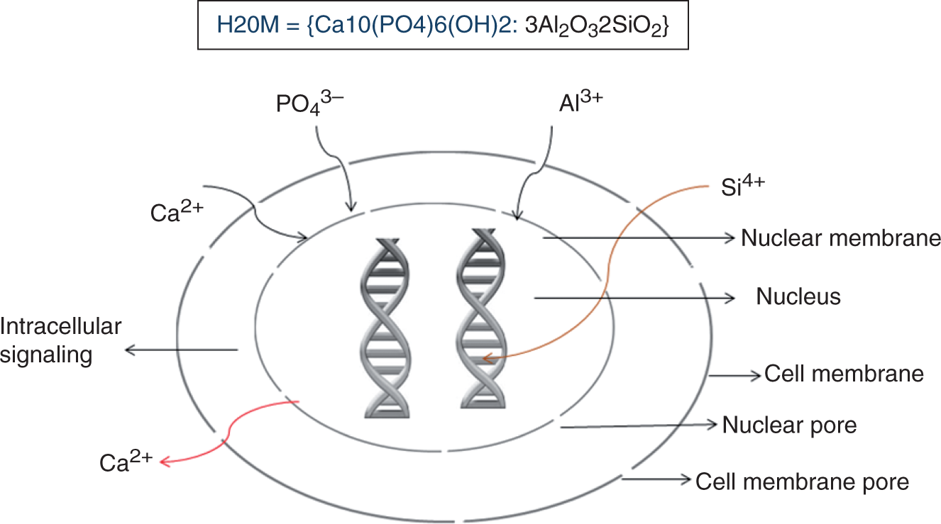
Conclusions
The results of the present investigation revealed the cytotoxic and genetic profiling of hFOB cells when treated with submicron to nanometer particle eluates of H20M. The experimental results demonstrated that hFOB cells exposed to H20M eluates decreased apoptotic cell death and up-regulated the expression of apoptotic and bone cell markers. The present work demonstrated that eluates of H20M composites elicited dose- and time-dependent necrotic hFOB death. The apoptotic cell population of H20M-treated cells increased with increasing concentration and treatment time of H20M to hFOB cells. On the other hand, necrotic cell populations decreased with time of treatment and concentration of H20M. More interestingly, 50% of eluate concentrations were the optimum concentrations. Above and below 50% concentration H20M eluate treatment up- and down-regulated Bcl-2 and bone cell markers, respectively. It was also apparent that the mullite phase did not cause apoptotic cell death. This finding can be correlated with the fact that after the treatment of hFOB with H20M, the necrotic cell population decreased, whereas the apoptotic cell population increased; also, the ratio of pro-apoptotic/anti-apoptotic population decreased. In addition, key markers of bone cell differentiation were also up-regulated with the treatment of the novel submicron to nanometer bioceramic eluates. Collectively, such results support that H20M should be considered as novel orthopedic implant materials, that can be safe in bulk and biodegradable forms and the concern for the presence of mullite to cause apoptosis at molecular level can be ruled out.
Footnotes
Acknowledgment
Financial support from the Hermann Foundation and the Indo-U.S. Center for Biomaterials for Healthcare is acknowledged.