
Abstract
This investigation describes the synthesis and in vitro evaluation of cationic hydrogel sub-microparticles based on polydimethylaminoethylmethacrylate for oral insulin delivery. Polymerization of dimethylaminoethylmethacrylate was carried out in aqueous medium with potassium persulfate as the initiator. Quaternization of the resulting hydrogel was carried out to introduce cationic surface groups and the derivatization was confirmed by zeta potential measurements, nuclear magnetic resonance and infrared spectroscopies. Swelling behavior of these particles was evaluated for dependence of pH. Insulin-loaded particles were subjected to in vitro release experiments at gastric and intestinal pH. Moreover, cytotoxicity evaluation showed that both polydimethylaminoethylmethacrylate and its quaternized derivative were non-toxic to Caco-2 and L929 cell lines. The presence of quaternary ammonium groups improved the cationic charge and enhanced the mucoadhesive properties of the hydrogel. Confocal microscopic observations showed that these sub-microparticles were capable of opening tight junctions between the Caco-2 cells and thus increased the paracellular permeability. The above studies suggest that cationic hydrogel sub-microparticles can act as a good candidate for oral insulin delivery.
Keywords
Introduction
In recent years, polymeric hydrogel nano/microparticles are gaining attention for biomedical and pharmaceutical applications, due to their biocompatibility and ability to alter their volume/surface properties in response to external stimuli such as pH, temperature, ionic strength, and electric field. Hydrogels can also be engineered to exhibit bioadhesiveness to facilitate drug targeting, especially through mucus membranes, for non-invasive drug administration. Hydrogel systems possess excellent chain flexibility and mobility which enable them to inter-diffuse across the mucosal barriers. 1
The transport of hydrophilic molecules via the paracellular pathway is severely restricted by the presence of tight junctions that are located at the luminal area of adjacent epithelial cells. It has been suggested that cationic macromolecules are capable of displacing divalent ions from electronegative sites on the tight junctions. 2 Tight junctional membrane requires coordination with cations to maintain their dimensional stability. They retain fixed negative charges and a relatively modest alteration in the concentration of specific species of ions within the volume of the pore could result in substantial alterations in a tight junction, leading to loosening or opening of the pore. Further, it can be expected that the positively charged particles would improve the mucoadhesion through electrostatic interaction with mucus glycoproteins.
Polydimethylaminoethylmethacrylate (PDMAEMA) has been proved to be an efficient gene-transfer agent due to its ability in binding DNA by electrostatic interactions. 3 PDMAEMA is an interesting polymer, as the tertiary amine function allows fixing of active substances onto resulting polymers. 4 N, N-dimethylamino-2-ethylmethacrylate was chosen as the monomer in this study because of the excellent biocompatibility, hydrophilicity, and pH and temperature sensitivities of its polymer, poly (2-(dimethylamino)ethyl methacrylate). 5–7 To date, by far the greatest demand has been for the controlled delivery of insulin. Herein, we describe the synthesis of PDMAEMA hydrogel sub-microparticles and their quarternized derivative for oral insulin delivery. Our approach involves the modification of PDMAEMA hydrogel sub-microparticles to yield quaternary groups on the surface. Cationic surface charge could enhance the interaction of the sub-microparticles with the mucus gel layer and may induce opening of tight epithelial junctions across the intestinal membrane.
Materials and methods
Materials
Dimethylaminoethylmethacrylate (DMAEMA), ethyleneglycoldimethacrylate (EGDMA), 3-(4, 5-dimethylthiazol-2-yl)-2, 5-diphenyl-tetrazoliumbromide (MTT), Pluronic F-127, chymotrypsin, benzoyltyrosine p-nitroanilide (BTPNA), fluorescein isothiocyanate (FITC), rhodamine phalloidin, and mucin from porcine stomach were obtained from Sigma–Aldrich. Potassium persulfate was obtained from BDH Chemicals and human insulin, human insulin ELISA kit and calcium kits were purchased from Eli Lilly, Mercodia, and Enzyme Technologies Private Ltd, Mumbai, India, respectively. Eudragit L100-55 was a gift from Rohm Pharma, India. Caco-2 cells (human epithelial colorectal adenocarcinoma cells) and L929 cells (mouse fibroblast cells) were obtained from National Centre for Cell Sciences, Pune, India. Fetal bovine serum (FBS), Dulbecco’s modified eagle medium (DMEM), Hank’s balanced salt solution (HBSS) was from Gibco Chemicals.
Synthesis and characterization of PDMAEMA hydrogel particles
PDMAEMA hydrogel particles were prepared by free radical polymerization in aqueous medium. Monomer DMAEMA (2 mL) mixed with cross-linking agent, EGDMA (800 µL) was added to 100 mL of distilled water containing Pluronic F-127 (0.05%). Thereafter, free radical initiator, potassium persulfate was added to the reaction mixture and the whole content was stirred at 70℃ for 6 h. The resulting white suspension was centrifuged at 10,000 r/min and washed several times with distilled water and dried under vacuum.
Synthesis of quaternized PDMAEMA
Quaternization reaction of PDMAEMA was conducted using methyl iodide (CH3I) as the quaternizing agent. Typically, 100 mg of PDMAEMA was dispersed in 40 mL phosphate buffer (pH 7.4). Then, a defined amount of CH3I (e.g. 0.5 mL, 1 mL) was added dropwise and stirred for 18 h at room temperature. Thereafter, the quaternized polymer was recovered by filtration and dried under vaccum.
Particle size and zeta potential analysis
The hydrodynamic diameter and surface charge density of the particles were measured using Zetasizer Nano of Malvern Instruments. These measurements were performed in triplicate. Zeta potential at different values of pH (4–10) was measured using dynamic light scattering equipped with MPT titrator.
Scanning electron microscopy
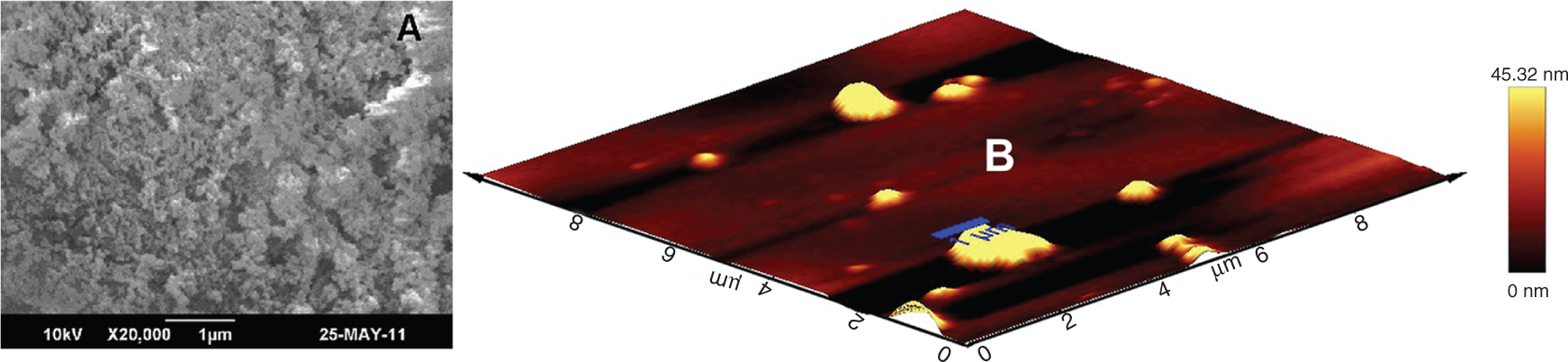
The surface morphology of the quaternized particles was studied using scanning electron microscope (SEM; HITACHI S-2400, Hitachi, Japan). Samples were mounted on metal stubs using double-sided adhesive tape coated with gold under vacuum and then examined. The surface topography and size of the quaternized derivative was analyzed using atomic force microscopy (AFM; WITEC confocal Raman microscope system with atomic force microscope extension, Germany).
Nuclear magnetic resonance and infrared spectroscopies
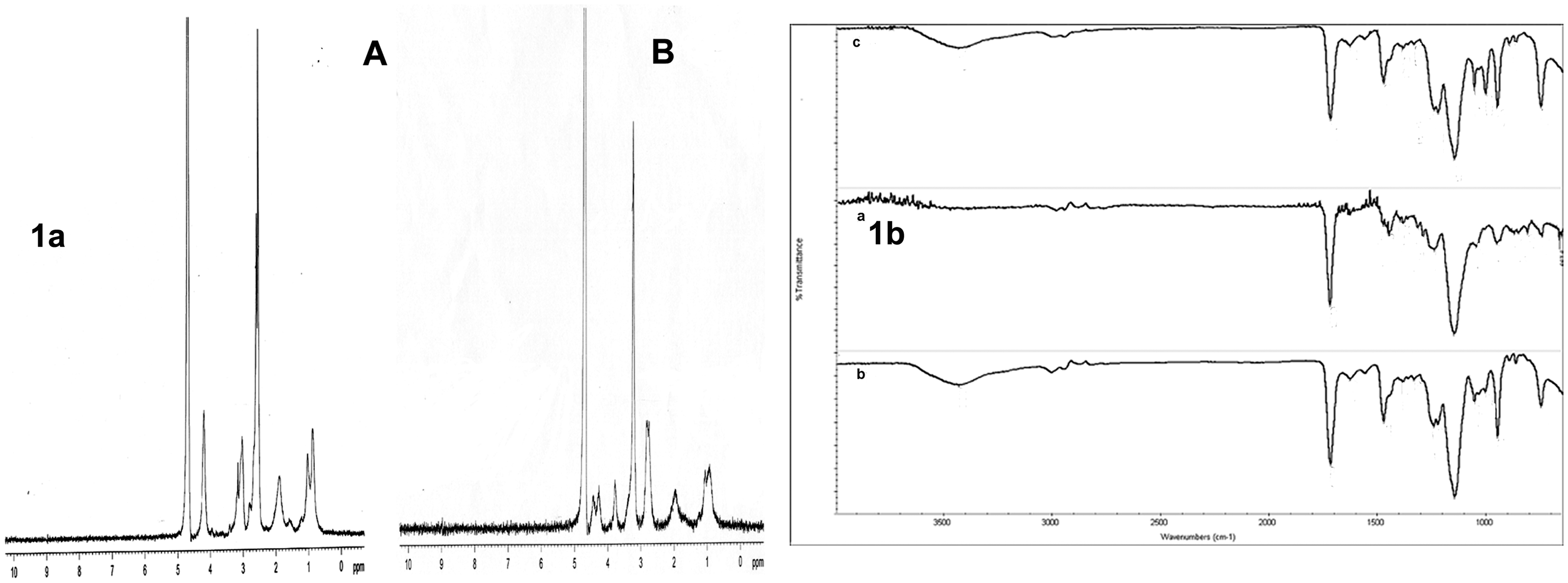
As the hydrogel particles are insoluble in deuterated solvents,1H-NMR (nuclear magnetic resonance) spectral analysis was carried out by polymerizing DMAEMA without using EGDMA (cross-linking agent) under the same experimental conditions.1H-NMR spectra of the polymer and its derivative were measured in D2O using 300 MHz spectrometer (Bruker Avance DPX 300) with trimethylsilane as an internal standard. Fourier transform infrared (FT-IR) spectra of PDMAEMA and quaternized PDMAEMA (QPDMAEMA) were recorded in 4000–400 cm−1 region using NICOLET 5700 FT-IR spectrophotometer.
Swelling studies
Swelling studies of the hydrogel particles were carried out separately at a pH of 1.2 and 6.8. A known amount of dried test (PDMAEMA and QPDMAEMA) samples were suspended in buffer solutions of the respective pH. At specific time intervals, the samples were removed from the buffer solution. Excess water on the surface was removed and the weight of swollen particles determined.
8
The degree of hydration of the samples was calculated using the following equation:

Calcium-binding studies
About 5 mg of particles were dispersed in 1 mL of 1 mM CaCl2 solution and incubated for 1 h. The particles were then centrifuged at 7000 r/min. CaCl2 solution without polymer was used as the standard. The calcium-binding efficiency of the particles was evaluated with 200 µL supernatant using a calcium assay kit. 9
Chymotrypsin inhibition study
The chymotrypsin inhibition assay was performed using the chromogenic substrate BTPNA. Particles (5 mg) were dispersed in Tris–HCl buffer (pH 7.8) to get a final concentration of 0.5% w/v solutions. Mixture of BTPNA, polymer dispersion, and 40 U of chymotrypsin solution (in 10 mM HCl) was incubated at 37℃ for 30 min and the enzymatic action stopped by the addition of 1% trichloroacetic acid solution. Supernatant was analyzed by measuring the absorbance at 405 nm spectrophotometrically (Varian Cary 50 Conc). Control was the same without polymer, and percentage of inhibition was calculated relative to control chymotrypsin which was taken as 100%. 10
Insulin loading and release studies
In order to evaluate the potential application of the polymer to oral insulin delivery, insulin loading and release properties of the sub-microparticles were examined. Drug loading was performed by diffusion filling method. 11 A known weight of dried particles, say 100 mg, was kept in 1 mL of insulin solution (100 IU/mL) for remote loading. After 24 h, particles were taken out and excess insulin solution was gently wiped off. Loaded particles were kept for drying at low temperature (2–4℃). Due to the high swelling nature of the matrix in acidic media, insulin-loaded particles were coated with Eudragit L100-55 (Eudragit L100-55 was dissolved in 2-propanol with the help of triethylcitrate). 5% Eudragit L100-55 was used for this study. About 500 µL of 5% Eudragit L100-55 was dropped into 50 mg of insulin-loaded particles and the particles were dried at 2–4℃.
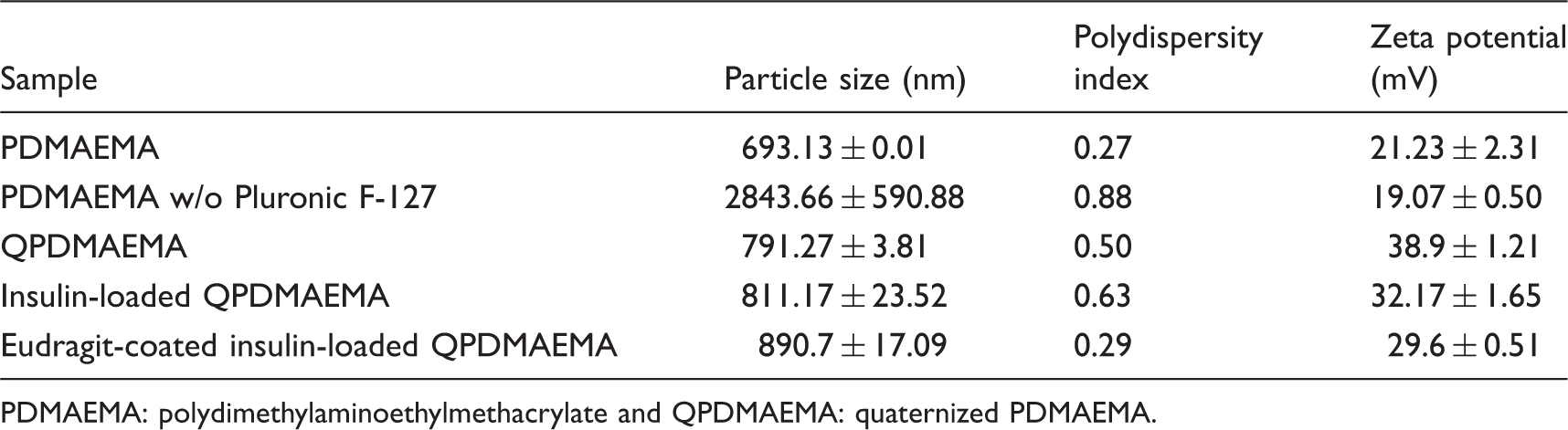
Release studies were carried out under sink conditions. Loaded particles (20 mg) were suspended separately in 20 mL of buffer with a pH of 1.2 and 6.8. This was then kept in a shaker at 37℃ (50 r/min). At specified intervals of time, aliquot of sample (200 µL) was withdrawn and insulin content estimated by Lowry method. The dissolution medium was replaced with fresh buffer to maintain the total volume after each withdrawal. The amount of insulin in the test solution was calculated from the insulin standard maintained during the assay. 12 Particle size and zeta potential of QPDMAEMA, insulin-loaded QPDMAEMA and Eudragit-coated insulin-loaded QPDMAEMA were also analyzed.
Analysis of released insulin
Biological activity of loaded insulin in the particles was investigated using ELISA (DAKO Cytomation) technique. About 100 mg of insulin-loaded particles was suspended in phosphate buffer (pH 7.4) for 24 h. After specified intervals, an aliquot of sample was withdrawn, and ELISA was done as per the standard procedure. Results were obtained by reading the optical density at 450 nm using microplate reader (Finstruments microplate reader). Circular dichroism (CD) spectra of the released insulin were recorded on JASCO-J-810 spectropolarimeter equipped with JASCO PTC-423S Peltier type temperature control system using a 1 cm cell, at a speed of 100 nm/min, response time of 0.25 s, and bandwidth of 1 nm.
Hemolysis
Whole blood was collected from a healthy volunteer and anti-coagulated with sodium citrate (ratio of blood to anticoagulant taken was 9:1). Erythrocytes (RBC) were isolated by centrifuging whole blood at 700 r/min for 10 min. The erythrocytes were washed thrice with saline before use. Particles, both PDMAEMA and QPDMAEMA were mixed with RBCs and then incubated for 2 h at 37℃. The supernatant was spinned off at 1500 r/min for 5 min. Hemoglobin release was monitored spectrophotometrically at 541 nm. TritonX-100 and 0.9% NaCl were taken as the positive and negative controls, respectively. 13
The hemolysis rate was calculated as follows

Cytotoxicity
Cytotoxic evaluation of the polymer was carried out in L929 and Caco-2 cells. Caco-2 cells were seeded into a 24-well plate containing DMEM culture medium with 20% FBS. L929 cells were seeded into a 48-well plate containing 10% DMEM culture medium. The seeded plate was then incubated for 24 h at 37℃ in a CO2 incubator with a humidified 5% CO2/95% air atmosphere to allow the cells adhere to the well. DMEM medium without sample was used as positive control and phenol as the negative one. DMEM culture medium (2 mg/mL containing samples along with positive and negative controls was added to each well. After incubation for an additional 24 h at 37℃, the samples were carefully removed and MTT reagent (0.2 mg/mL) was added to each well and incubated for 3 h. The reagent was then removed and 200 µL dimethyl sulfoxide (DMSO) was added to dissolve the formazan crystals. The absorbance of each solution was read at 570 nm using an automated microplate reader. Furthermore, concentration dependent (0.25–1.5 mg/well) cytototoxic evaluation of both modified and unmodified samples were carried out on L929 cells.
Mucoadhesion studies
The animal experiments were done as per the requirements of the Animal Ethics Committee of Sree Chitra Institute for Medical Sciences and Technology. Animals were housed in rooms at controlled temperature and relative humidity. Mucoadhesion studies were carried out on freshly excised rat intestinal tissue. The rats were under fasting before euthanasia by cervical dislocation. The intestinal tissue of about 5 cm was taken out, flushed with normal saline to remove the luminal contents, and cut open longitudinally. The tissue was mounted and fixed on a semi-cylindrical polypropylene support and washed with saline to remove free mucin. A known amount of particles was spread on the intestinal tissue and kept in humid conditions for 5 min. It was then washed with phosphate buffer saline (PBS) at a rate of 10 mL/min for 20 min. The dislodged particles were collected and dried. The percentage of mucoadhesion was calculated by comparing the weight of particles adhered to the weight of particles applied. 14
Mucoadhesion testing was also conducted in vitro using using a texture analyzer (TA.XT plus, Stable Micro Systems, UK) with 0.05 N load cell equipped with mucoadhesive holder. Each sample was attached to the base of an aluminum probe (using double-sided adhesive tape) fixed to the mobile arm of the texture analyzer. The probe was lowered at a rate of 1 mm/s until contact with the tissue was made. The tissue was equilibrated for 15 min at 37℃ before placing onto the holder stage of the mucoadhesive holder. The probe was subsequently withdrawn at a specified test speed. 15 Using the texture analyzer, the maximum force required to separate the probe from the tissue (i.e. maximum detachment force; Fmax) could be detected directly from Texture Exponent 32 software. Work of adhesion (Wad) was calculated from the area under the force versus distance curve. Triplicate determinations were made.
Adsorption studies on mucin
Mucin stock solutions with a concentration of 2 mg/mL in phosphate buffer (pH 7.4) were prepared. Hydrogel particles (10 mg), modified and unmodified, were dispersed in the above mucin solutions, vortexed, and shaken at 37℃ for 30 and 60 min, separately. Then, the dispersions were centrifuged at 4000 r/min for 2 min and supernatant was used for the measurement of free mucin content. Around 200 -µL aliquot was taken, and the protein estimation was done by Lowry method. The absorbance of mucin was measured by colorimetry at a wavelength of 750 nm. The amount of mucin adsorbed by the particles was determined as the difference between its initial concentration and the concentration found in the dispersion after incubation and centrifugation. The calculations were made on the basis of mucin standard curves. 16 The interaction of mucin was further confirmed by differential scanning calorimetry (DSC) where thermograms were performed on a Q 20 DSC thermal analyzer (TA Instruments, USA).
Cellular uptake studies using FITC-labeled insulin
Insulin was covalently labeled with FITC as described below. Briefly, 0.1 mg of FITC was dissolved in 200 µL of DMSO. This solution (50 µL) was added to about 1 mL of insulin dissolved in 200 µL of sodium carbonate–bicarbonate buffer. The solution was then kept for incubation in the dark for 2 h. The reaction was quenched with the addition of 50 µL of 1 M ammonium chloride solution. The above solution was dialyzed against water for 3 h at 37℃ with subsequent changes of water at intervals. Cell-uptake studies were performed with Caco-2 cell monolayers. FITC-labeled insulin was loaded into particles. FITC insulin-loaded particles were incubated with Caco-2 cells for 4 h. 13 The cells were then washed with PBS thrice. The cells were fixed with 250 µL of 1% paraformaldehyde for 20 min at room temperature. The entry of the FITC-labeled insulin into the cells was assessed with the help of fluorescence microscope (Leica DMIRB, Germany).
Tight junction visualization
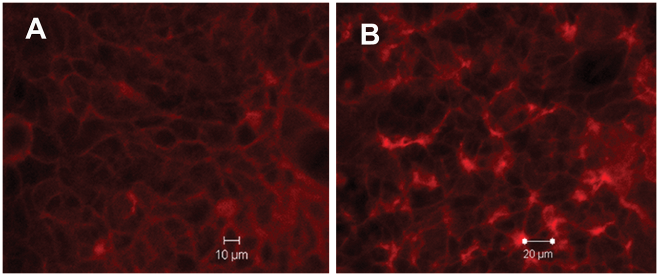
Caco-2 cells (passages 22–28) were grown in incubator at 37℃ under 5% CO2. Cells were maintained in T-75 flasks using MEM supplemented with 20% FBS,1% non-essential amino acids, 10,000 U/mL penicillin, and 10,000 µg/mL streptomycin. Cells were passaged at 80–90% confluency using 0.25% trypsin/EDTA solution. The cells were maintained under incubation conditions as mentioned above and used for transport experiments 6 days post-seeding. 17 Medium was replaced with HBSS transport medium, and cells were equilibrated at least for 2 h before the experiments. Cells were treated with particles at a concentration of 1 mg/mL for 1 h. The medium was removed and the cells were washed thrice with PBS. The cells were fixed with 250 µL of 1% paraformaldehyde for 20 min at room temperature. Then, the cells were permeabilized using 0.2% Triton X-100 in blocking solution, made of 1% (w/v) bovine serum albumin (BSA) in PBS, for 20 min. The permeabilized cells were then washed twice with PBS and incubated with 250 µL of 1% BSA for 30 min. For actin filament visualization, the blocking solution was removed and cells were incubated with 200 µL of rhodamine phalloidin solution (0.2 µg/mL) for 20 min at room temperature. The cells were washed with PBS and dried overnight at 4℃. Images were obtained using Carl Zeiss LSM Meta 510 inverted confocal laser scanning microscope (Carl Zeiss, Germany), equipped with He/Ne laser.
Statistical analysis
The data obtained from different assays were compared for statistical significance by one-way analysis of variance test using SPSS (Version 11, SPSS Inc., USA). All results were expressed as mean ± standard deviation. Differences were considered to be significant at a level of p < 0.05.
Results and discussion
The use of hydrophilic monomers for hydrogel synthesis allows for a reaction to proceed without phase separation or the use of organic solvents, resulting in a less harsh environment for the encapsulation of fragile biological macromolecules. Cross-linking can be achieved during polymerization, or afterwards using chemical or physical methods. Monomer and cross-linking agent, along with the initiator, undergo copolymerization at high temperature, resulting in the formation of water-insoluble cross-linked network
18
(Scheme 1). Cross-linked hydrogels are suitable candidates for drug-delivery applications; however, a high degree of cross-linking can result in restricted chain mobility and loss of functionality.19,20 Optimal bioadhesive properties can be achieved by controlling the degree of cross-linking. Pluronic F-127, non-ionic surfactant having a hydrophile–lipophile balance (HLB) value of 18–23 was used in this study.
21
This HLB value favors solubility in water at room temperature. Pluronic F-127 was added to improve the compatibility and to minimize the particle size of the polymer, thereby minimizing aggregation. As evident from particle size analysis, the size of PDMAEMA was found to be higher in the absence of Pluronic F-127 (Table 1). In this investigation, quaternization of the polymer was carried out by reaction with methyl iodide to increase the cationic nature of the polymer. Derivatization was confirmed by NMR and IR spectra.1H-NMR (Figure 1(a)) confirmed the successful quaternization of PDMAEMA to form strong cationic network. The peak at 4.435 ppm confirmed the quaternization of PDMAEMA. The peaks at 0.88, 1.88, 2.53, 2.58, 3.22, 3, 4.1 and 4.435 ppm were attributed to (C(CH3)PDMAEMA), (CH2 PDMAEMA), (N(CH3)2PDMAEMA), (O–CH2–CH2–N(CH3)2) PDMAEMA, (N(CH3)3), (CH2N(CH3)3), (O–CH2–CH2–N(CH3)2 PDMAEMA, and (O–CH2–CH2–N(CH3)3. IR spectrum (Figure 1(b)) showed the evidence of the introduction of the quaternary ammonium group on polymeric backbone, at 1476 cm−1 (C–H bending of trimethylammonium group). In addition to the confirmation by FT-IR and NMR spectroscopy, the increase in zeta potential may also be taken as an indication of quaternization. Zeta potential confirmed the increase in positive charge density of the derivative (38.9 ± 1.21 mV) as compared to unmodified polymer (21.23333 ± 2.31 mV). Zeta potential measurements showed an iso-electric point at a pH of 7.79, with the particles having a high positive charge at low pH as a result of the strong protonation of the weakly basic PDMAEMA shell (Figure S1). The negative zeta potentials were observed at pH values of 8–11. This may be caused by deprotonation of groups in the sub-microgels that leads to the overall negative charge. The results demonstrated that the prepared particles were able to maintain electrostatic stabilization at both low and high pH values. Particle size and zeta potential of PDMAEMA prepared in the presence and absence of surfactants, QPDMAEMA, insulin-loaded QPDMAEMA, and Eudragit-coated insulin-loaded QPDMAEMA determined at a pH of 6.8 are presented in Table 1. All the formulations mentioned in Table 1 exhibited positive charge at intestinal pH. Zeta potential of Eudragit-coated insulin-loaded particles were measured at intestinal pH and was found to be 29.5 mV. These data indicate that enteric coating have been eroded on dispersion of these particles in alkaline condition, retaining the positive surface charge at intestinal pH. Zeta potential plays an important role in particle uptake because the surface of the intestinal mucosa is negatively charged owing to the presence of sialic acid moieties. It has been reported that particles with a positive surface charge, such as chitosan, are usually attracted by the intestinal mucosa which helps in increasing the intestinal absorption of the encapsulated drug. SEM and AFM micrographs also confirmed the particle size of the quaternized derivative (of about 1 µm). The particles appeared to be of spherical morphology in both AFM and SEM (Figure 2).
Preparation of QPDMAEMA from DMAEMA. QPDMAEMA: quaternized PDMAEMA and DMAEMA: dimethylaminoethylmethacrylate. (a) NMR Spectra of PDMAEMA (A) and QPDMAEMA (B) and (b) IR spectra of PDMAEMA (A) and QPDMAEMA obtained by adding 0.5 mL (B) and 1 mL (C) of methyliodide. PDMAEMA: polydimethylaminoethylmethacrylate and QPDMAEMA: quaternized PDMAEMA. SEM (A) and AFM (B) images of QPDMAEMA. SEM: scanning electron microscopy; AFM: atomic force microscopy; and QPDMAEMA: quaternized PDMAEMA. Particle size and zeta potential of PDMAEMA, prepared in the presence and absence of surfactants, QPDMAEMA, insulin-loaded QPDMAEMA, and Eudragit-coated insulin-loaded QPDMAEMA. PDMAEMA: polydimethylaminoethylmethacrylate and QPDMAEMA: quaternized PDMAEMA.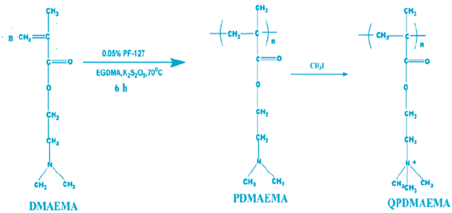
PDMAEMA is a weak cationic polyelectrolyte in aqueous solutions. At low pH, electrostatic repulsions between the protonated tertiary amine groups lead to PDMAEMA swelling.
22
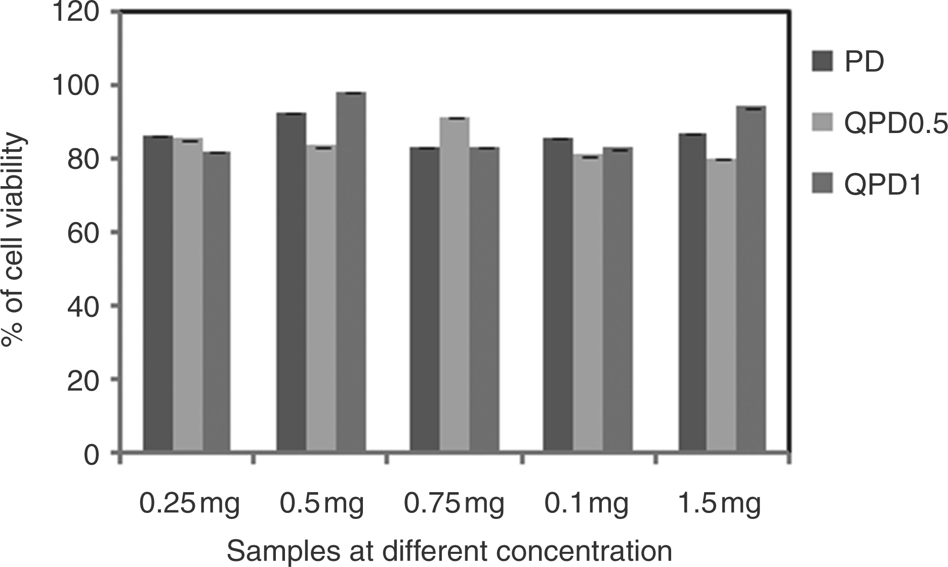
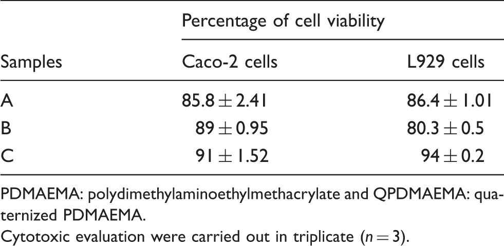
In contrast, at high pH, most of the amino groups are deprotonated and neutral. As a result, interactions between non-polar groups in a polar solvent (hydrophobic interactions) are increased, which leads to a more compact PDMAEMA conformation. The sub-microparticles of QPDMAEMA exhibited significant swelling with time. Due to the presence of basic tertiary amine pendants on the polymer structure, the swelling behavior of the prepared gel can be affected by pH. At a pH of 1.2, the degree of swelling increased significantly due to ionization of tertiary amine groups. QPDMAEMA 1 exhibited high percentage of swelling at a pH of 1.2 due to the increase in positive charge density of the particles as well as due to hydrogen bonding (Figure 3). The degree of swelling was lower at neutral and alkaline pH. The non-cytotoxic nature of quaternized particles has been established through MTT assay, which makes it suitable for oral administration. The percentage viability Caco-2 and L929 cells is presented in Table 2. Both the modified and unmodified particles were found to be non-toxic at a concentration of 0.25–1.5 mg/well in L929 cells (Figure 4). Cytotoxic evaluation of the polymer showed that quaternized particles are good candidates for the delivery of protein drugs. QPDMAEMA 1 was used for further studies and was abbreviated as QPDMAEMA. At physiological pH, hemolytic activity of sub-microparticles was 1. 49 ± 0.05%. Hemolysis assay further confirmed the blood compatible nature of polymers.
Swelling studies of QPDMAEMA obtained by adding 1 mL (QPDMAEMA 1) methyliodide at pH 1.2 and 6.8. QPDMAEMA: quaternized PDMAEMA. Concentration dependent cytototoxic evaluation of PDMAEMA and QPDMAEMA obtained by adding 0.5 mL (QPDMAEMA 0.5) and 1 mL (C) of methyliodide (QPDMAEMA 1). PDMAEMA: polydimethylaminoethylmethacrylate and QPDMAEMA: quaternized PDMAEMA. Cytotoxic evaluation of PDMAEMA (A) and QPDMAEMA obtained by adding 0.5 mL (B) and 1 mL (C) of methyliodide on Caco-2 and L929 cells. PDMAEMA: polydimethylaminoethylmethacrylate and QPDMAEMA: quaternized PDMAEMA. Cytotoxic evaluation were carried out in triplicate (n = 3).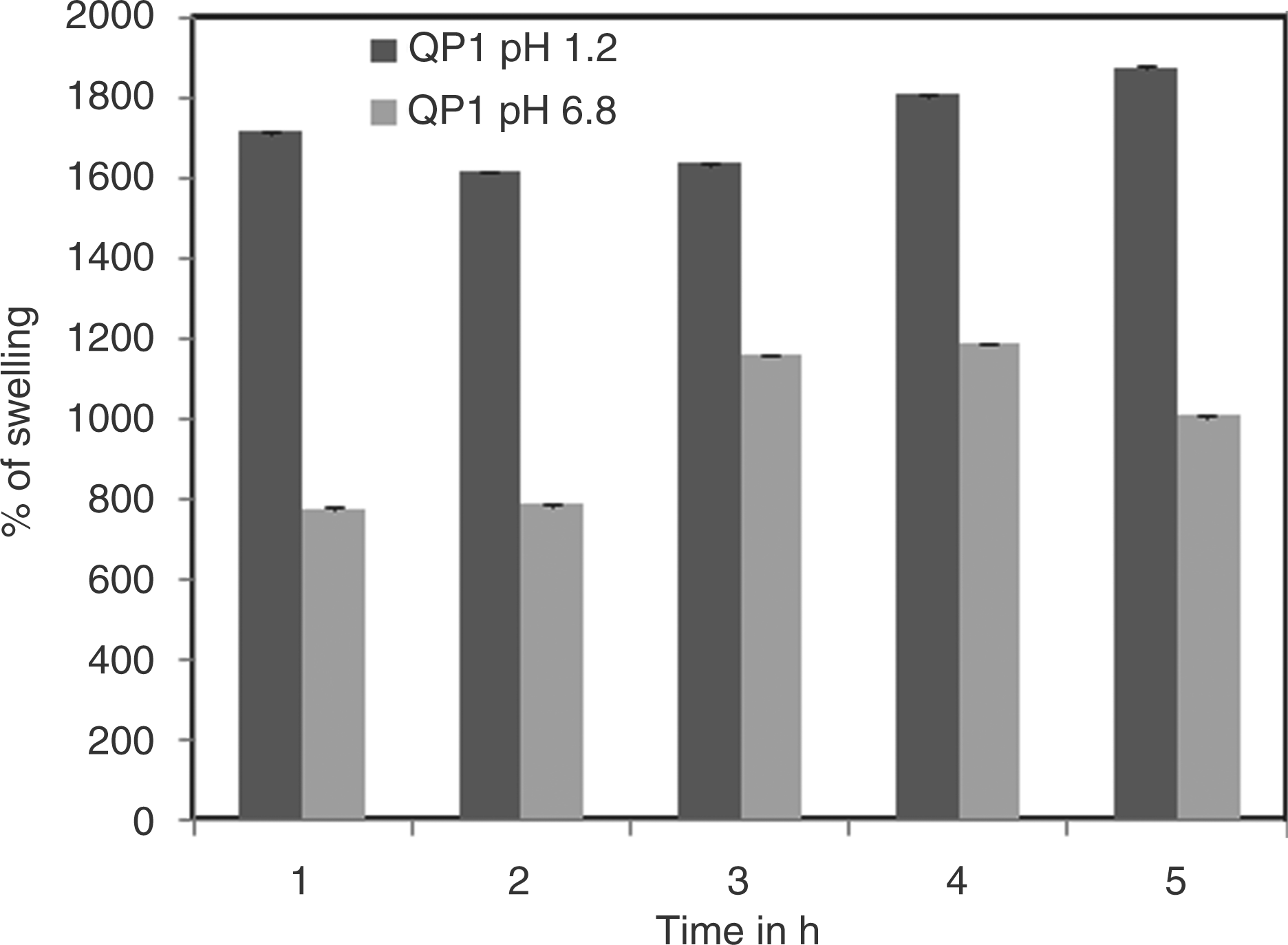
Hydrogel based delivery devices enhance mucoadhesive interactions by their inter-diffusion across the mucosal surfaces. The major prerequisites for mucoadhesive delivery systems are good wetting and/or swelling of the mucoadhesive polymers. Further, the mucoadhesion involves the penetration or the inter-diffusion of the mucoadhesive polymer chains into the crevices of the mucosal surface and also the entanglement with the mucus chains.
23
On a molecular level, mucoadhesion is a result of van der Waals’ forces, electrostatic attraction, hydrogen bonding, and hydrophobic interactions. The literature suggests that the initial interaction between the polymer and the biological surface is through electrostatic interaction followed by mechanical interlocking of the polymer chains.
24
Hydrogels display excellent chain flexibility and are considered to be an interesting system for mucoadhesive delivery systems. The higher mucoadhesive properties of quaternized derivative resulted from the synergistic effects of interpenetration of chains into the mucus, hydrogen bonding, and electrostatic interaction between positive charged QPDMAEMA and anionic glycoproteins present in the mucus layer (Figure 5).
Mucoadhesion studies of PDMAEMA (A) and QPDMAEMA. PDMAEMA: polydimethylaminoethylmethacrylate and QPDMAEMA: quaternized PDMAEMA.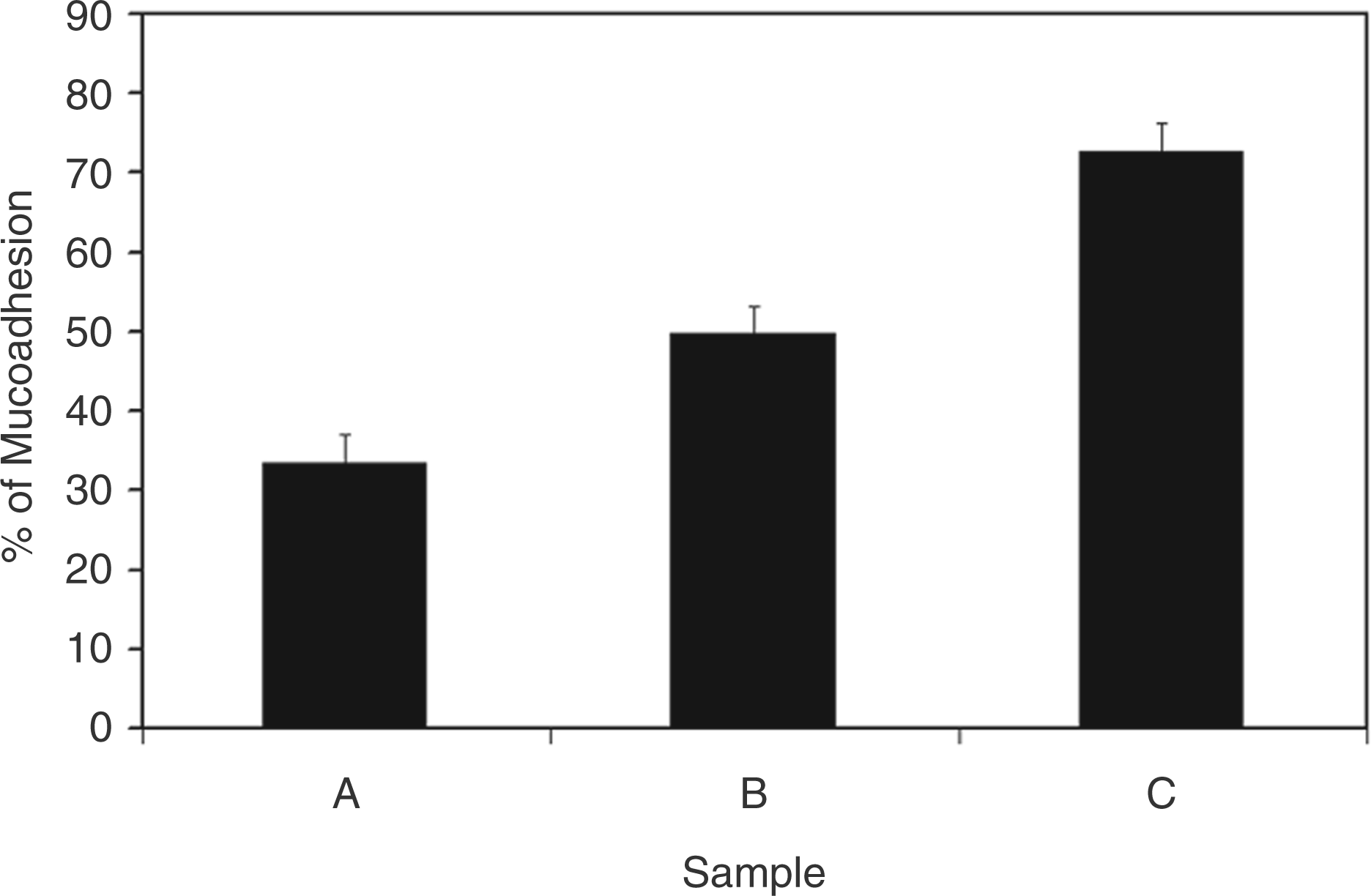
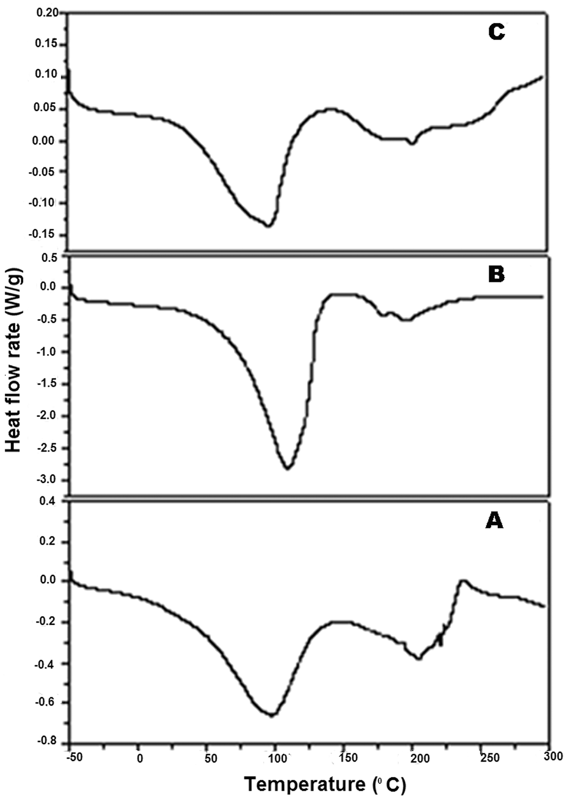
Surface charge density of polymers is important for electrostatic behavior during the adhesion process. The mucoadhesive nature of QPDMAEMA particle was further confirmed using the texture analyzer. Based on the physiological condition in the gastrointestinal (GI) tract after oral administration, mucoadhesive samples could not be forced to attach directly to the mucosa. So, the contact force employed was kept at the lowest, i.e. 0.05 N. The maximum detachment force (Fmax) and work of adhesion (Wad) are depicted in (Figure 6(a) and (b)). The maximum detachment force for PDMAEMA (A) and QPDMAEMA was calculated from the force versus distance curve. Fmax and Wad were higher for QPDMAEMA 1. This can be attributed to the electrostatic interaction of the positive charge of −NH3+ with the negatively charged sialic acid residues of the mucus, which further improved the particulate transport across the intestinal mucosa. Quaternized derivative with high positive charge density could establish specific bioadhesive interactions with mucosal tissues because of their ability to inter-diffuse across the mucus network. This is also evident from the adsorption studies of mucin. A strong interaction between the hydrogel and mucin was detected. It was found that the amount of mucin adsorbed increased with time and concentration of mucin. In DSC thermograms, the endothermic peak at about 210℃ for mucin, that characterized its melting, was also observed for QPDMAEMA/mucin physical blend with a slight shift. This evidenced that some amount of mucin had been conjugated onto QPDMAEMA particles through electrostatic interactions.
25
The shift in endothermic peaks confirmed the interaction of mucin with the polymer (Figure 7).
The plot of maximum detachment force and work of adhesion for PDMAEMA (A) and QPDMAEMA obtained by adding 0.5 mL (B) and 1 mL (C) of methyliodide tested with small intestinal mucosa) from the mucoadhesive test using texture analyzer. PDMAEMA: polydimethylaminoethylmethacrylate and QPDMAEMA: quaternized PDMAEMA. DSC thermograms of mucin (A), mucin complexed with PDMAEMA (B) and QPDMAEMA (C). PDMAEMA: polydimethylaminoethylmethacrylate and QPDMAEMA: quaternized PDMAEMA.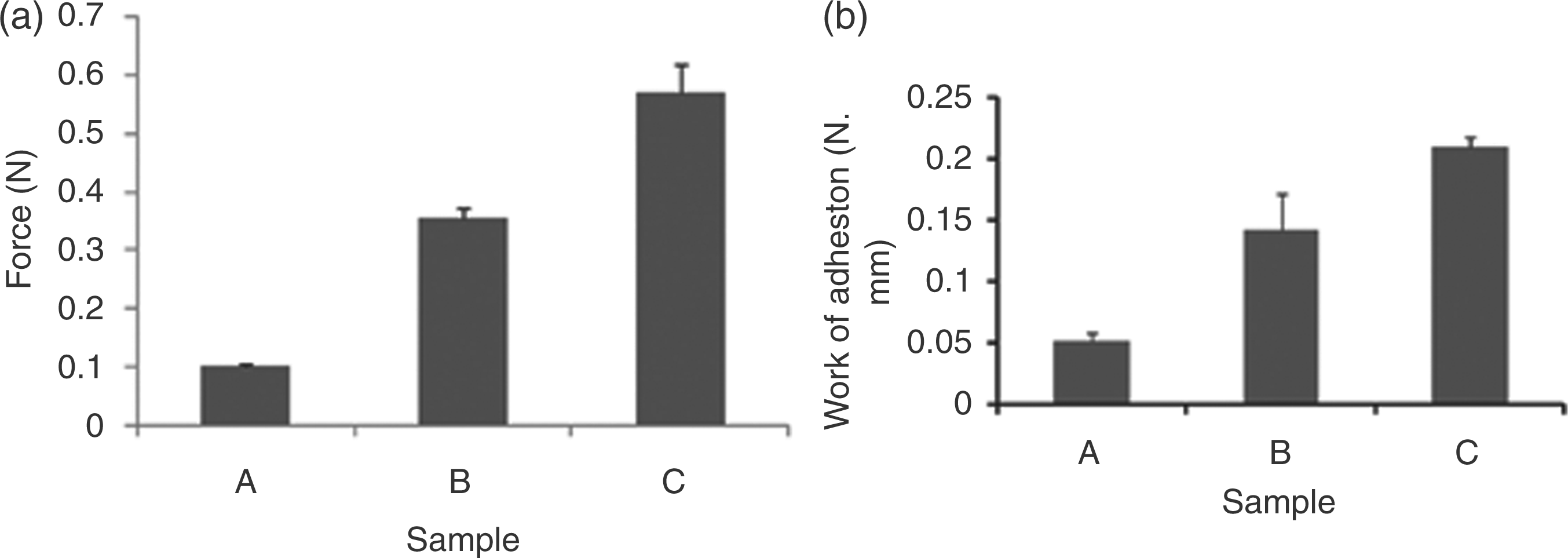
Drug loading was achieved by diffusion filling method (remote loading) in which the dried particles were exposed to insulin. By this method, a higher concentration of insulin was achieved inside the particles without affecting its biological activity, as evident from ELISA studies. In vitro release studies of the insulin-loaded, quaternized particles, were carried out at gastric and intestinal pH. QPDMAEMA particles showed a burst release at acidic and alkaline conditions due to the swelling nature of these particles, followed by diffusion of insulin. For oral protein delivery systems, minimal release in gastric pH is appropriate as it may saves the loaded insulin and increases the bioavailability compared to that of a matrix which does not exhibit this property. Therefore, the particles were coated with Eudragit L100 to prevent the release of insulin at gastric pH. Eudragit L100 is a pH responsive anionic polymer which is soluble in intestinal fluid. Zeta potential of Eudragit-coated, insulin-loaded particles indicated that the enteric coating have been eroded on dispersion of these particles in alkaline condition, retaining the positive surface charge at intestinal pH. As evident from the release profile, enteric-coated (Eudragit L100-55) particles protected insulin from degradation by gastric juice and allowed it to be released in the region of the GI tract of pH > 6. Eudragit-coated particles exhibited slow release of insulin at a pH of 1.2, which may be, probably due to decreased water entrance (no swelling) and unfavorable diffusion of insulin. At intestinal pH, enteric coating dissolved and about 62% of insulin is released in the first hour. It is suggested that the release of insulin is controlled by the swelling followed by diffusion. A sustained release of insulin was achieved by coating these particles with Eudragit L100 in the intestinal fluid.
26
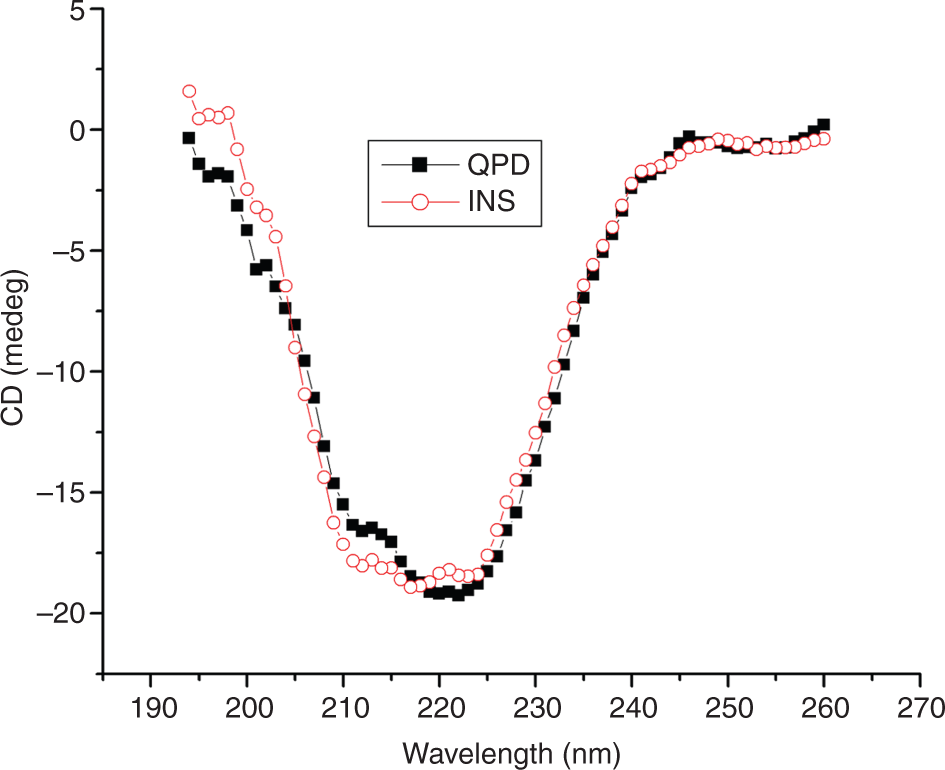
Figure 8 shows the release profile of Eudragit coated quaternized polymer at a pH of 1.2 and 6.8. Gastric retention time of particles on oral administration has been noted to be ∼2 h. In this investigation, about 15% of insulin is released in the second hour at gastric pH whereas 75% of insulin is released at intestinal pH in the second hour. CD studies proved that the released insulin were capable of maintaining the conformation of insulin (Figure 9).
Release studies of Eudragit L100 coated (Eudragit-coated QPDMAEMA) and non-coated QPDMAEMA at pH 1.2 and 6.8. QPDMAEMA: quaternized PDMAEMA. CD spectra of native insulin (□ INS) and insulin released from insulin-loaded QPDMAEMA (▪ QPDMAEMA). CD: circular dichroism and QPDMAEMA: quaternized PDMAEMA.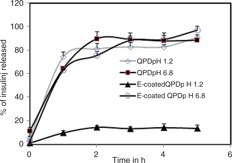
The particles exhibited a weak chymotrypsin inhibitory effect at neutral pH (7% for unmodified and 11% for modified polymer). This may be attributed to the formation of complexes with divalent metal ions like calcium, as evident from calcium-binding studies. Luessen et al.
27
suggests the role of some non-specific interactions such as van der Waals and electrostatic interaction of polymer with enzymes for inhibition process. Sajeesh et al.10 have reported that mucoadhesive polymeric particles with weak inhibitory effect may reduce the proteolytic attack in the small intestine and can localize the delivery systems onto the intestinal wall without exposing the active ingredient directly to the intestinal fluids.
10
In vitro uptake studies proved that the particles are capable of crossing the intestinal cell wall (Figure 10). It is reported that calcium ions are essential for the tight junction integrity. Smith et al.
28
have reported that cationic polymers, such as chitosan, are able to reversibly open the tight junctions between enterocytes, allowing the transport of macromolecular drugs. Calcium chelators such as chitosan can disturb the cell–cell adhesion phenomena by depleting the concentration of extracellular calcium ions, and this may in turn lead to the uptake of the particle. Its protonated form can interact with the epithelial tight junctions, inducing a redistribution of actin filaments and of the tight junction protein ZO−1.
29
The amount of calcium that binds to the sub-microparticles was determined from the free concentration of calcium before and after binding. Results show that the calcium-binding capacity of the modified derivative was 18.31 ± 1.76%. Literature suggests that the incubation of Caco-2 epithelial cells in calcium-free solution rapidly increased paracellular permeability, indicating a decreased tightness of a junction
30
The tight junctions present in the intestinal epithelial cells are linked by the presence of divalent ions like calcium and magnesium. As discussed earlier, calcium-binding capacity of quaternized particles helps in the opening of tight junctions that can act as a driving force for insulin absorption through the intestinal wall through paracellular pathway.
31
Thus, the binding of intracellular calcium by QPDMAEMA may cause the opening of a tight junction, and thus, the permeability of drugs may increase (Figure 11). Figure 11 confirmed the transport of large hydrophilic compounds via the paracellular route as well as the mechanism of action of the polymer in which redistribution of the cytoskeletal F-actin is provoked, which leads to the opening of the tight junctions. The cationic polymer can interact with anionic sialic acid residues on the surface of the epithelial cells which contains fixed negative charges. Cationic polymers are able to reversibly open the tight junctions between enterocytes, allowing the transport of macromolecular drugs.
32
He et al.
33
had proposed a salt bridge effect for the interaction of positively charged chitosan with the negatively charged mucus glycoprotein, but subsequently it was demonstrated that positive charge on the surface of chitosan could give rise to a strong electrostatic interaction with mucus or with a negatively charged mucosal surface. In addition to this, residual positive charge could induce opening of the epithelial tight junctions. It was observed that trimethylated chitosan provoked a redistribution of the cytoskeletal F-actin, a phenomenon that appeared to correlate well with the opening of epithelial tight junctions.
34
Previous reports shows that absorption enhancers can induce structural separation of the tight junctions in Caco-2 cells and parallel changes in F-actin distribution. The control cells showed continuous perijunctional F-actin rings required for the maintenance of tight junction integrity. QPDMAEMA showed a total disruption of F-actin distribution. Thus, QPDMAEMA having a high positive charge modulates opening of tight junctions by actin filament dislocation. Therefore, the development of quaternized hydrogel particles could be a promising strategy in developing specific bioadhesive interactions with the intestinal mucosa for efficient delivery of insulin.
Fluorescent micrographs of effect of FITC-labeled insulin PDMAEMA (A) and QPDMAEMA (B) on Caco-2 cells. FITC: fluorescein isothiocyanate; PDMAEMA: polydimethylaminoethylmethacrylate; and QPDMAEMA: quaternized PDMAEMA. Confocal micrographs of effect of rhodamine phalloidin labeled PDMAEMA (A) and QPDMAEMA (B) on Caco-2 cells. PDMAEMA: polydimethylaminoethylmethacrylate; and QPDMAEMA: quaternized PDMAEMA.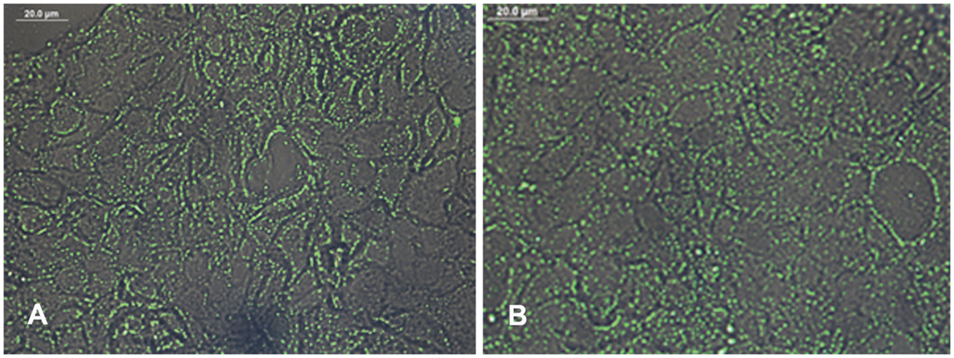
Conclusions
Cationic hydrogel sub-microparticles based on synthetic polymer PDMAEMA for oral delivery of insulin was successfully synthesized. QPDMAEMA was found to be non-toxic. Moreover, this matrix, capable of retaining positive charge at intestinal pH, enhanced mucoadhesion through electrostatic interactions, leading to increase in retention time in the intestine. It exhibited calcium-chelating property which helped in improving the permeability of intestinal cells by loosening the tight junctions. Thus, cationic modification of synthetic polymers could be an efficient strategy for successful oral delivery of proteins across mucosal barriers.
Footnotes
Acknowledgements
Authors thank Dr K. Radhakrishnan, Director, and Dr G.S. Bhuvaneswar, Head, BMT Wing, SCTIMST, Thiruvananthapuram, for providing facilities. The authors also thank CSIR for Senior Research Fellowship and Dr M.R Rekha, Mr Willi Paul, Mr Suresh Babu, and Dr Ajay Ghosh for cell culture, AFM, texture analyzer, IR, and CD studies.
Funding
This study was supported by the Department of Science and Technology, Government of India through the project ‘Facility for nano/microparticle based biomaterials – advanced drug delivery systems’ #8013, under the Drugs & Pharmaceuticals Research Program and Council of Scientific and Industrial Research, India.
References
Supplementary Material
Please find the following supplemental material available below.
For Open Access articles published under a Creative Commons License, all supplemental material carries the same license as the article it is associated with.
For non-Open Access articles published, all supplemental material carries a non-exclusive license, and permission requests for re-use of supplemental material or any part of supplemental material shall be sent directly to the copyright owner as specified in the copyright notice associated with the article.