
Abstract
Conventional and silicone hydrogels as models for contact lenses were prepared to determine the effect of the presence of hyaluronic acid on lysozyme sorption and denaturation. Hyaluronic acid was loaded into poly(2-hydroxyethyl methacrylate) and poly(2-hydroxyethyl methacrylate)/TRIS – methacryloxypropyltris (trimethylsiloxy silane) hydrogels, which served as models for conventional and silicone hydrogel contact lens materials. The hyaluronic acid was cross-linked using 1-ethyl-3-(3-dimethylaminopropyl)-carbodiimide in the presence of dendrimers. Active lysozyme was quantified using a Micrococcus lysodeikticus assay while total lysozyme was determined using 125-I radiolabeled protein. To examine the location of hyaluronic acid in the gels, 6-aminofluorescein labeled hyaluronic acid was incorporated into the gels using 1-ethyl-3-(3-dimethylaminopropyl)-carbodiimide chemistry and the gels were examined using confocal laser scanning microscopy. Hyaluronic acid incorporation significantly reduced lysozyme sorption in poly(2-hydroxyethyl methacrylate) (p < 0.00001) and poly(2-hydroxyethyl methacrylate)/TRIS – methacryloxypropyltris (trimethylsiloxy silane) (p < 0.001) hydrogels, with the modified materials sorbing only 20% and 16% that of the control, respectively. More importantly, hyaluronic acid also decreased lysozyme denaturation in poly(2-hydroxyethyl methacrylate) (p < 0.005) and poly(2-hydroxyethyl methacrylate)/TRIS – methacryloxypropyltris (trimethylsiloxy silane) (p < 0.02) hydrogels. The confocal laser scanning microscopy results showed that the hyaluronic acid distribution was dependent on both the material type and the molecular weight of hyaluronic acid. This study demonstrates that hyaluronic acid incorporated as a wetting agent has the potential to reduce lysozyme sorption and denaturation in contact lens applications. The distribution of hyaluronic acid within hydrogels appears to affect denaturation, with more surface mobile, lower molecular weight hyaluronic acid being more effective in preventing denaturation.
Introduction
A major issue with soft contact lenses, both conventional and silicone hydrogels, is deposition of tear film components (proteins, lipids, mucins) onto the surface or into the matrix.1–3 Factors affecting protein deposition include porosity, surface charge, protein charge, hydrophilicity, and material chemical composition.4–15 These tear protein deposits can negatively affect transparency and comfort.16–18 Tear film deposits are also believed to play a role in the development of such complications as giant papillary conjunctivitis (GPC).19,20 These tear film proteins can undergo structural changes or become denatured when deposited onto contact lenses 21 and it has been theorized that the protein denaturation is related to the ability of the protein to act as an antigen in the development of GPC. 22 While it is thought that the ideal contact lens would minimize or possibly even eliminate any deposition onto the lens surface, 4 reducing protein denaturation may be as important in the prevention of undesired side effects.
Lysozyme, one of the main proteins in the tear film, is a low molecular weight (14.3 kDa), positively charged protein (isoelectric point pH = 11.1). Studies of lysozyme sorption and denaturation have shown that conventional poly(2-hydroxyethyl methacrylate)-based hydrogels deposit significantly more lysozyme than silicone-based hydrogel materials in vitro23,24 and in vivo25,26 but that silicone hydrogels exhibit a higher percentage of denatured lysozyme.24,25
Hyaluronic acid (HA) is a lubricating glycosaminoglycan that is found throughout the human body, including the cartilage of the knee 27 and the vitreous humor of the eye. 28 HA has been investigated in a variety of biomedical applications29–38 including various ophthalmic uses.39–46 Previous studies by our group have found that HA incorporated as an internal wetting agent has the ability to decrease sorption of the tear film proteins lysozyme, albumin and immunoglobulin G (IgG) onto model conventional hydrogel 47 and lysozyme onto model silicone hydrogel 48 contact lens materials in vitro. A recent study has also shown that an HA-containing contact lens solution prevents lysozyme denaturation. 49 Given the importance of denaturation in adverse contact lens effects, it is of interest to examine whether this incorporated HA affects denaturation of the sorbed protein and to examine the physical location of the wetting agent in the model lens materials, in order to correlate this to the observed protein interactions.
Materials and methods
Materials
2-Hydroxyethyl methacrylate (HEMA), ethylene glycol dimethacrylate (EGDMA), diaminobutane-4 (DAB-4) G1 dendrimer, benzoyl peroxide and 1-ethyl-3-(3-dimethylaminopropyl)-carbodiimide (EDC), 6-aminofluorescein, lysozyme (chick), and Micrococcus lysodeikticus were purchased from Sigma Aldrich (Oakville, ON, Canada). Methacryloxypropyltris (trimethylsiloxy) silane (TRIS) was purchased from Gelest (Morrisville, PA, USA). Irgacure was purchased from CIBA (Mississauga, ON, Canada). HA was purchased from LifeCore Biomedical (Chaska, MN, USA).
Hydrogel preparation
HEMA monomer was passed through a column packed with inhibitor remover to remove 4-methoxyphenol hydroquinone (MEHQ). The cross-linker, EGDMA (1% by weight), water (1:1 wt:wt HEMA), and the photoinitiator benzoyl peroxide (1% by weight and dissolved in THF) were added to the mixture under constant stirring. The mixture was transferred to a Teflon® mold and was placed in a 400 W UV chamber (Cure Zone 2 Con-trol-cure, Chicago, IL, USA) for 25 min to polymerize. Following polymerization, the formed hydrogels were placed in a 50℃ oven overnight to ensure complete reaction. Hydrogels were then placed in water for at least 24 h to swell and remove unreacted monomers. The hydrogels were cut into ¼” diameter discs and then dried for HA incorporation.
For pHEMA/TRIS hydrogels, the HEMA and TRIS were passed through separate columns packed with inhibitor remover and mixed to create a mixture that was 90% HEMA and 10% TRIS by weight. EGDMA (5% by weight) was then added to the HEMA/TRIS mixture followed by the photoinitiator, Irgacure (0.5% by weight), under constant stirring. The curing procedure was the same as for the pHEMA hydrogels. The resulting gels were similar to our previous work in terms of transparency and water uptake.
Hyaluronic acid incorporation
HA was covalently incorporated using dendrimers as in our previous studies.47,48 Briefly, HA of varying molecular weights (4.7, 16, 35, 132, and 910 kDa) and DAB-4 dendrimer (both 5 g/L) were dissolved in a 30:70 ethanol:water solution. Dried hydrogels were soaked in these solutions for 4 days at 4℃ to load the HA and DAB-4 into the gels. Control pHEMA/TRIS hydrogels were swollen in 30:70 ethanol:water to remove any unreacted silicone. Following loading, the hydrogels were dried and then placed in an EDC solution (1% by weight) for 24 h at 25℃ to allow for HA cross-linking within the hydrogel networks. Following cross-linking, the hydrogels were soaked in water for 48 h to remove any unreacted dendrimers and HA. The samples were then dried and stored for analysis.
Determination of total lysozyme
The total amount of sorbed lysozyme was determined using lysozyme labeled with iodine 125 (125-I). Lysozyme was labeled with 125-I using the iodine monochloride (ICl) method. Radiolabeled lysozyme solution was passed through two columns packed with AG 1-X4 (Bio-Rad, Hercules, CA, USA) to remove unbound 125-I. The columns were then rinsed with phosphate buffered saline (PBS) to ensure that all of the labeled lysozyme had been removed. Free iodide was determined using trichloroacetic acid (TCA) precipitation and the percentage of free iodide in a labeled lysozyme solution was typically less than 3%.
Lysozyme solutions (2 mg/ml, 2% labeled in PBS – pH 7.4) were prepared. The pHEMA hydrogels were incubated in 2 ml of lysozyme solution (2 mg/ml in PBS – pH 7.4) for 2 h in a water bath at 37℃; the pHEMA/TRIS gels were incubated for 2 weeks under the same conditions. The silicone hydrogels were incubated for a longer period of time due to the fact that silicone hydrogels sorb less protein than pHEMA-based hydrogels, as mentioned above, and these materials are typically approved for longer wear than conventional hydrogel materials. Following sorption, the hydrogels were rinsed 3 times for 5 min each in PBS to remove any loosely adherent protein. The hydrogels were then blotted dry with a Kimwipe and the radioactivity was determined using a Wizard 3 1480 Automatic Gamma Counter (Perkin Elmer, Waltham, MA, USA); radioactivity was converted to a protein amount using a standard curve.
Quantification of active lysozyme
The activity of deposited lysozyme on the hydrogels in vitro was determined using previously reported methods.24,50,51 The hydrogels were incubated under the same conditions as for the total lysozyme. Following sorption, the hydrogels were rinsed 3 times, 5 min each to remove loosely adhered protein. The hydrogels were then shaken in a 50: 50 acetonitrile (ACN): 0.2% trifluoroacetic acid (TFA) (v:v) solution in the dark for 24 h to extract the sorbed protein. 52 The extraction solution was aliquoted, dried using a Savant Speed Vac (Halbrooke, NY, USA), and then frozen and stored at –70℃. The amount of active lysozyme was determined using Micrococcus lysodeikticus. Extracted lysozyme was resuspended in buffer and then added to 1 ml of bacteria solution. Changes in the optical density were measured at 450 nm, using the cuvette component of a Multiscan Spectrum ELISA Plate Reader (Thermo Labsystems, Waltham, MA, USA), with respect to time. The changes correlate to the amount of active lysozyme in the solution. The percentage of active lysozyme was determined.
Determination of extraction efficiency
Following lysozyme quantification, hydrogels containing sorbed radiolabeled lysozyme were placed in vials containing a solution of 50:50 ACN:0.2% TFA. Hydrogels were shaken in this solution for 24 h to extract lysozyme. Following extraction, the radioactivity of the extraction solution was determined using the Wizard 3 1480 Automatic Gamma Counter. The extraction efficiency was determined using the following equation:
HA distribution
The distribution of HA within the gels was determined by labeling the HA with 6-aminofluorescein and examining the materials using a confocal laser scanning microscope.
The HA (5.1, 35, and 910 kDa) was labeled with a 6-aminofluorescein using EDC chemistry. Briefly, 200 mg of HA, 10 mg of 6-aminofluorescein, and 70 mg of EDC were dissolved in water and stirred for 24 h. The mixture was then dialyzed using a 3500 molecular weight cutoff membrane against MilliQ water for a minimum of 48 h for purification. The mixture was lyophilized and stored at −20℃ in the dark until use. The labeled HA was then loaded and cross-linked into pHEMA and pHEMA/TRIS hydrogels using the methods described above.
Prior to analysis, hydrogels were swollen to equilibrium in PBS (pH 7.4). The fluorescence intensity of the conjugated HA was determined with a Zeiss 510 confocal laser scanning microscope (Zeiss Inc., Toronto, ON, Canada), using the Argon Laser for excitation at 488 nm and a long pass filter of >505 nm to determine the emission intensity. Four scans were undertaken on each sample. The Zeiss custom software associated with the microscope and ImageJ (Bethesda, MD, USA) were used to produce an image of fluorescence intensity with respect to depth to show the HA distribution within the hydrogel in the swollen state. This method has been used previously to determine the location of proteins sorbed in/onto contact lenses.53,54
Statistics
The statistical significance of the results of the modified gels compared to the controls was determined using Student’s t-tests with 1-tail and unequal variance. The differences between the materials containing different molecular weights of HA were determined using single-factor ANOVAs.
Results and discussion
Protein sorption and denaturation
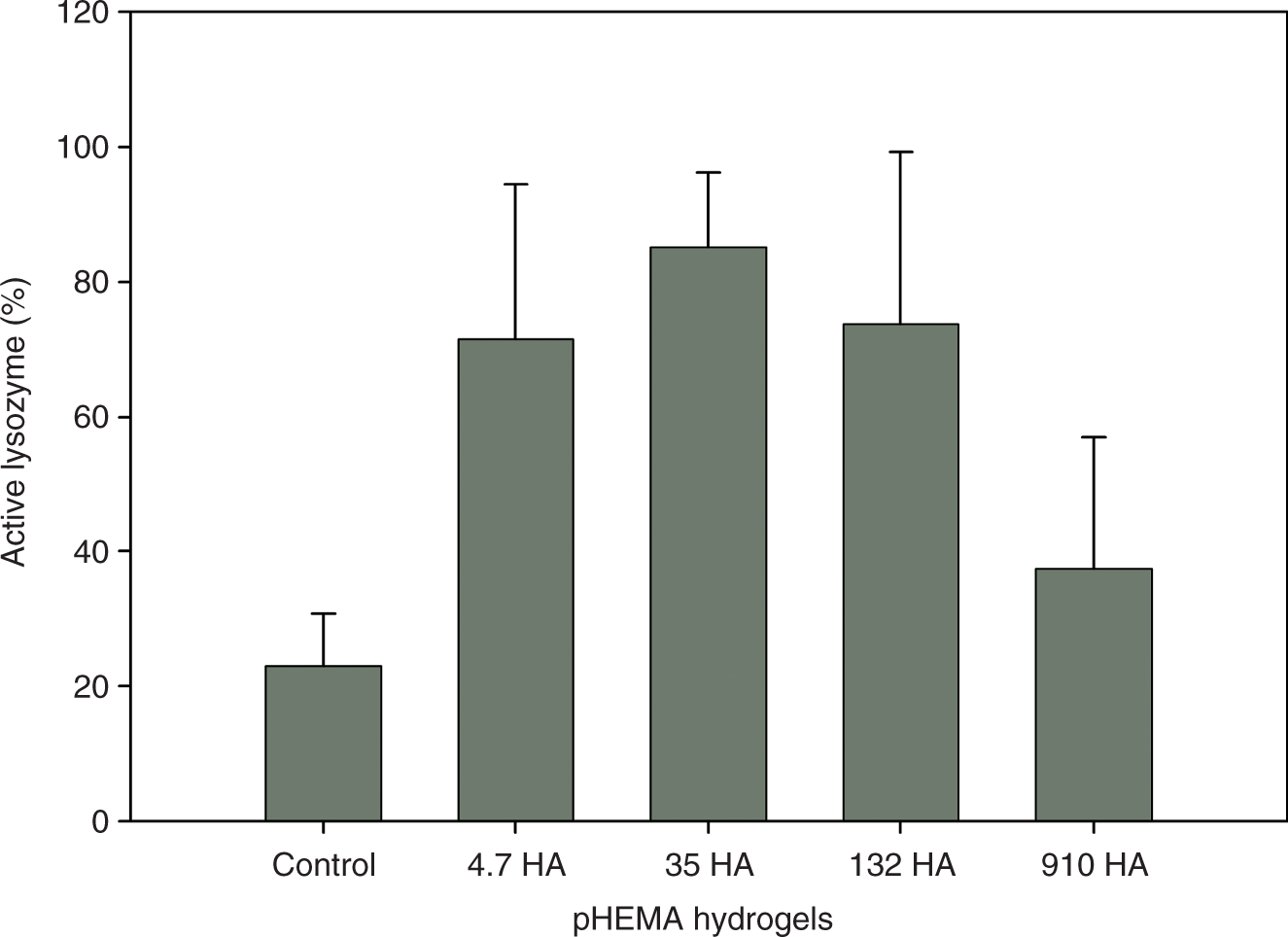
The results of this study show that HA-modified pHEMA hydrogels deposit significantly less lysozyme (p < 0.00001) than control pHEMA hydrogels (Figure 1), taking up approximately 20% of the control. In general, the molecular weight of HA was not found to have a significant (p > 0.348) effect on the amount of protein associated with the materials. This work demonstrates that in addition to sorbing less protein, the incorporation of HA leads to significantly decreased denaturation of lysozyme on the surface of pHEMA hydrogels (p < 0.005) (Figure 2), with the exception of 910 kDa HA modified materials.
Mean (±SD) amount of lysozyme sorbed onto poly(2-hydroxyethyl methacrylate) (pHEMA) hydrogels following 2 h of in vitro lysozyme loading (n ≥ 3). The hyaluronic acid (HA)-containing materials sorbed significantly less lysozyme than the controls (p < 0.00001), with no significant molecular weight effect (p > 0.348). Mean (±SD) amount of active lysozyme sorbed onto poly(2-hydroxyethyl methacrylate) (pHEMA) hydrogels expressed as a percentage of the total lysozyme sorbed following 2 h of in vitro loading (n ≥ 3). The hyaluronic acid (HA)-containing materials, with the exception of the 910 kDa HA, had a significantly higher percentage of active lysozyme compared to the controls (p < 0.005).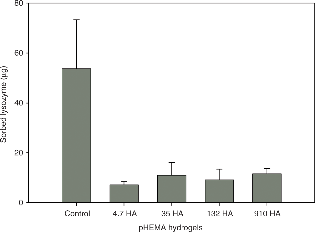
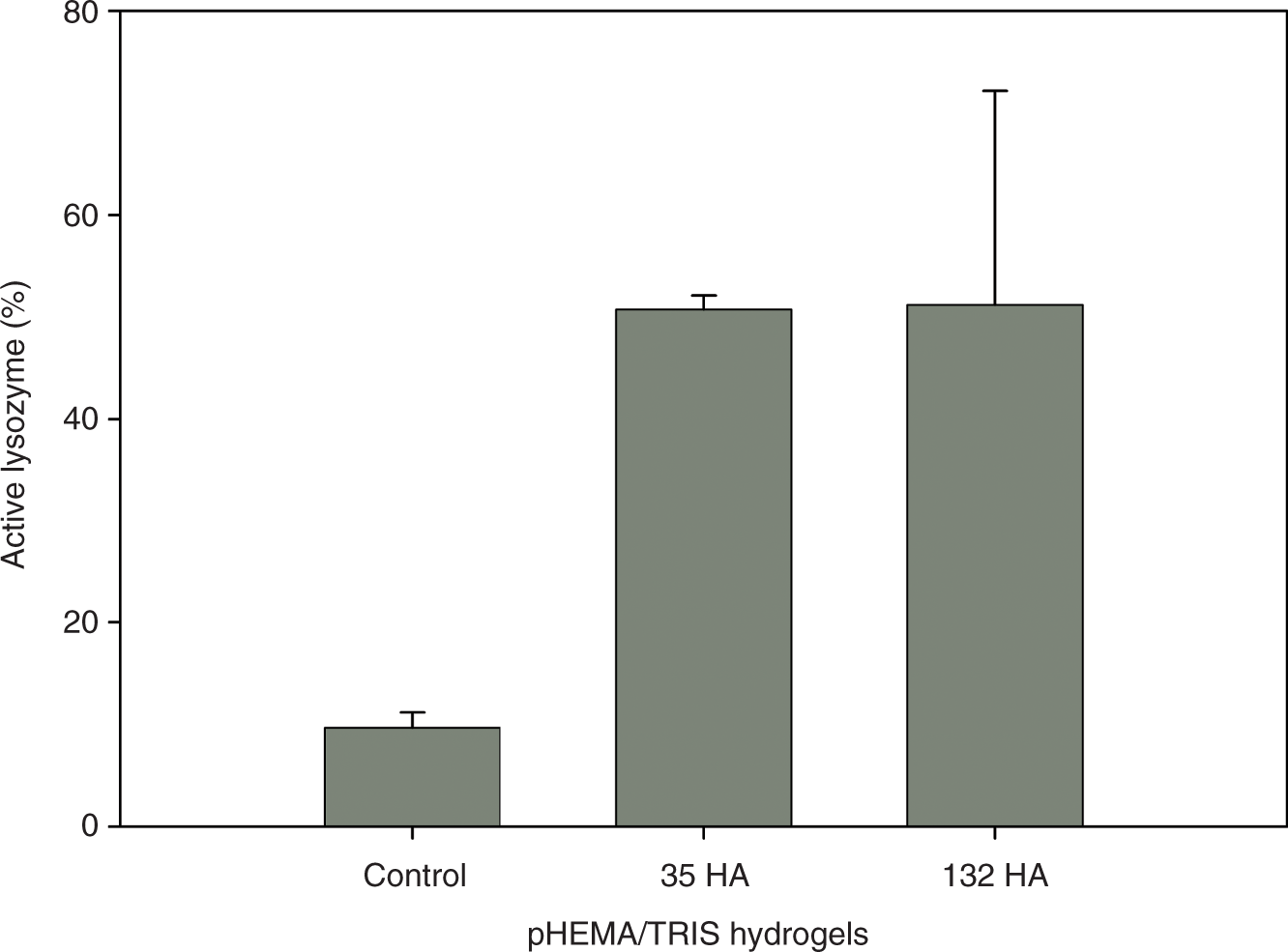
As was seen with the pHEMA hydrogels, the results for the pHEMA/TRIS hydrogels revealed that HA-modified silicone hydrogels sorb significantly less lysozyme than controls after a 2-week in vitro loading period (p < 0.0002) (Figure 3), with the HA-modified hydrogels sorbing approximately 16% of that associated with the controls. The molecular weight of HA had no effect on the amount of lysozyme sorption onto pHEMA/TRIS hydrogels (p > 0.899). The results of the lysozyme denaturation study showed that the presence of HA significantly decreased lysozyme denaturation (p < 0.009) (Figure 4), with no difference between the different HA molecular weights (p > 0.962). Surprisingly, the percentage of active lysozyme on the control pHEMA/TRIS gels was approximately 50% of that of the control pHEMA gels when a lower percentage would seem more likely given the much longer loading time. However, previous studies have shown that protein sorption kinetics are affected by the type of contact lens18,23,55,56 and it is likely that the sorption kinetics are different for these two materials. Also, during the 2-week loading period for the pHEMA/TRIS gels, it is possible that the denatured protein present on the hydrogel surface hindered interactions between the hydrogel surface and additional lysozyme, preventing further denaturation.
Mean (±SD) amount of lysozyme sorbed onto poly(2-hydroxyethyl methacrylate)/TRIS – methacryloxypropyltris (trimethylsiloxy silane) (pHEMA/TRIS) hydrogels following 2 weeks of in vitro lysozyme loading (n ≥ 5). The hyaluronic acid (HA)-containing materials sorbed significantly less lysozyme than the controls (p < 0.0002). The molecular weight of the HA did not appear to affect lysozyme sorption (p > 0.899). Mean (±SD) amount of active lysozyme sorbed onto poly(2-hydroxyethyl methacrylate)/TRIS – methacryloxypropyltris (trimethylsiloxy silane) (pHEMA/TRIS) hydrogels expressed as a percentage of the total lysozyme sorbed following 2 weeks of in vitro loading (n ≥ 5). The hyaluronic acid (HA)-containing materials had a significantly higher percentage of active lysozyme compared to the controls (p < 0.009). The molecular weight of the HA had no effect on lysozyme denaturation (p > 0.962).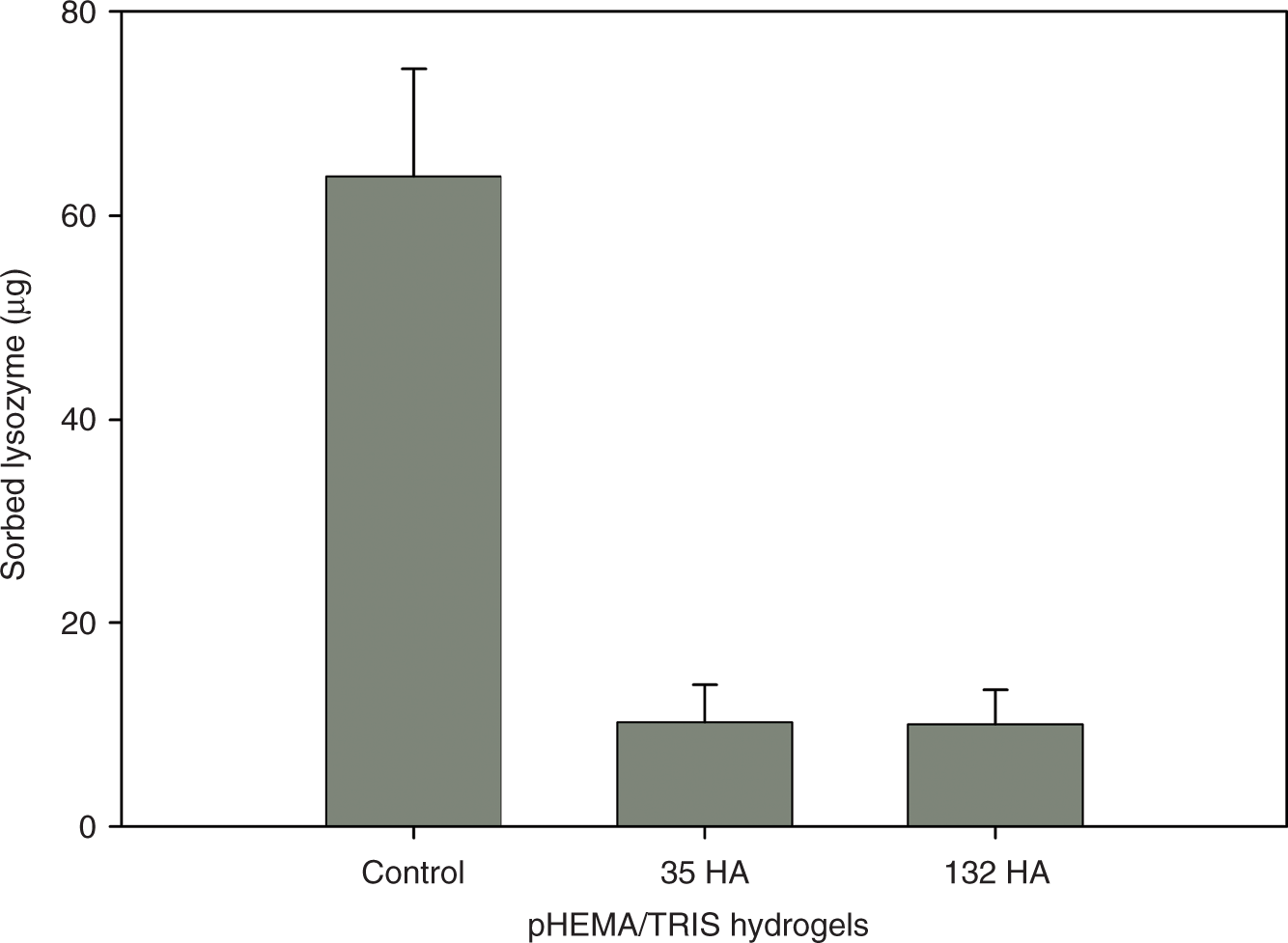
Previous studies have shown that the incorporation of cross-linked HA improves hydrophilicity and decreases lysozyme sorption in pHEMA 47 and pHEMA/TRIS 48 hydrogels. The decreased lysozyme sorption onto the HA-modified hydrogels compared to controls seen in this study was therefore expected. Protein denaturation has a strong correlation with the hydrophobicity of the material 25 so the increased hydrophilicity of the HA-modified hydrogels likely explains the decrease in protein denaturation. Our previous studies have shown that the incorporation of HA increases the equilibrium water content in these materials;47,48 lower water content contact lenses are associated with a greater degree of denaturation. 26 While the molecular weight of HA does not appear to affect the amount of lysozyme sorption, it does appear to have a slight effect on lysozyme denaturation. While the differences in the hydrophilicity of these materials as determined by water uptake were not shown to be significantly different in our previous work, it seems reasonable to hypothesize that subtle differences in hydrophilicity associated with different molecular weights of HA as well as differences between the location of the HA in the material likely contributed to the denaturation differences observed. Therefore, studies of the distribution of HA in the materials were performed.
Extraction efficiency
The results of the extraction efficiency study showed, not surprisingly, that lysozyme was extracted more efficiently from pHEMA hydrogels than from pHEMA/TRIS hydrogels (Figure 5). In general, the presence of HA had no effect on extraction efficiency for pHEMA hydrogels (p > 0.05). The extraction efficiency was not affected by the molecular weight of HA (p > 0.069). With the model silicone hydrogel materials, the addition of HA had no effect on extraction (p > 0.05) nor was there a molecular weight effect (p > 0.41). Although the extraction efficiency was not 100%, it is unlikely that the unextracted lysozyme would have any effect on the activity results. Due to the difficulty of removal, it is very likely that this remaining lysozyme is highly denatured and would therefore actually decrease the percentage of active lysozyme.
Mean (±SD) percentage of lysozyme extracted from poly(2-hydroxyethyl methacrylate) (pHEMA) (n ≥ 3) and poly(2-hydroxyethyl methacrylate)/TRIS – methacryloxypropyltris (trimethylsiloxy silane) (pHEMA/TRIS) (n = 10) hydrogels following a 24-h extraction process. The addition of hyaluronic acid (HA) in pHEMA hydrogels had no effect on the extraction efficiency (p > 0.05), with no molecular weight effect (p > 0.069). The presence of HA in pHEMA/TRIS hydrogels had no effect on the extraction efficiency (p > 0.05) and neither did the molecular weight of HA (p > 0.41).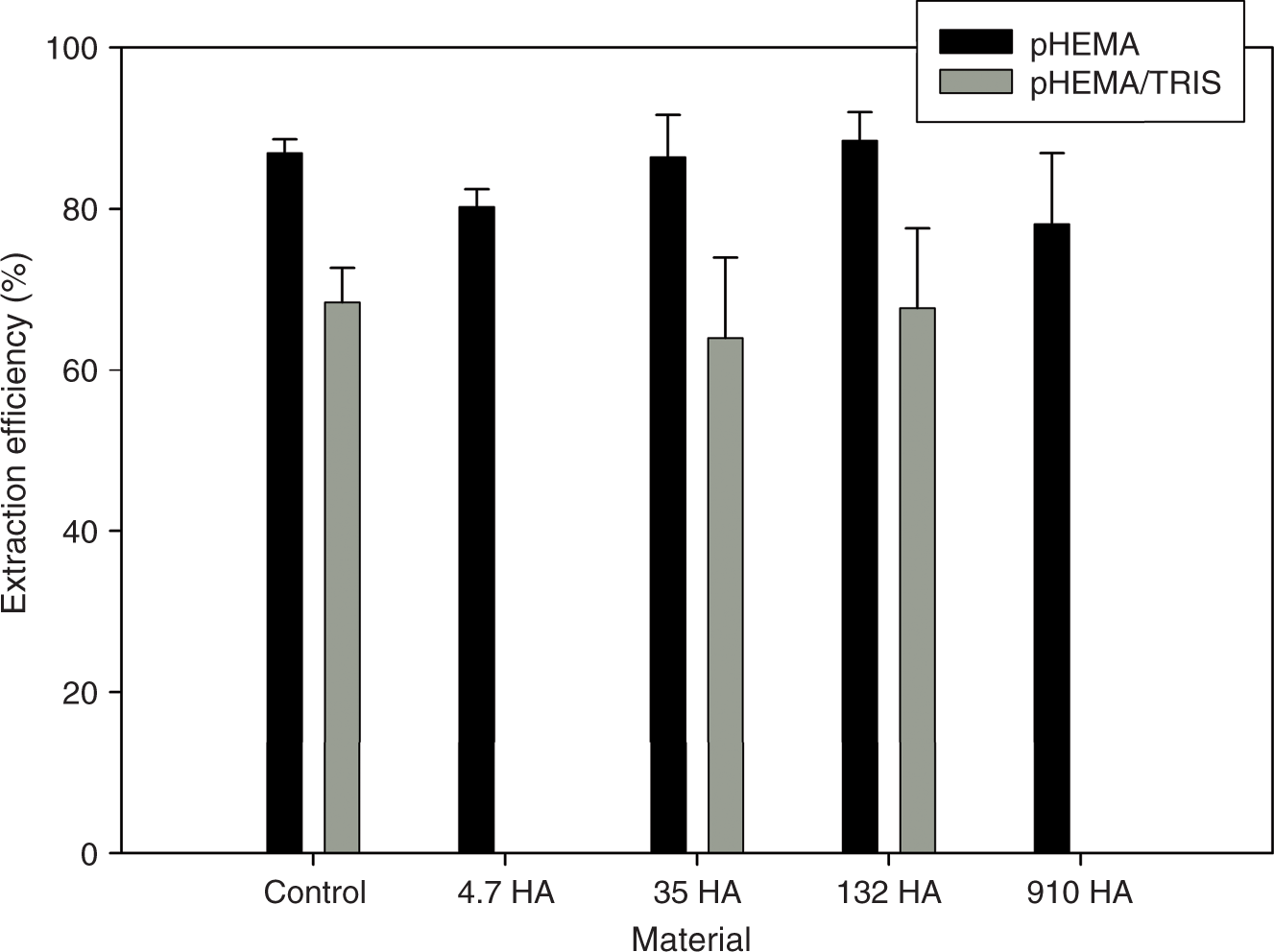
HA distribution
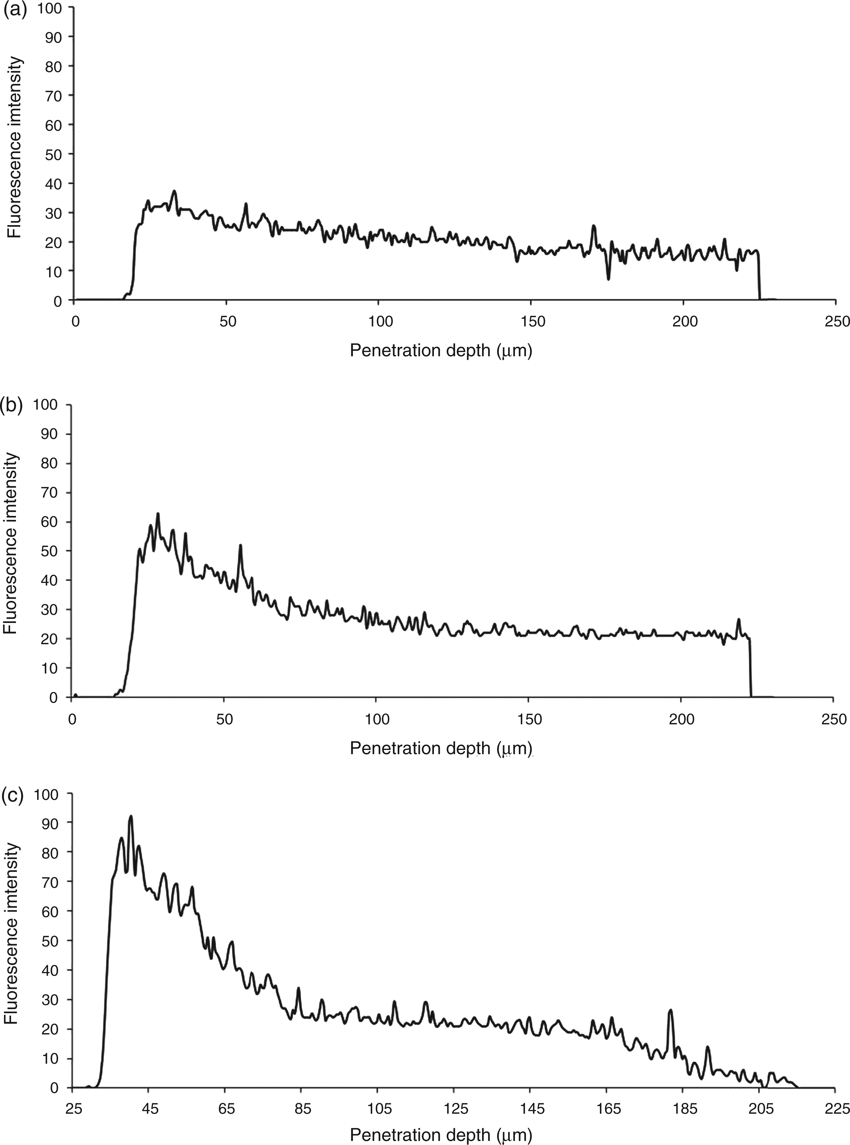
The HA distribution within the hydrogels was found to depend on both the material and the molecular weight of HA. For the pHEMA hydrogels, 5.1 and 910 kDa HA were distributed evenly throughout the hydrogel, whereas 35 kDa HA was present in greater amounts at and near the upper surface compared to the bulk. The HA distribution in pHEMA hydrogels is seen in Figure 6. In the pHEMA/TRIS hydrogels, the 5.1 kDa HA was distributed throughout the hydrogel, whereas 35 kDa and 910 kDa HA were distributed primarily in the surface region with some penetration into the hydrogel. The HA distribution in pHEMA/TRIS hydrogels is seen in Figure 7.
(a)–(c) Fluorescence intensity confocal laser scanning microscopy (CLSM) measurements through poly(2-hydroxyethyl methacrylate) (pHEMA) hydrogels containing 5.1, 35, and 910 kDa hyaluronic acid (HA). The fluorescence intensity at a given depth is representative of the amount of HA located at that depth. All three materials show evidence of HA being located in the bulk of the hydrogel for the depth investigated, with the 35 kDa HA showing an increased concentration of HA located at the surface, indicated by a stronger fluorescence intensity measurement at the surface. (a)–(c) Fluorescence intensity confocal laser scanning microscopy (CLSM) measurements through poly(2-hydroxyethyl methacrylate)/TRIS – methacryloxypropyltris (trimethylsiloxy silane) (pHEMA/TRIS) hydrogels containing 5.1, 35, and 910 kDa hyaluronic acid (HA). As with pHEMA hydrogels, the fluorescence intensity at a given depth is representative of the amount of HA located at that depth. The scans of all three materials provide evidence that HA is located in the bulk of the hydrogel, with the 35 kDa and 910 kDa HA showing accumulation at the surface, indicated by a higher fluorescence intensity measurement at the surface.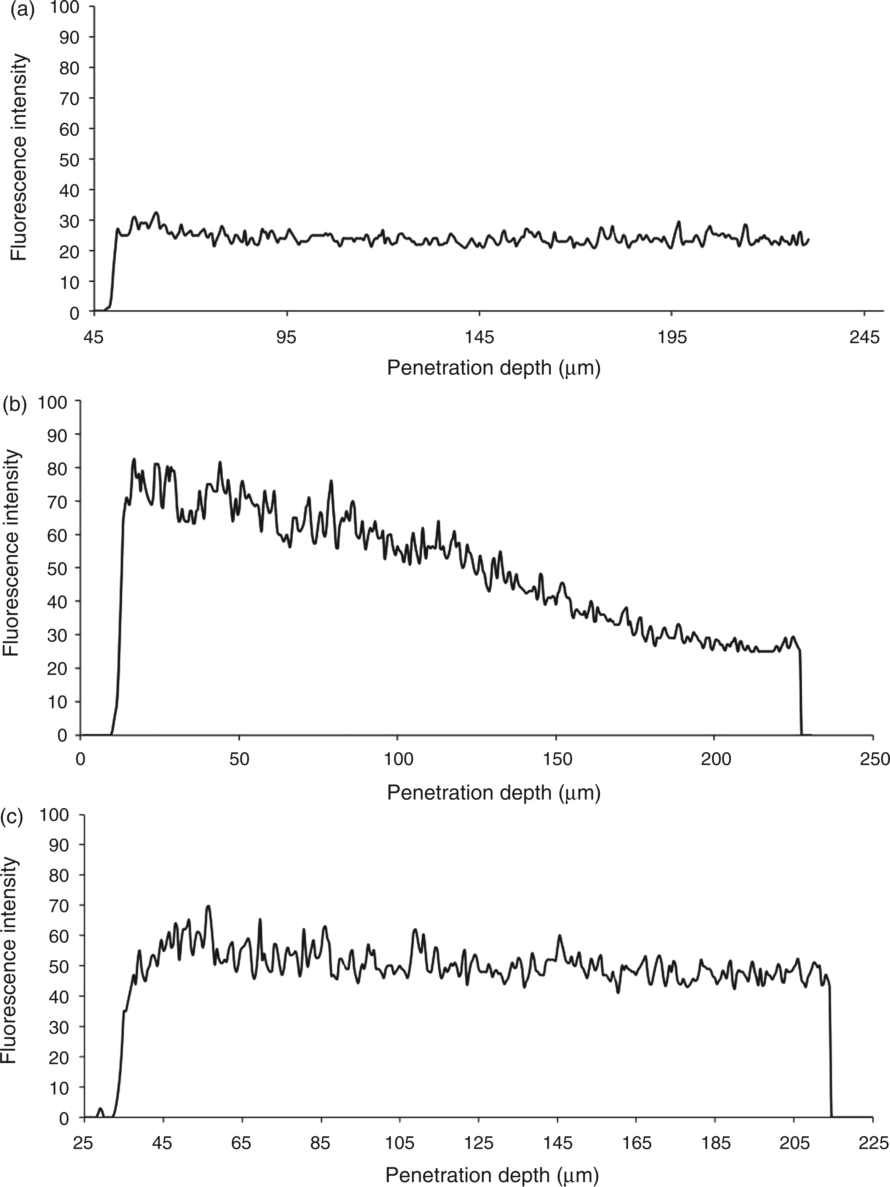
The distribution of HA appeared to be affected by both the material and the molecular weight of HA, although the differences were surprisingly less than what might have been expected based on previous studies using fluorescently labeled proteins.53,54 Clearly, the HA, even relatively high-molecular-weight HA, is more mobile and can penetrate these materials more effectively than proteins. Furthermore, previous work has shown that very high molecular weight HA can be released from model pHEMA hydrogels 47 and poly(vinyl alcohol) (nelfilcon) lenses 57 for prolonged time periods, demonstrating the mobility of even very high-molecular-weight HA chains within a hydrogel in an aqueous environment. Additionally, porous hydrogel matrix structures have been shown to favor protein matrix penetration 54 compared to lower water content materials where protein sorption occurs primarily as surface adsorption. 12 Although the pHEMA gels used in these studies have a lower water content and are therefore expected to be less porous than high water content lenses, these results suggest that penetration of HA into these materials is not problematic. Even more surprisingly, the penetration of HA into the silicone materials was also not seen to be an issue. While there was slightly more, particularly the high-molecular-weight, HA observed near the surface of the pHEMA/TRIS gels, the differences are relatively small. It is expected that the mobility of the HA once cross-linked into the gel and the amount of HA at the surface that will ultimately determine the level of protein sorption and denaturation. However, the nature of confocal laser scanning microscopy (CLSM) studies make quantification of the total amount of HA associated with the materials and the amount of HA at the different locations in the matrix impossible. Future studies will focus on attempting to quantify the amounts of HA associated with the different materials.
Hydrophilic polymer chains such as poly(ethylene oxide) (PEO) are believed to reduce protein adsorption by means of steric hindrance.
58
Increasing the number of the chains, as well as the mobility, has been shown to improve their ability of block protein from interacting with the surface.10,59 Therefore, it is believed that that HA prevents protein from attaching to the surface and becoming denatured using this same mechanism that has been postulated for PEO. Based on our protein sorption results, all HA-containing materials contain sufficient HA at the surface to reduce protein sorption. The denaturation results suggest that the mobility of the HA is also important in preventing lysozyme denaturation. In the cases where the HA is highly mobile and/or accumulates at the surface, HA denaturation is significantly decreased, as seen with 4.7 and 35 kDa HA in pHEMA hydrogels. Furthermore, when HA is distributed throughout the material but is immobile as might be expected with the high molecular weight of HA, there is less of a decrease in denaturation, as seen with 910 kDa HA in the pHEMA hydrogels. This concept is depicted schematically in Figure 8. Overall, lower, more mobile molecular weights of HA are more effective in preventing lysozyme denaturation and likely reducing the risk of the development of such conditions as GPC.
Schematic diagram showing that low-molecular-weight, mobile hyaluronic acid (HA) can interact with lysozyme at the surface of a contact lens and prevent denaturation (left) whereas the high molecular weight is too sterically hindered within the hydrogel to interact with the lysozyme to prevent denaturation (right).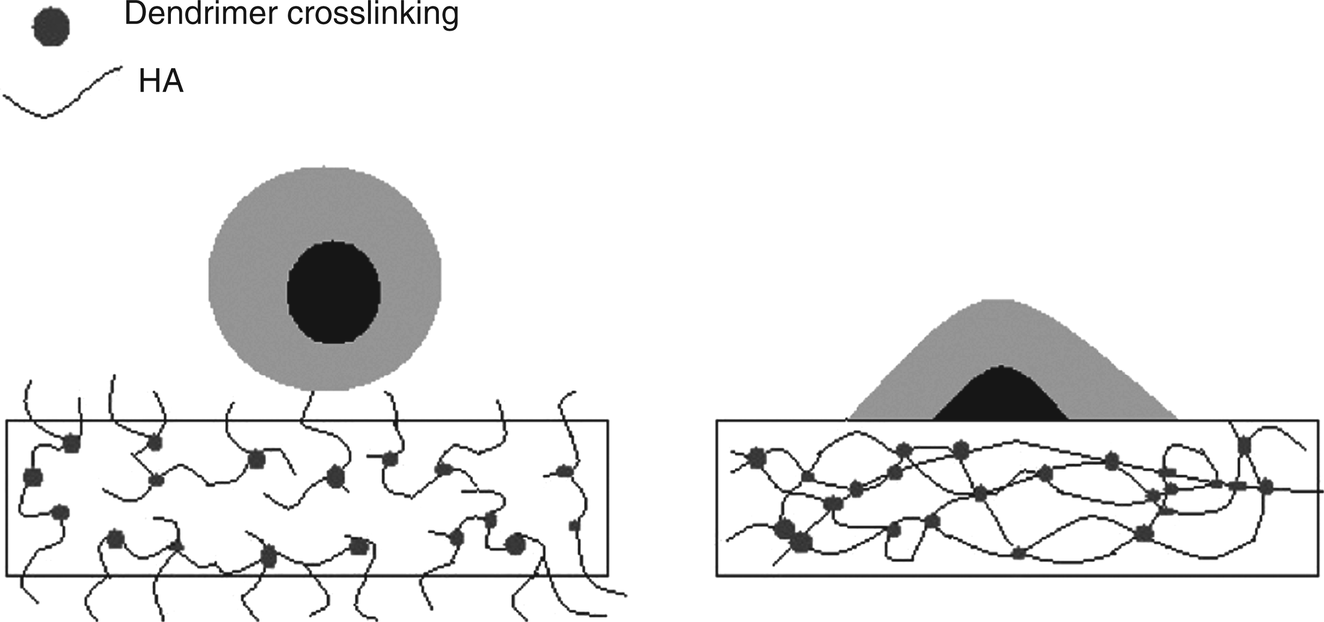
Conclusions
The results of this study showed that HA incorporation into hydrogel materials has the ability to decrease lysozyme sorption and denaturation in model conventional and silicone hydrogel contact lenses. The molecular weight of HA does not appear to have an effect on the quantity of lysozyme sorption but can affect the denaturation of lysozyme in pHEMA hydrogels. CLSM studies showed that despite a relatively even distribution of HA within all of the materials, there were differences in denaturation, suggesting that mobility of the polymer chains is important as would be expected based on studies using PEO. In conclusion, covalent incorporation of low molecular weight HA has potential to be used to prevent lysozyme sorption and denaturation and may be useful in contact lens applications.
Footnotes
Acknowledgements
Funding support from NSERC and 20/20 NSERC Ophthalmic Materials Network is gratefully acknowledged.
Funding
This work was funded by NSERC and 20/20 NSERC Ophthalmic Materials Network.