
Abstract
Electrospun polycaprolactone and poly(lacto-co-glycolide) membranes were loaded with biphasic calcium phosphate powder to facilitate osteoconductivity. Different concentrations of biphasic calcium phosphate powder were added to the polymer solution, and successful loading was confirmed by X-ray diffraction analysis, transmission electron microscope, and scanning electron microscope with energy-dispersive spectroscopy visualization. The effect of the added biphasic calcium phosphate on the polymer membrane was investigated in terms of the material’s tensile strength and strain, in vitro cytocompatibility, and in vivo tissue regeneration. It was observed that the tensile strength of the membranes increased with the addition of the biphasic calcium phosphate powder. Immersion in simulated body fluid solution for seven days leads to the formation of apatite-like deposits in the fibers, which further improved the mechanical stability. Moreover, proliferation and adhesion of osteoblast-like cells were more apparent upon the addition of the biphasic calcium phosphate powder as seen with the increasing cell density from (3-(4,5-dimethylthiazol-2-yl)-2,5-diphenyltetrazolium bromide assay and micrographs from scanning electron microscope and confocal microscopy. Sample membranes were also implanted to investigate the membrane’s ability to regenerate bone in a rat calvarium. Histological staining and micro-CT histomorphometric analyses showed neo-bone formation in the implanted rat skull.
Introduction
Bone biomaterials are studied extensively with the aim of developing suitable materials to direct tissue regeneration, guide repair, or replace damaged tissues. Different materials such as metals, ceramics, and polymers have been utilized as biomaterials and scaffolds for bone tissue engineering depending on the desired application and target placement. These materials have distinguishing characteristics that can be advantageous, but sometimes accompanied by compromising qualities. Bioceramics, like bioactive glass and calcium phosphates, resemble the mineral phase of natural bone, 1 which renders it to be biocompatible and display good bone-bonding adhesion. 2 The mechanism of the osteconductivity of calcium phosphates lies in its ability to accommodate its surface, the formation of carbonate hydroxyapatite, which accounts to 65% of total bone mass.3,4 However, ceramics are brittle and have low mechanical strength. Moreover, there is a limited choice of fabrication methods that can be utilized for ceramics compared to their polymer counterparts.
Polymers, on the other hand, are easily manipulated, and their material properties can be controlled according to the application. Polycaprolactone (PCL) is a biodegradable polymer that has been used extensively in the fabrication of tissue engineering scaffolds. 5 It is known to possess considerable mechanical strength and biocompatibility. Several studies have attested implanting PCL scaffolds producing very minimal or non-existent inflammatory symptoms.6–8 Poly(lacto-co-glycolic acid (PLGA) is another biodegradable polymer that degrades by hydrolysis of ester bonds. The lactic acid and glycolic acid by-products of ester bond hydrolysis are excreted by natural metabolic pathways. 9 PLGA has high cost and mechanically weaker than PCL, such that it is combined to increase degradation rates of the latter. These polymers, however, do not have such excellent osteconductivity compared with ceramics. For these reasons, composites of these two systems are developed to obtain the optimum properties for bone tissue engineering purposes.
Biphasic calcium phosphate (BCP) is widely used in tissue engineering and has proven its efficiency in bone tissue regeneration in the clinical setting.10,11 BCP is a biodegradable ceramic, 12 composed of a balanced rate of hydroxyapatite, which is relatively more stable in body fluids, and β-tricalcium phosphate, a more soluble phase 13 of the calcium phosphate ceramic. Since the dissolution rate of the β-tricalcium phosphate is too fast for bone bonding, combination of the hydroxyapatite can achieve favorable bioresorbability. 14
Several studies on polymer/bioceramic composites have been published, which have used different methods of fabrication including salt-leaching, 15 extrusion, 16 solid–liquid phase separation, 17 the 3-D printing method, 18 and encapsulation in microspheres. 19 Electrospinning also represents a considerable portion of scaffolds fabricated for bone tissue engineering20,21 since it is known to provide favorable substrata for cell anchorage and growth due to the nanofibrous nature compared to dense substrates. 22 It employs the application of a high-voltage power source to a capillary through which a solution passes, resulting in a spraying of fibers, which are gathered by a grounded collector. 23 The resulting membrane has been repeatedly proposed as a supporting scaffold for cell growth since the morphological structure of the electrospun membrane resembles the extracellular matrix. 24
In this study, an electrospun PCL/PLGA membrane optimized in our laboratory 25 was loaded with BCP powder to introduce a bone-bonding component. The successful loading of BCP powders was verified by scanning electron microscopy (SEM) with energy-dispersive spectroscopy (EDS) profiles, transmission electron microscopy (TEM), and X-ray diffraction (XRD) studies. Moreover, we demonstrated how the loading of BCP powders affected the mechanical strength and biocompatibility of polymer membranes by using both in vitro cell cultures and in vivo bone regeneration by implantation into a rat calvarial bone.
Materials and methods
Materials
PLGA (85:15), PCL (Mn 80,000), tetrahydrofuran (THF, minimum 99%), and dimethylformamide (DMF, 99%) were purchased from Sigma-Aldrich, USA. Dimethylsulfoxide (DMSO, 99.0%) and methylene chloride (MC, 99%) were from Samchun Pure Chemical Co., LTD, Korea. (3-(4,5-Dimethylthiazol-2-yl)-2,5-diphenyltetrazolium bromide (MTT) solution was from Sigma. Fetal bovine serum, penicillin/streptomycin (P/S), phosphate-buffered saline (PBS), and 0.5% trypsin–EDTA (TE) were purchased from GIBCO (Carlsbad, CA). Dulbecco’s modification of Eagle’s media (DMEM) was from Hyclone (Logan, UT). The water used in the entire experiment was deionized through Milli-Q System (Millipore, USA). All other chemicals used were of reagent grade.
Fabrication of BCP-loaded PCL/PLGA nanofiber membrane
PCL/PLGA solution was prepared following a study previously published in our laboratory. 26 PCL and PLGA were separately dissolved in a cocktail of DMF: THF: MC (40:40:20). PCL was dissolved in the solvent to prepare a 12% (w/v) solution and while PLGA was dissolved in the solvent to prepare a 10% (w/v) solution. The resulting solutions were then combined in an 80:20 (10% PCL solution: 12% PLGA solution) ratio and mechanically stirred for 12 h. Biphasic tricalcium phosphate (BCP) powder was added to the PCL/PLGA solution in different mass ratios of 10, 30, and 50% (w/w) relative to the amount of the solute. BCP was synthesized through microwave hydrothermal method as previously reported. 27 Briefly, Ca(OH)2 (99.995%, Aldrich Chemical) and H3PO4 (85–87%, Dongwoo Fine Chemicals, Korea) were weighed to coincide with the stoichiometric HAp with a molar ratio of Ca/P = 1.67 and then used as precursors. The precursors were mixed homogeneously in distilled water and its pH being regulated with ammonia solution. The solution was kept in a microwave oven for 25 min, and the resulting precipitate was washed thoroughly with de-ionized water before drying. Sonication was done using ultrasonic water bath (Power Sonic 410, Korea) for 4 h to distribute the BCP powder in the solution. The temperature was regulated at 37℃. The solution was electrospun immediately after sonication. Electrospinning parameters were set at an injection rate of 0.5 ml/h using a 19G needle and a collecting distance of metallic cylindrical collector from needle tip at 15 cm. Voltage supply was set at 20 kV, and the collecting drum was rotated at 100 rpm.
Characterization
Fiber microstructure of the sample membranes was visualized using field emission electron microscope (JEOL, Japan). Average fiber diameter was measured using 100 samples across different membrane sites, and the frequency distribution was plotted for each membrane sample. Verification of successful BCP loading was examined using SEM with accompanying EDS. Representative membrane fibers of neat P0 and P50BCP membrane were visualized using TEM (JEOL, Japan) as well as the loaded BCP powder. Chemical composition of the BCP powder and sample membranes were also examined using XRD (Miniflex II, Desktop X-ray Diffractometer, Rigaku, Japan). The X-ray diffraction angle 2θ was from 0.00° to 60.00°.
Mechanical strength
The ability of the fabricated membranes to resist breakage during application of stress was determined to be its tensile strength. Membranes were cut into rectangular strips with dimensions of 3 mm (L) × 22 mm (H) × W, where W is the cross-sectional area of the membrane measured through SEM and its accompanying image analysis software. The membranes were attached in a cardboard frame with an adhesive, and the sides of the cardboard frame were cut prior to application of stress. Sampling was done at several points of the membrane to obtain proper representation of the mechanical values. The frame was anchored to the 1-kg load cell of the universal testing machine (Unitech TM, R & B, Korea) as stress is applied at a rate of 1 mm/min. Stress and strain are computed and plotted by the accompanying Helio-X software.
Membranes were also immersed in PBS (Sigma) solution and simulated body fluid (SBF) and prepared following the previously published procedure. 28 After 7 days of immersion, membranes were dried and tested following the procedure mentioned above. SEM micrographs and EDS profiles of P50BCP as representative membrane were also taken after 7 days of immersion. Reported values were the average of six trials with standard deviation.
Material cytotoxicity and cell proliferation by MTT assay
The cell line used was MG-63 osteoblast-like cells obtained from Korean Cell Line Bank and was maintained using a standard protocol. 29 To test the material cytotoxicity of the samples, ISO 10993-5 was followed with minor modifications. BCP-loaded PCL/PLGA membranes were sterilized with 70% ethanol for 30 min and then thoroughly rinsed with PBS. Samples were then immersed in DMEM in a fixed sample/media ratio for 24 h at 37℃, 5% CO2 with shaking at 100 rpm. Consequently, MG63 cells, 104 cells/ml, were seeded in 24-well tissue culture plates and incubated at same conditions without agitation. After 24 h, the cells were treated with extract solutions obtained from respective samples combined with fresh DMEM at different ratios relative to extract solution content (0, 25, 50, and 100%). The treated cells were then incubated for another 24 h.
To check for cell proliferation, BCP-loaded PCL/PLGA membranes were sterilized with 70% ethanol for 30 min and then thoroughly rinsed with PBS. The sterilized membranes were placed at the bottom of the plates with sizes completely fitting the whole area of the well, thereby having a diameter of 15 mm. The membranes were pre-conditioned with DMEM for another 30 min prior to seeding. MG-63 osteoblast-like cells were harvested, and the cell population was adjusted to 104 cells/ml and was seeded into the membranes. Cell-seeded membranes were incubated at 37℃, 5% CO2 for 1, 3, 5, 7, 9, and 11 days with media replacement every 2 days.
The cell density was quantified using MTT assay. MTT solution (5 mg/ml) was added to the wells and re-incubated for 4 h. DMSO was added to dissolve the formed formazan salts, and the plates were shaken for 1 h. Cell density was read using an ELISA reader (TECAN 150, Turner Biosystems) at 595 nm. Experiment was done in three trials.
Cell adhesion behavior and proliferation via microscopy
Sterilization of the samples and cell seeding of MG-63 osteoblast-like cells on the membranes were done as previously stated. Cell density changes were examined after incubation for 1, 5, and 11 days. Immunofluorescence staining of MG-63 cells grown on the scaffold with 4',6-diamidino-2-phenylindole (DAPI) was done to stain the cell nucleus and fluorescein isothiocyanate labeled-phalloidin to stain the actin filaments of the cell cytoskeleton. The cell-seeded constructs were then viewed with confocal laser scanning microscope (Fluoview 1000, Olympus) using multiple diode laser with pre-installed fluorescence filter cubes: ultraviolet (360–370 nm), blue (460–495 nm), and green (530–550 nm). Fluorescence wavelength was selected (DAPI, 405 nm and FITC, 473 nm), and filter settings were adjusted using the fluorophore database in the FV10i software. Images were analyzed using the accompanying FV10-ASW 3.0 Viewer software.
In vivo bone regeneration
Male Sprague–Dawley rats, 300–350 g and 9 weeks of age, were properly anesthetized prior to surgery. The hair on the rat cranium was thoroughly shaved and sterilized with 70% ethanol and povidone iodine. An incision was made on the parietal part of the cranium, and two 5-mm holes were drilled in left and right of the saggital border using a trephine drill with continuous saline washing to prevent tissue dehydration. Sample membranes (P0 and P50BCP) were placed over the drilled parietal bone after sterilization and sterile saline washing. No membranes were implanted for control rats. The subcutaneous tissue was closed, and the overlying skin was re-sutured. Five rats were used for each sample. Laboratory animals were kept and handled according to the procedures set by the Soonchunhyang Institutional Animal Care and Use Committee.
Four weeks after implantation, the animals were sacrificed, and the entire portion of the defected skull was removed. The samples were immersed in 10% formalin at room temperature for tissue fixation. The harvested grafts were covered with paraffin, and each bone graft was attached to the sample stage of SkyScan Desktop Micro-CT 1172 (Aartselaar, Belgium). Together with the paraffin cover, the samples measure at around 3 mm. X-ray radiographs of the samples were taken with the micro-CT machine with a source voltage of 60 kV and a current of 167 mA. The sample was rotated at 360°, during which the images were acquired every 0.4°. The resulting images were further analyzed using SKyScan CTAn software. Micro-CT scanning was done to observe new bone formation in the defect area of the specimens with focus on the parallel direction of the coronal aspect of the defect. The harvested samples were then decalcified, dehydrated, and embedded in paraffin for histological staining with hematoxylin and eosin.
Statistical analyses
Experimental values were reported as average mean of four replicates with standard deviation unless otherwise stated. Statistical analyses were done using SPSS Version 17.0. ANOVA (Analysis of variance) and consequent t-tests (Tukey’s and Bonferroni) were done to determine statistical significance. A value of p
Results
Characterization of samples
TEM and SEM micrographs of BCP powder used are shown in Figure 1(a) and (b), respectively. The representative EDS profile of the powder showed data comparable to that in a previous study.
27
The particle size of the powder was estimated to be in the range of 60–100 nm.
SEM (a) and TEM (b) micrographs and EDS profile of BCP powder loaded in PCL/PLGA membrane.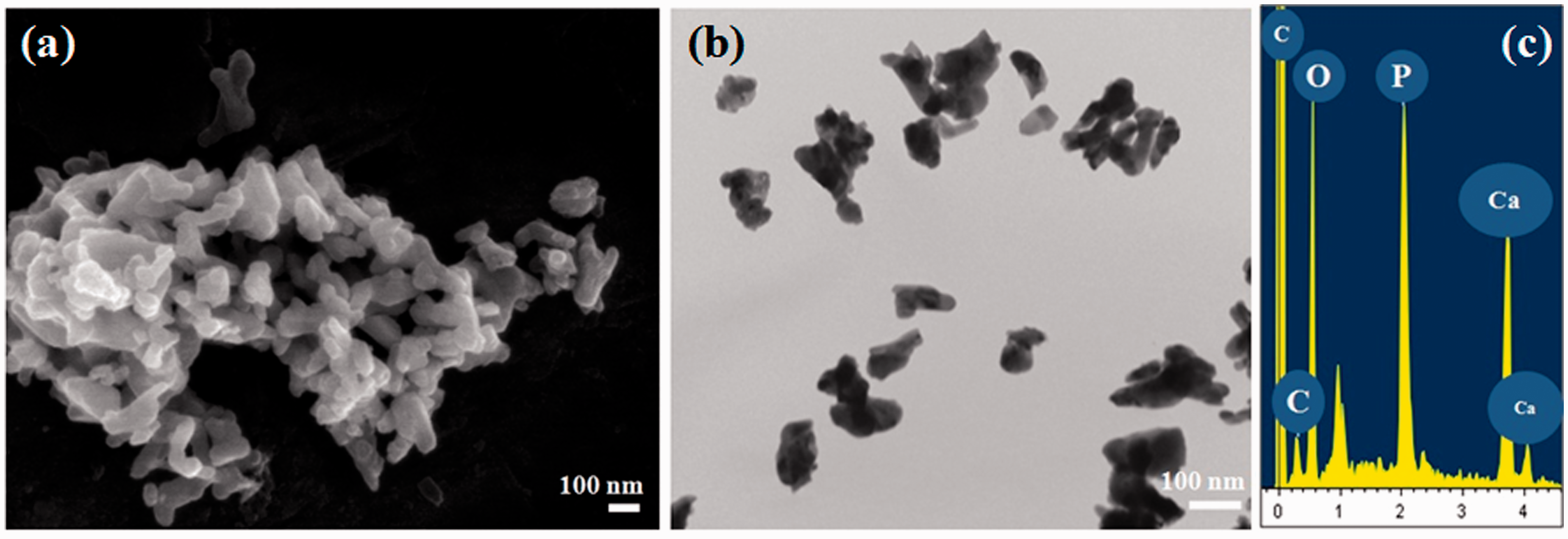
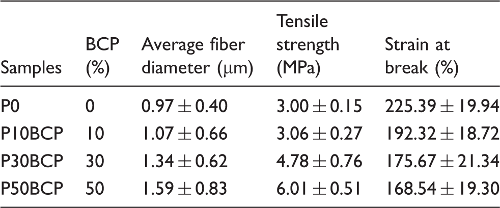
The fiber microstructure of the neat PCL/PLGA membrane and the BCP-loaded membranes is shown in Figure 2. BCP loading in the membranes produced significant morphological changes, specifically in the fiber diameter of the membranes. Prior to loading, more than 50% of the P0 fibers were between 0.5 to less than 1.0 µm. Inclusion of BCP powder produced a shift in the peaks of the fiber diameter frequency particularly with the P30BCP and P50BCP membranes, wherein the majority of the fiber diameters ranged from 1.0 to less than 1.5 µm. The average fiber diameter of the sample membranes is as follows: P0 (0.975 ± 0.402 µm) < P10BCP (1.074 ± 0.664 µm) < P30BCP (1.348 ± 0.623 µm) < P50BCP (1.591 ± 0.830 µm) (Table 1).
SEM micrographs (1) and corresponding fiber diameter frequency (2) of P0 (a), P10BCP (b), P30BCP (c), and P50BCP (d). Composition and characterization of neat and BCP-loaded PCL/PLGA membranes.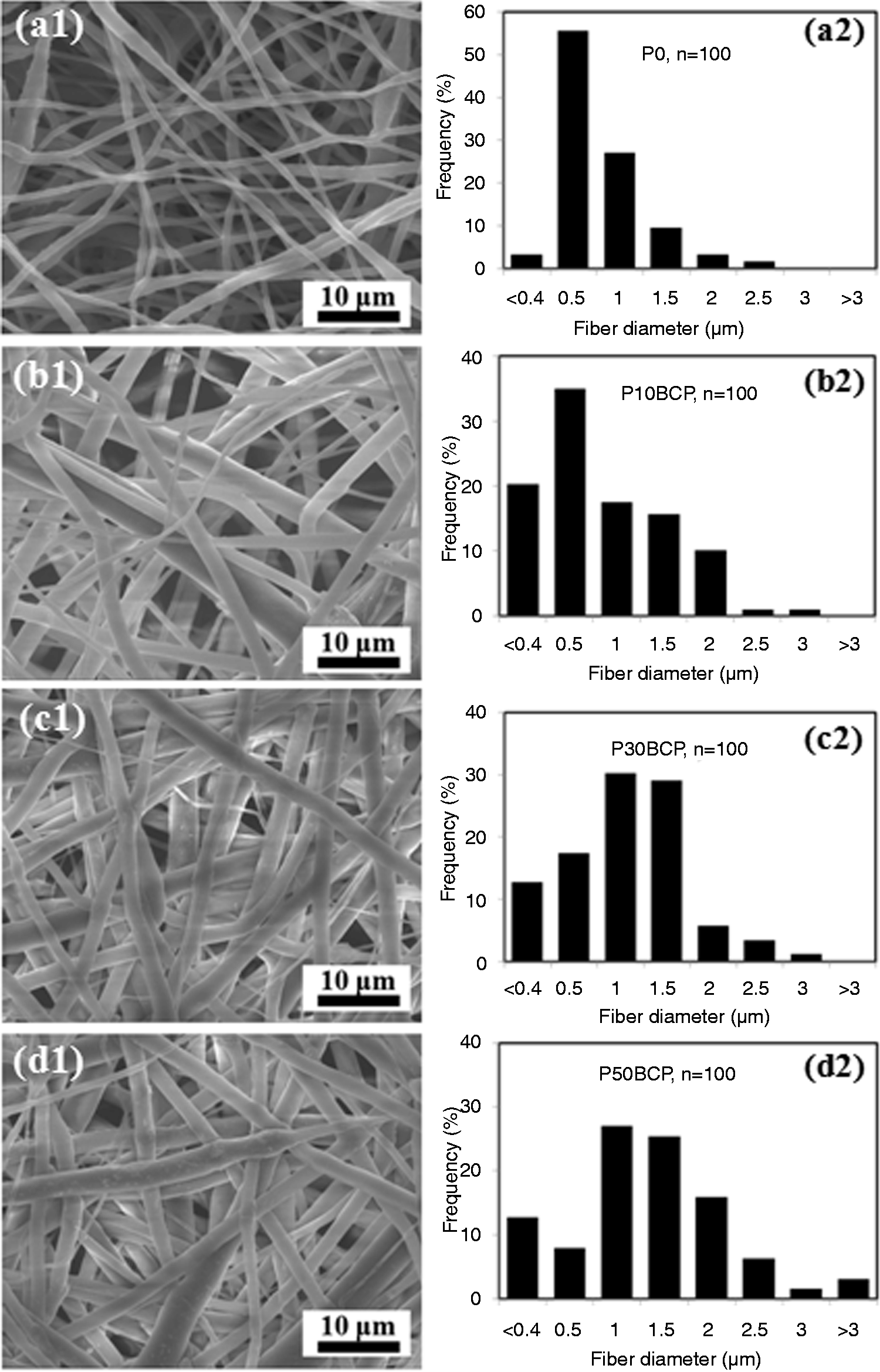
High magnification of the membrane fibers revealed rougher surface after the addition of BCP. Figure 3(a) shows a SEM micrograph of a representative fiber of P50BCP with particle deposits on the external surface of the membrane fiber. The EDS profile of this membrane (Figure 3(a), inset) revealed Ca and P peaks, which were attributed to the presence of BCP. The microstructures of the fabricated scaffolds were visualized using bright-field TEM technique. Figure 3(b1) and (b2) distinguished between the electron diffraction patterns of P0 (neat PCL/PLGA) and P50BCP membrane fibers. The P0 fibers showed diffused ring pattern while the BCP-loaded membrane was characterized by sharp rings with nanocrystalline particles deposited on the examined fiber. Further magnification of a BCP particle deposited on a fiber using a high-resolution TEM technique confirmed agglomeration of β-TCP and HAp nanocrystalline particles ranging from 5 to 20 nm in diameter (Figure 3(b3)). Electron diffraction pattern of the BCP particle confirmed the presence of HAp (211) and β-TCP (0210) on ring 1 and β-TCP (220) on ring 2 (inset).
SEM micrograph of a representative fiber of P50BCP showing rough external texture due to BCP deposition (a) with corresponding EDS reading with elevated P and Ca peaks (inset). TEM micrographs of representative fibers of P0 (b1) and P50BCP showing particle deposits on a fiber (arrows) (b2). High-resolution TEM image of a deposit in the fiber showed typical of a BCP particle (b3) with agglomeration of HAp and β-TCP nanoparticles confirmed by the electron diffraction pattern (inset) showing HAp and β-TCP (ring 1) and β-TCP (ring 2).
Successful loading of BCP particles onto the PCL/PLGA membranes was also confirmed by XRD analysis. Figure 4 shows the XRD patterns of the loaded BCP powder and sample membranes. The P0 membranes displayed PCL peaks that can be observed in P10BCP, P30BCP, and P50BCP. BCP peaks were not detected in the P10BCP membrane, which may be due to the low concentration of BCP. BCP peaks were noted in the P30BCP and P50BCP membranes, with peak intensities increasing in proportion to the concentration of the BCP content.
X-ray diffraction profiles of BCP powder (a) and sample membranes: P50BCP (b), P30BCP (c), P10BCP (d), and P0 (e).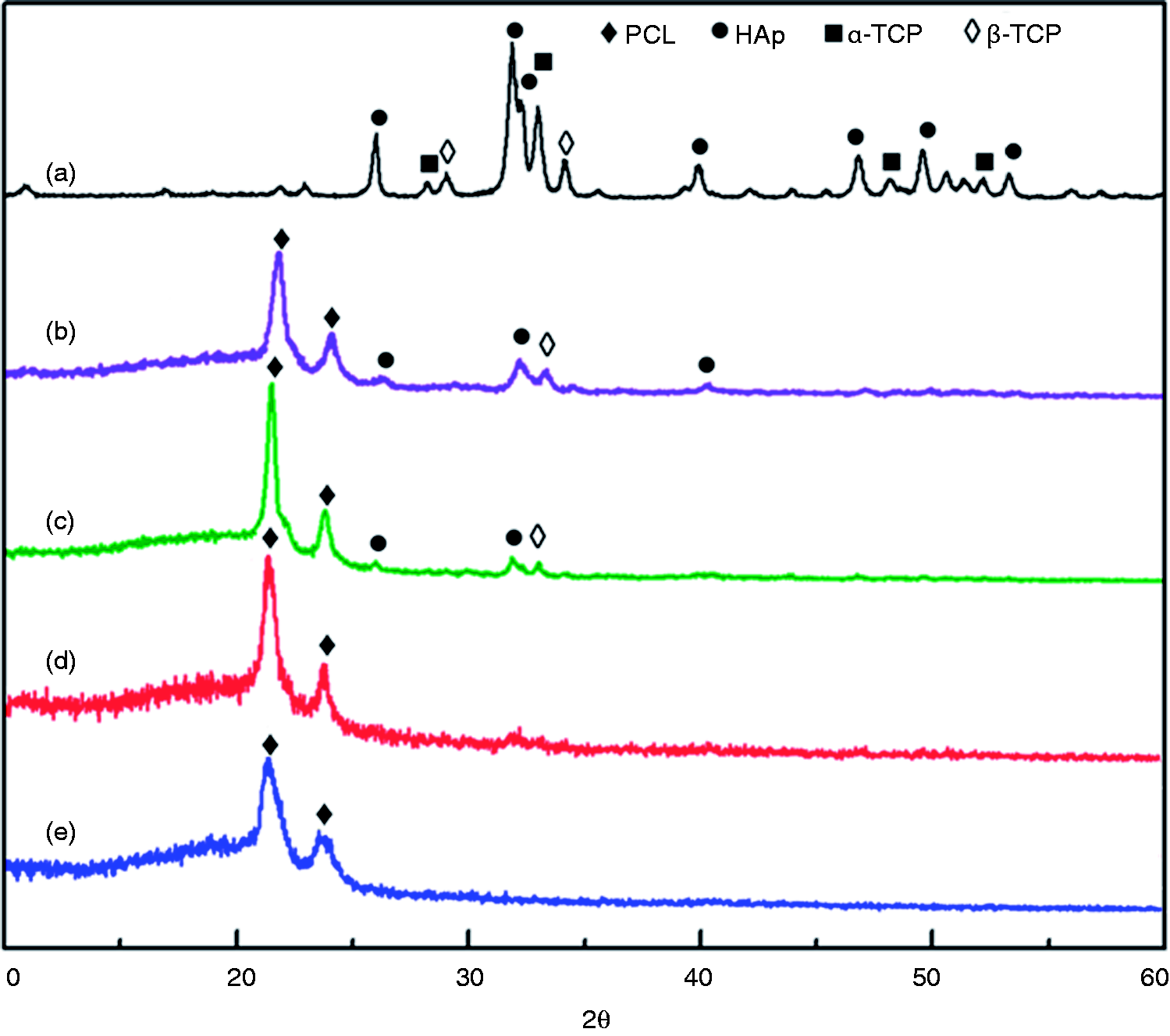
Mechanical properties
The tensile strength and strain values of the sample membranes were determined using a universal testing machine before and after immersion in PBS and SBF solution. Prior to immersion, it was observed that the addition of BCP to the membranes generally increased the tensile strength but delimited the membrane elongation. The values were as follows: P0 (3.001 ± 0.147 MPa, 225.390% ± 19.939), P10BCP (3.064 ± 0.273 MPa, 192.318% ± 18.720), P30BCP (4.779 ± 0.764 MPa, 175.672% ± 21.335), and P50BCP (6.098 ± 0.509 MPa, 168.544% ± 19.302). The tensile strength values of P0 and P10BCP were not statistically significant (p
Microscopic images of P50BCP membrane as a representative sample showed that there was no significant change in the fiber microstructure after 7 days of immersion in PBS (Figure 5(c)), with an EDS profile similar to that measured prior to immersion (Figure 3(a), inset). Immersion in SBF solution after 7 days produced apatite-like deposition on the fiber surfaces (Figure 3(d) and (e)). The EDS profiles of the membrane fibers showed Ca and P peaks with higher intensities (Figure 3(d), inset).
Tensile strength (a) and strain (b) of membranes before and after immersion in PBS and SBF solution for 7 days. SEM micrographs of P50BCP membrane immersed in PBS (c) and SBF solution after 7 days (d) with more general view of apatite-like formation in the membrane fibers (e). (*) represents statistically significant difference (p < 0.05) among sample readings.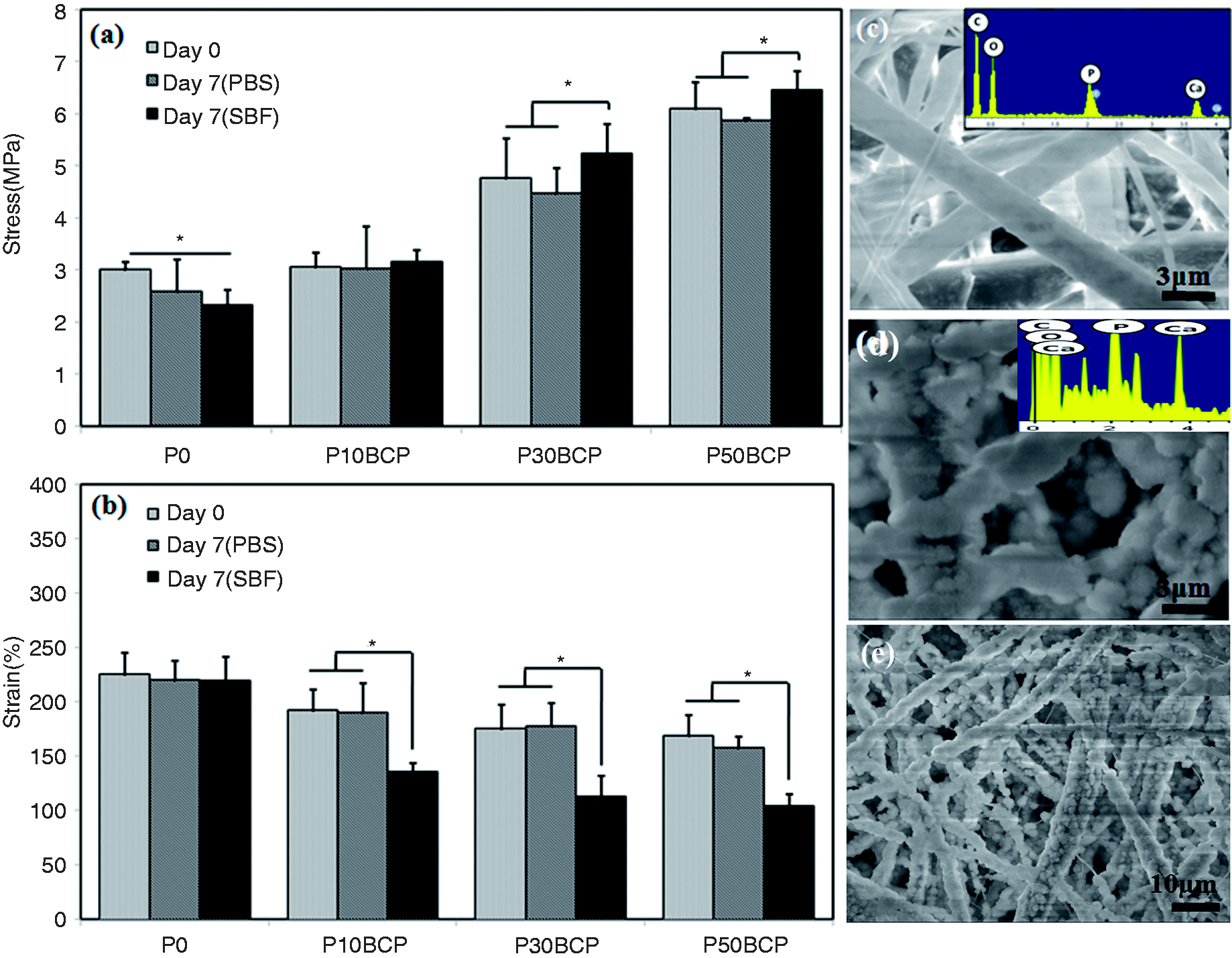
Material cytotoxicity and cell proliferation by MTT assay
Cell quantization was determined using MTT assay by equating the cell density with that of the formazan salts formed as a product of mitochondrial enzyme conversion of living cells.
30
Figure 6(a) shows how the cell viability was affected by the treatment of solutions extracted from the samples. It was observed that there was no statistically significant variation between values (p Material cytotoxicity depending on the percentage of extract solution used for treatment (a) and cell proliferation (b) profiles of MG-63 osteoblast like cells on P0, P10BCP, P30BCP, and P50BCP sample membranes. (*) represents statistically significant difference (p < 0.05) among sample readings per day. Confocal micrographs of MG63-osteoblast like cells grown on P0 and P50BCP after 1, 5, and 11 days of culture using low magnification (left frames) and high magnification (right frames). Scale bar is at 50 µm.

Figure 6(b) depicts the cell proliferation behavior of MG63 osteoblast-like cells grown on a neat PCL/PLGA membrane and on sample membranes with increasing BCP concentration after 11 days of incubation. The growth behaviors apparently increased in all samples with prolonged incubation time, and all samples had similar growth rate patterns. The cell growth rates for each sampling period were statistically different among samples after 3 days of incubation (*, p
Cell adhesion and proliferation behavior
Following the results of material cytotoxicity and cell proliferation rates measurement, P0 and P50BCP were used to demonstrate the cell adhesion and proliferation behavior on the membrane scaffolds (Figure 7). Low magnification images of the membrane sections were obtained to show a large surface area of cell growth and high magnification to provide a closer view of cell growth behavior on the scaffolds. All micrographs generally represented several sections of the scaffold for a given incubation period. Staining the cellular nuclei with DAPI visibly showed increasing cellular density during culture, and filamentous actin staining with phalloidin-labeled FITC demonstrated changes in cell morphology during scaffold attachment. On day 1, cell growth was apparent on both membranes. The cells were observed to be attached in the membranes via what seems to be cytoplasmic extensions. But it was noted that cells were significantly denser in the P50BCP compared to the sparse growth of cells in P0.
Further incubation shows that the cells proliferated in the fabricated material. Increased cell density was observed in both samples on the fifth day. However, it was noted that cells occupied almost the entire scaffold surface in P50BCP. The P0 surface was also covered with cells on the 11th day of incubation, which may have been due to the limited area of cell growth.
In vivo bone regeneration
Prior to extraction, all rats appeared to be in stable condition. No external signs of inflammation or infection were noted throughout the period of observation. Figure 8 shows the rat calvarial bones harvested four weeks after implantation. A thin membrane, the periosteum, was seen to envelop the defected part of the skull bone for all samples. Signs of internal hemorrhage were also observed and seen to be more prominent in the control rat (Figure 8(a1)) and for the defect with P0 (neat PCL/PLGA) (Figure 8(b1)) compared to the defect implanted with P50BCP (Figure 8(c1)). Bleeding was attributed to the trauma from surgery but was not seen to adversely affect the grafts. Both membrane samples, however, were seen to adhere to the native tissues and were kept in place.
Harvested rat calvarium with no implant (a), with P0 (neat PCL/ PLGA) (b), and with P50BCP (PCL/PLGA with 50% BCP) (c) showing gross morphology before extraction after four weeks (1), two-dimensional micro-CT scanning micrograph (2), and reconstructed three-dimensional image (3). Histomorphometric analyses showing the increasing percentage bone volume (BV/TV) (e) of the extracted grafts (*p < 0.05; n = 4). Harvested rat calvarial bone with no implant (control), gross morphology (a1), and magnified sections of the adjacent region of the cranial defect (a2) and loose connective tissue in the defect region (a3) stained by hematoxylin and eosin method.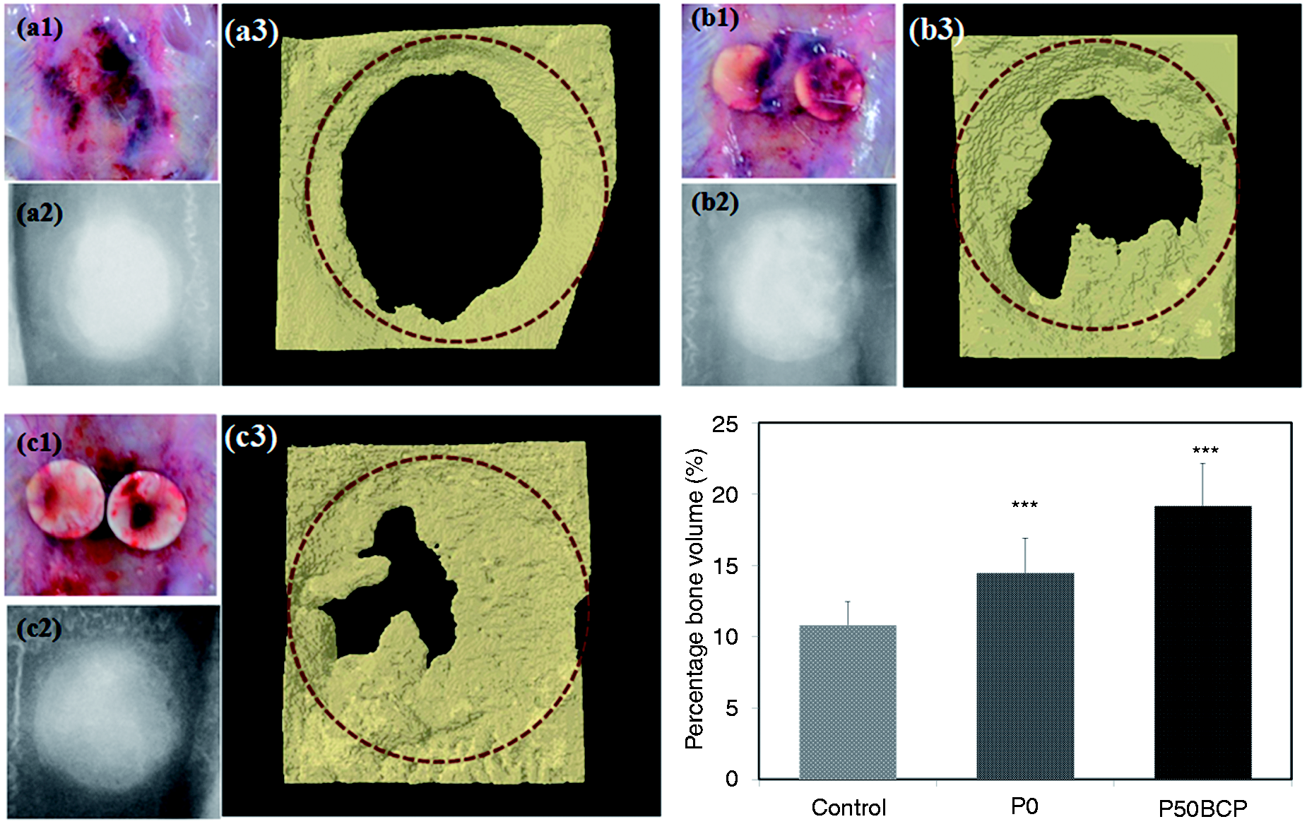
Micro-CT micrographs showed representative defects with the formation of new tissues on the defect region. Growth apparently formed from the periphery first and progressed toward the central region. The P50BCP implanted defect (Figure 8(c2)) showed a relatively larger dark region representing tissue formation compared with P0 (Figure 8(b2)) and the control (Figure 8(a2)). Three-dimensional reconstructions of the images showed clearer representations of the tissue growth over the defect regions (Figure 8(a3), (b3), and (c3)). Micro-CT histomorphometric analysis showed that the percentage bone volume (BV/TV) values in the defected regions with P50BCP had significantly increased values (p < 0.05) compared with P0 and the blank control. The percentage bone volume (BV/TV) values were consistent with the total bone values: P50BCP, 19.18 ± 2.51 mm3 > P0, 14.516 ± 2.23 mm3 > control, and 10.83 ± 1.51 mm3 (Figure 8(d)).
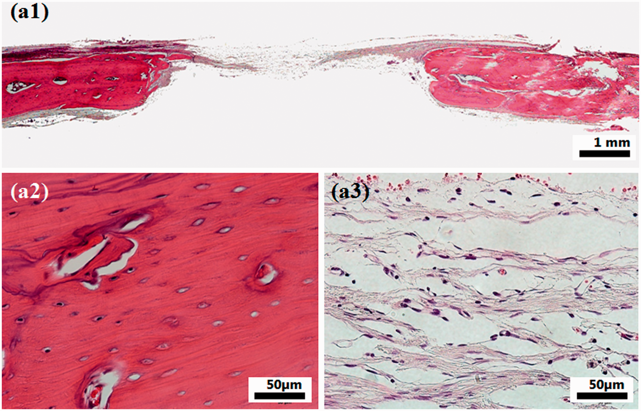
In order to illustrate the quality of the tissue growth in the defected area implanted with P50BCP, tissue sections were obtained and compared with the control defect. Without an implant, only the periosteum membrane was seen to grow over the defect (Figure 9(a1)). Higher magnification imaging reveals loose connective tissue regenerating as a natural means of repair to protect the defect underneath (Figure 9(a3)). Implantation with P50BCP showed significant tissue growth within the defect region (Figure 10(a1)). Detailed micrograph of the tissue growth on the defect region showed increased cellular activity (Figure 10(a3)) compared to that of the control (Figure 9(a3)). Other parts of the defect region showed dense tissue formation (NB) (Figure 10(a4)) with morphology similar to that of the native bone in the adjacent region of the cranial defect (Figure 10(a2)). Another significant observation is the possible blood vessel formation (BV) within the defect area (Figure 10(a4) and (a5)), which may have aided in more pronounced tissue growth.
Harvested rat calvarial bone implanted with P50BCP: gross morphology (a1) and magnified sections of the adjacent region of the cranial defect (a2), tissue growth in the defect region (a3). Detailed sections showed dense tissue formation with similar morphology with the pre-defect region (a4) and possible blood vessel formation (a5).
Discussion
The loading of the bioceramic component into the biopolymer membrane was done to provide an osteoconductive component to an established nanofiber membrane scaffold of considerable strength. The ceramic–polymer combination has been used in order to take advantage of the properties of the two components, which are complimentary to each other.
The morphology of the fabricated membranes in this study was compared to those of membranes fabricated earlier in our laboratory. The fiber diameter of the PCL/PLGA membrane was relatively smaller than that from a previous study 25 but relatively larger than that in another study using the same solvent combination. 31 DMF and THF organic solvents were used in combination to dissolve the PCL and PLGA polymer. MC was added to the cocktail as this facilitated the polymers dissolvability during lower room temperatures to produce smoother and more uniform fibers. The cocktail was similarly used in a previous study done in our laboratory. 32 The fiber diameter of the membranes prior to loading mostly ranges from 0.5 to less than 1.0 µm. This can enable the BCP nanoparticles, which measures between 0.1 and 0.3 µm, to be completely encapsulated by the nanofibers. In the study by Ba Linh et al., 33 it was observed that the electrospun fibers and BCP nanoparticles incorporated in the fibers assume a positive charge via the positive electrode from a high-voltage power supply connected in the nozzle during the electrospinning process. Due to this, the positively charged BCP was being repulsed by the also positively charged surface of the fibers, thus pushing the nanoparticles inward the fibers and embedding it in the middle. 33 The solid material (BCP particles) that was originally in the solution was distributed to the fibers during electrospinning. As the solvent evaporated, the solid particles remained with the polymer as this was not dissolved, but its mass was added in the starting solution with a fixed solvent volume. It can be inferred that the variation of the solvent also affected the fiber morphology and that the addition of the BCP powder augmented the diameter size of the membrane fibers. BCP addition produced a significant increase in the fiber diameter compared to the membrane fibers of the neat PCL/PLGA membrane (P0). The increase in mass of the solution translated to a relative amplification of fiber diameter size. Thus, increasing the BCP content significantly increased the fiber diameter of the membranes as the highest peaks were shifted from 0.5 µm for P10BCP to a range of 1.0–1.5 µm for P50BCP (Figure 2(b2) and (d2)). Similar results were obtained from a previous study by Jose et al., 34 wherein the addition of a ceramic component resulted to an increase of fiber diameter size.
Verification of the BCP loading was done by XRD studies, and visualization by TEM and EDS analysis of the granules was attached to the membrane surface. Analysis of the results sufficiently proved the successful loading of the BCP particles onto the membrane fibers. EDS data showed the presence of Ca and P peaks, which were non-existent in the neat PCL/PLGA membrane. Some peaks were left unlabeled as were only after the presence of BCP. It was deemed by the authors that the presence of Ca and P peaks in the EDS spectra of the BCP-loaded membranes and its absence in the spectra of the neat PCL/PLGA membrane are enough evidence that the BCP was successfully incorporated to the electrospun fibers. In the case of TEM, the lath-like structure of the BCP granules at high magnification was similar to previous published data on hydroxyapatite, tricalcium phosphate, and BCP.35–37 XRD profiles of the membrane samples displayed peaks of the hydroxyapatite phase and the β-tricalcium phosphate phase of the BCP on P30BCP and P50BCP. While these peak intensities may not have been evident on the P10BCP, the relative amount of BCP on this sample may not have been sufficient to be detected by XRD analysis.
The addition of a ceramic component to the polymer-based scaffolds has been done to improve their mechanical properties. In a study by Tan et al., 38 in which he observed the strength of PCL membranes based on fiber diameter, it was observed that fibers with a smaller diameter had higher strength. But in this study, P50BCP, which displayed the wider fiber diameter, registered the highest strength. Due to this result, it was assumed that it was the addition of BCP that affected the mechanical properties of the membranes. Previous studies had shown that the increasing the ceramic component in these polymer–ceramic composites corresponded to an increase in the strength values.39–42 In this study, the tensile properties of the membranes were also noted to improve when the BCP component was added. The strength values were also seen to increase with higher BCP concentration. Consequently, the degree of elongation was delimited due to the increasing stiffness of the material with the addition of the ceramic to the polymer solution. The BCP deposits made the membranes more rigid and thereby reduced their ability to elongate. The addition of BCP provided reinforcement to the membrane fibers, which required a greater amount of force to break. The noted reduction in the elongation of the BCP-loaded membranes was attributed to the immobilization of the particles when force was applied to the membrane.
Immersion of the membranes into PBS and SBF provided more details of the behavior of the scaffolds compared to a dry state. PBS has similar pH, osmolality, and ion concentrations to the human body. Likewise, the composition of the SBF solution was also similar to that of human plasma and has been used to predict the in vivo bone bioactivity of bone tissue engineering scaffolds by the formation of an apatite layer.43,44 Consequently, alteration in the tensile strength values of the membranes was noted to be affected depending on the presence of the ceramic component and relative amount that was loaded in the membrane samples. In the case of the neat P0 membrane, reduction of the tensile strength was attributed to the mass lost during immersion. PLGA has a relatively faster rate of degradation by hydrolysis than PCL and was seen to show significant degradation in 7 days. 34 In contrast with the ceramic-loaded membranes, the BCP reinforcement appeared to be existent even after fluid immersion, which can be attributed to the hydroxyapatite phase of the powder. The tensile properties were relatively maintained with slight reduction compared to the dry state testing. SBF immersion of the ceramic-loaded membranes produced significant morphological alterations with the deposition of apatite-like layers on the fiber surfaces (Figure 5(d) and (e)). This additional mineral-based layer created further reinforcement in the membrane fibers that may have impeded the effects of the polymer hydrolysis.
The elemental composition of BCP includes calcium and phosphate functional groups, which were generally biocompatible, bioactive, and osteoconductive, as they also exist in the physiological environment. A previous study reported that some serum proteins such as fibronectin and vitronectin can be adsorbed easily onto the calcium phosphate surface, rendering it bioactive and biofunctional. 45 This property was highly useful in the field of implant technology, as it meant that the implant readily integrated with the native host tissues by facilitating osteoblast cell adhesion. The results of the experiment showed cell material deposition on the membrane surface during a short period of incubation indicating that the cell has recognized adhesion points on the scaffold surface in a relatively limited period of time. This explains why the cells adhered better in the P50BCP and P30BCP compared to P0BCP and P10BCP as higher concentration of BCP means more recognition sites for the cells. In addition, BCP degradation products enhance bone formation when released in an aqueous solution 45 and also improve as well as increasing the hydrophilic property of the material, which further supports cell adhesion and division. 46 This is increasingly beneficial to hydrophobic polymers such as PCL and PLGA.
Bone regeneration is the measure of a scaffold’s efficiency as a bone tissue engineering scaffold. Many scaffolds have been fabricated with the intention of being used as a bone-regenerating scaffold or for guided bone tissue formation. The scaffold fabricated in this study not only showed favorable mechanical properties but also displayed evidence for encouraging new bone formation in a rat model. The addition of BCP in the membrane facilitated the osteoconductivity of the material to the native bone tissue. BCP enhanced the regenerative activity of osteoblast cells in repairing bone defects as was observed in previous studies. The bone formation pattern observed in the extracted samples showed regeneration starting on the defect edge and eventually moving inward, which was consistent with the results of a study by Jegal et al. 47 The formation of blood vessel-like structures (BV) in the tissue formed in the defect area implanted with P50BCP was non-existent in the control rat. This may had been responsible for the tissue regeneration in the defect region implanted with P50BCP. However, further studies are recommended to verify these microvasculature-like structures since no study has provided significant data on the efficiency of the BCP to induce angiogenesis unlike with bioactive glasses and silicates.
The stated results validated the potential of the PCL/PLGA membranes to be incorporated with BCP. The addition of BCP was not only able to increase the mechanical property of the membrane but also improved the osteoconductivity of the fabricated material. It is planned to conduct further study regarding more in-depth analysis of the fabricated material and the possibility to further improve it and use it in other applications. Together with this plan, the scaffold will be further characterized as there is a need to understand better its properties such as biodegradation behavior and loading efficiency of BCP. Also, more in-depth in vivo analysis may be needed to better quantify new bone formation such as histomorphometry and using bigger defect size (8 mm).
Conclusion
The loading of BCP particles onto the PCL/PLGA membrane fibers was successfully carried out using the electrospinning method and was verified by SEM and TEM micrographs, EDS, and XRD analyses. BCP loading was seen to affect the mechanical properties of the membrane as the stress values increased with increasing BCP concentration but the elongation was reduced, which was attributed to the fiber heterogeneity caused by the particle loading. The in vitro biocompatibility of the membrane was enhanced by BCP loading as evidenced by the increased cell proliferation rate and the more evident cell morphological changes during the prolonged incubation period. BCP loading on the membranes was also noted to influence faster tissue regeneration in vivo when implanted in the rat calvarium. The percentage bone volume was significantly higher compared to the tissue growth on the defects without BCP loading and on the control defect. In addition, histological sections of the defects showed that the tissue formed over the P50BCP-implanted defect was bone-like in nature.
Footnotes
Funding
This research received no specific grant from any funding agency in the public, commercial, or not-for-profit sectors.
Acknowledgments
This work was supported by Mid-Career Researcher Program through NRF grant funded by the MEST (NO 2009-0092808), Republic of Korea and partially supported by Soonchunhyang University Research Fund. The authors also acknowledge the contributions of Mr. Shin Woo Kim in the in vivo surgeries, Mr. Dong Woo Jang and Miss Ji Hyun Kim for the assistance in obtaining the Micro-CT data, and Prof. Kim Chang-Jin of Pathology Lab for histology consultation.