
Abstract
Purpose:
The objective of this study was to investigate the therapeutic potential of poly(lactic-co-glycolic acid) (PLGA) microspheres loaded with recombinant human growth and differentiation factor-5 (rhGDF-5) on the disc degeneration induced by needle puncture in a rat caudal disc model.
Methods:
The rhGDF-5-loaded PLGA microspheres were prepared by the water-oil-water double-emulsion solvent evaporation method, and release kinetics was determined over 42 days. Rats that underwent 21-G needle puncture at rat tail discs were injected with rhGDF-5/PLGA microspheres at four weeks after needle injury. At eight weeks after the injection, disc height, glycosaminoglycans content, and DNA content of the discs were evaluated. In addition, gene expression analysis of aggrecan, collagen type I, and collagen type II in the rat nucleus pulposus was measured by real-time polymerase chain reaction. Rat discs were also assessed by histology using hematoxylin and eosin stain.
Results:
Encapsulation of rhGDF-5 in PLGA microspheres guaranteed a sustained release of active rhGDF-5 for more than 42 days. The injection of GDF-5/PLGA microspheres resulted in a statistically significant restoration of disc height (p < 0.01), improvement of sulfated glycosaminoglycan (p < 0.05), DNA content (p < 0.05), and significantly increased mRNA levels of collagen type II (p < 0.01), and the differentiation index (the ratio of collagen type II to collagen type I, p < 0.01). In addition, rhGDF-5/PLGA microspheres treatment also improved histological changes induced by needle puncture.
Conclusions:
The results of this study suggest that injection of rhGDF-5 loaded in PLGA microspheres into rat tail discs may be as a promising therapy strategy to regenerate or repair the degenerative disc.
Keywords
Introduction
Low back pain, which is often related to intervertebral disc (IVD) degeneration, affects approximately 80% of the adult population and has become a major cause of disability and suffering. Furthermore, it causes a significant socioeconomic burden worldwide. IVD degeneration is considered to be a multifactorial process involving mechanical, genetic, systemic, and biological factors. The IVD consists of an annulus fibrosus (AF) that encloses the nucleus pulposus (NP), a highly viscous gel-like tissue that produces aggrecan and other proteoglycans (PG), providing the disc with its critical water-retaining characteristics. Disc degeneration often starts with cellular and biochemical changes in the NP and AF, resulting in an imbalance between anabolism and catabolism of disc tissues. NP, the main constituent of the IVD, plays a major role in maintaining normal function of the IVD. As a consequence, any cellular, biological, or biochemical changes in the NP ultimately deteriorate disc function, leading to disc degeneration. Unfortunately, because of the absence of blood supply in the inner AF and NP, IVD tissues have little potential for self-repair. Thus, treatment methods for the degenerated IVD vary from the conservative, including medications, steroid injection,1,2 and physical therapy, 3 to surgery. Importantly, most treatments are aimed at treating symptoms, rather than at preserving disc structures or functions. The challenge of developing a therapeutic method that is able to restore disc functions has therefore been a long-standing aim of researchers. Recently, biological repair or regeneration of the degenerative IVD has been pursued.
One of the most advanced biological therapeutic approaches to regenerate or repair a degenerated disc is the injection of a recombinant growth factor,4,5 such as bone morphogenetic protein (BMP-2, -7), insulin-like growth factor-1, basic fibroblast growth factor, platelet-derived growth factor, platelet-rich plasma, and transforming growth factor beta. Growth and differentiation factor-5 (GDF-5), another member of the BMP family, is important for the development of skeletal system, especially for the cartilage-like tissue. It was also found to stimulate PG and type II collagen expression in mouse IVD cells.6,7 Furthermore, recombinant human GDF-5 (rhGDF-5) enhances cell proliferation and matrix synthesis and accumulation by both bovine NP and AF cells. 8 Furthermore, the injection of GDF5 protein into rabbit discs can promote cell proliferation. 9
Growth factors are biologically active molecules capable of stimulating cellular growth, proliferation, and differentiation in an endocrine or paracrine fashion. 10 However, the half-life and solubility of the factors, the proper carrier, etc. are issues that need to be taken into consideration.
In recent years, use of biodegradable microspheres for controlled local drug delivery became a valuable approach to overcome the potential serious side effects arising from life-long, systemic administration of therapeutic agents. 11 Among different types of biodegradable microspheres, those composed of poly(lactic-co-glycolic acid) (PLGA), a biodegradable and biocompatible polymer, have received great amount of interest. The composition of the PLGA allows the control of degradation rate and drug release kinetics. Due to its unique properties, PLGA copolymers have been used in controlled delivery of many proteins,12,13 drugs, and other factors.
In the present study, we synthesized PLGA microspheres (PMs)-packaged rhGDF-5, which were injected into in vivo in a rat model of IVD degeneration, to evaluate whether the controlled long-term release of rhGDF-5 can repair the degradation of the IVD.
Materials and methods
rhGDF-5-loaded PMs
PMs encapsulating growth factor rhGDF-5 (PeproTech Inc, USA) were prepared by double-emulsion solvent evaporation technique. 14 Briefly, 1 mg of the PLGA (lactic acid (LA): glycolid acid ratio (GA) 75:25, Shandong Medical Instruments Institute, China) polymer was dissolved in 1 ml of methylene chloride (DCM) as oil phase (O). Growth factor rhGDF-5 was dissolved in deionized water as the first water phase (W1). The W1 solution was then emulsified in oil phase to form the primary W1/O emulsion via homogenization at 8000 r/min for 30 s. The resulting emulsion was added dropwise to 100 ml of an aqueous phase containing 1.6% (w/v) polyvinyl alcohol (PVA) (Sigma-Aldrich, St. Louis, MO, USA) (external phase, W2) and homogenized for 5 min at 1000 r/min. The resulting W1/O/W2 emulsion was stirred at 300 r/min for 2 h at room temperature with a Laboratory Mixer-JJ-1 A (Changzhou, China) to allow DCM to evaporate completely. The microspheres were then isolated by filtration by using 0.8 µm membrane filter (Millipore, HVLP), washed three times with distilled water, and vacuum dried. Final products were stored at 4℃ in desiccators.
Morphology
The microspheres were immobilized on a copper stub and coated with gold by an Ion Sputte (HCP-2, HITACHI, Japan), and the voltage for morphologic analysis is 5.0 kV.
Morphology of microspheres was conducted using scanning electron microscopy (SEM; JSM-6360LV, JEOL, Japan).
Kinetics of in vitro release of rhGDF-5
The release kinetics of rhGDF-5 in vitro was determined. 10 mg of PLGA microspheres were suspended in 1 ml of phosphate-buffered saline solution (PBS, pH, 7.4) in polypropylene tubes placed in a shaker bath (37℃) at 100 rpm. The samples were centrifuged and supernatant was collected at predetermined time intervals. Fresh replacement media was added to resuspend the microspheres. The rhGDF-5 content of supernatant collected for each interval was determined by using the enzyme-linked immunosorbent assay. Using the accumulated release rate, the microsphere’s accumulated release kinetic curve was also obtained.
Experimental animals
Male Sprague-Dawley (SD) rats (three months old) were obtained from the animal center of Beijing University. Experiments were performed in accordance with the Guide for the Care and Use of Laboratory Animals, and the experimental protocols were approved by the university committee on the use and care of animals at the University of Beijing.
Surgical technique
Anesthesia was achieved and maintained by intraperitoneal injection of 3.5% chloral hydrate. A previously described animal model for IVD degeneration to puncture the caudal disc in rats by using a 21 gauge needle was used. 15 The needle was then inserted into the middle of the disc through the AF into the NP of levels 4–5, 5–6, and 7–8 (L4/L5, L5/L6, and L7/L8) of the caudal spine, rotated at 180°, and held for 5 s. Level 6–7 (L6/L7) of the caudal spine remained undisturbed and was used as the control level. Four weeks after puncture injury using the 21-G needle, PMs alone or PLGA loaded with rhGDF/5 was slowly injected into the NP of L5–L6 or L7–L8 using a microsyringe attached to a 31-G needle.
Radiographic analysis of disc height
Radiographic of rat tails disc were assessed at each time point by Micro-CT (Aloka Latheta LCT 200, Japan). Data are reported as the IVD height expressed as the disc height index (DHI): DHI = IVD height/adjacent vertebral disc height.16,17 Change in the DHI of injected discs was expressed as percentage DHI (% DHI) and normalized to the measured preoperative IVD height: % DHI = (postoperative DHI/preoperative DHI) × 100. 12
Biochemical analyses
Entire NP (the gelatinous tissue) was isolated from each level discs. The dissected NP was digested with papain (125 µg/ml in sterile PBS with 5 mM cysteine, HCl, and 5 mM disodium ethylenediaminetetraacetic acid (Na2EDTA), pH 6.0) at 60℃ for 24 h. The proteoglycan, mainly sulfated glycosaminoglycan (sGAG), and the DNA contents in the digested solution were assayed using the dimethyl-methylene blue method 18 and Hoechst dye 33258 method, 19 respectively.
Histological examination
After radiographic analysis, six discs from each group were harvested for histological studies. The IVDs were excised from the vertebral body disc-vertebral body unit, and each IVD was fixed in 4% paraformaldehyde for seven days at 4℃ and then decalcified in 0.5 M EDTA, embedded in paraffin, sectioned, and assessed by conventional histology. Midsagittal sections (5 µm) of each IVD were stained with hematoxylin and eosin (H&E).
Quantitative real-time polymerase chain reaction
Total RNA was extracted from the NP using the trizol method. Reverse transcription was carried out using the SuperScript First-Strand Synthesis Kit (Invitrogen). Type I and II collagens, aggrecan, and glyceraldehyde 3-phosphate dehydrogenase (GAPDH) gene expressions were quantified by real-time polymerase chain reaction (PCR) using ABI 7700 fluorescent quantitation instruments (Applied Biosystems). A positive standard curve for each primer was obtained using real-time PCR with a serially diluted cDNA sample mixture. The quantitative gene expressions shown in our study were calculated using standard samples and normalized using GAPDH. The amounts of mRNA expression were presented as ratios to the intact control group.
Statistical analysis
All data were expressed as mean ± standard error. The significance of differences among the means of data for gene expression, biochemical parameters, and height index were analyzed using one-way analysis of variance and the two-tailed paired t-test. A p-value of less than 0.05 was considered statistically significant.
Results
Microsphere morphology and rhGDF-5 release kinetics
rhGDF-5/PMs were prepared via double-emulsion techniques. The morphologies, demonstrated by SEM, are shown in Figure 1(a) and (b). SEM micrographs showed that the microspheres were spherical in shape, have a smooth surface, with diameters of 40–55 um and a mean particle size of 50 um. The profiles of rhGDF-5 release from PLGA-based microspheres over 40 days are shown in Figure 1(c). The curve of growth factor from microspheres showed a significant initial burst release during the first five days, and the growth factor release continued steadily thereafter. In microspheres, the cumulative release rate of the growth factor reached about 80% after 42 days.
Scanning electron microscope image of growth differentiation factor-5/PLGA microspheres (a and b) and in vitro cumulative release profile of growth differentiation factor-5/PLGA microspheres (c).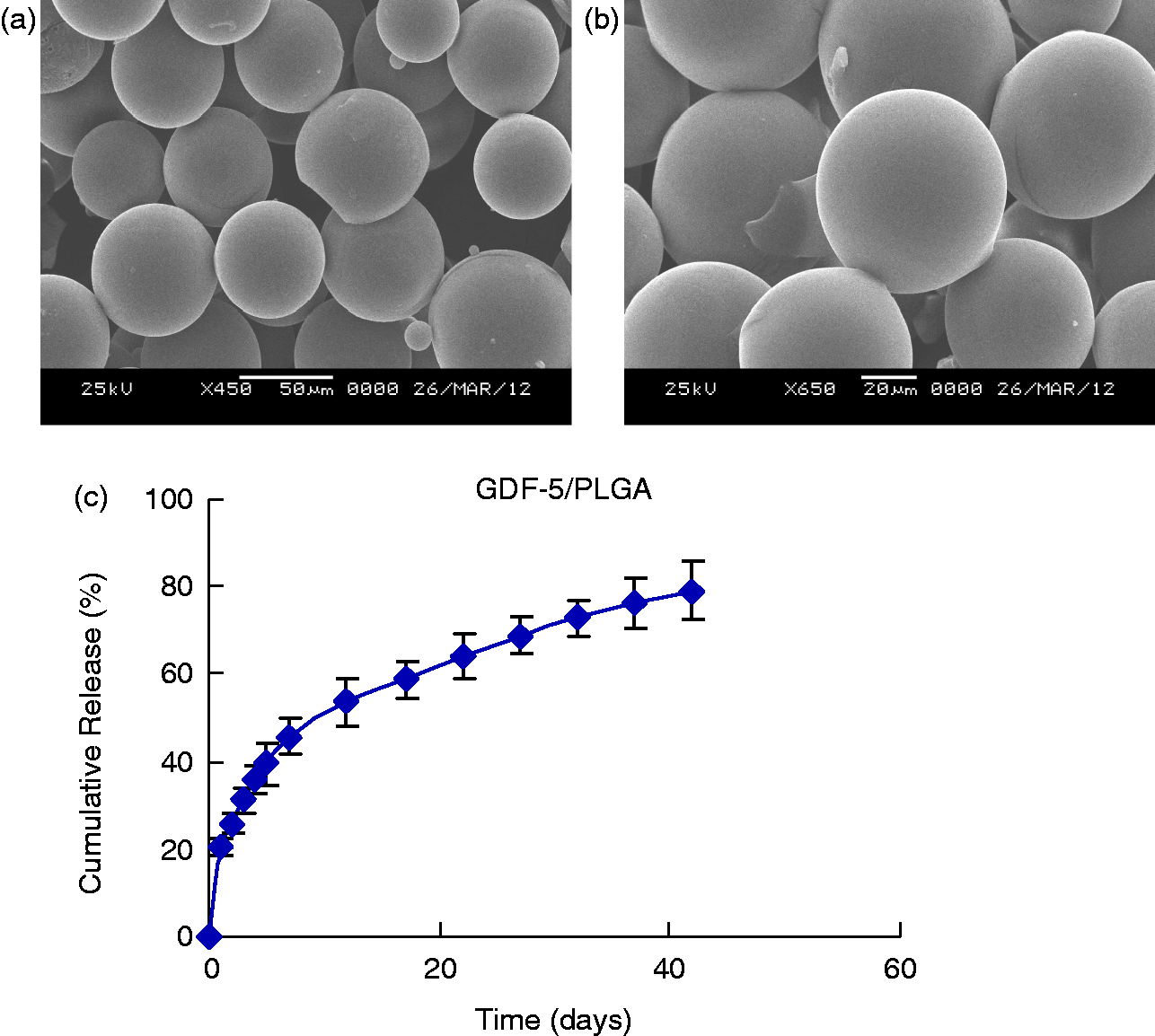
Radiographic and disc height assessment
In all rhGDF-5/PMs groups, PMs groups, and puncture groups, radiographs taken four weeks after the initial anular puncture showed a significant narrowing of disc height compared with that of the non-punctured disc (Figure 2(b)). Treatment with rhGDF-5/PMs significantly affected the post-injection disc height (p < 0.01). Two weeks after the rhGDF-5 injection, the percent DHI was similar to that of the puncture group. However, four weeks after the injection of rhGDF-5/PMs, the disc height began to recover compared with the discs injected with PMs group and puncture group (Figure 2(b), p < 0.05). Eight weeks after the injection, rhGDF-5/PMs induced restoration of disc height to the level approaching 90% of the non-punctured disc, also showing a significant recovery of disc height compared with the PMs alone treated group (Figure 2(a) and (b), p < 0.01). The injection of PMs alone treated group did not induce restoration of disc height over the same time period (Figure 2(a) and (b)).
(a) Lateral radiographic of 5/6, 6/7, and 7/8 discs at 4 weeks (after the anular puncture) and 8 weeks (after the injection of PMs alone and GDF-5/PLGA). (b) changes in the intervertebral disc (six discs per group) the percentage of height index (DHI). *p < 0.05 and **p < 0.01, when PLGA-GDF/5 treatment four weeks and eight weeks compared with puncture group. +P < 0.05 and ++P < 0.01, compared with PMs alone injection group. GDF: growth and differentiation factor; PLGA: poly(lactic-co-glycolic acid); PMs: PLGA microspheres.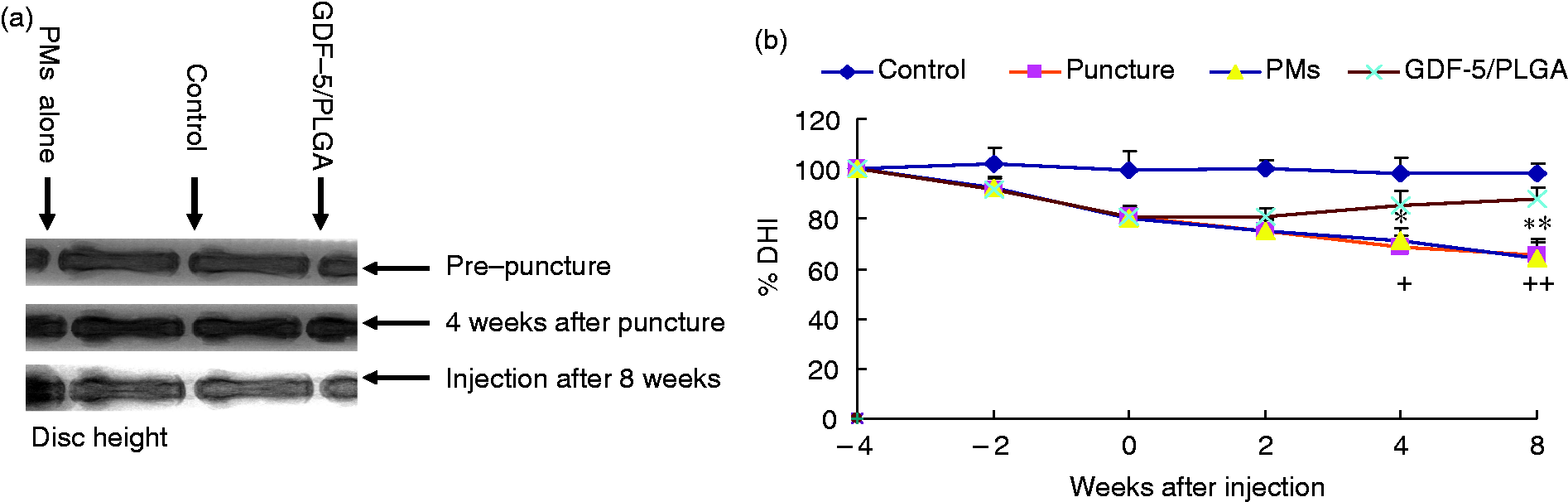
Biochemical analyses
DNA content in the puncture groups and PMs alone treated group was significantly decreased when compared with that of the intact control (Figure 3(a), p < 0.05). Eight weeks of treatment with GDF-5/PMs resulted in an increase in the DNA content that had decreased due to needle puncture (Figure 3(a), p < 0.05). The sGAG content in the PMs-treated discs decreased significantly as compared with the intact control group (Figure 3(b), p < 0.05). Treatment with GDF-5/PMs increased sGAG content and showed a significant difference with other groups (Figure 3(b), p < 0.05); however, it was not statistically significant with the control group.
(a) DNA and (b) sGAG content in the NP of different experimental groups (six discs per group) at eight weeks after injection. *p < 0.05, when PLGA-GDF/5 treatment eight weeks compared with puncture injection group. +p < 0.05, when compared with puncture with PMs alone injection group. #p < 0.05, when compared with control group.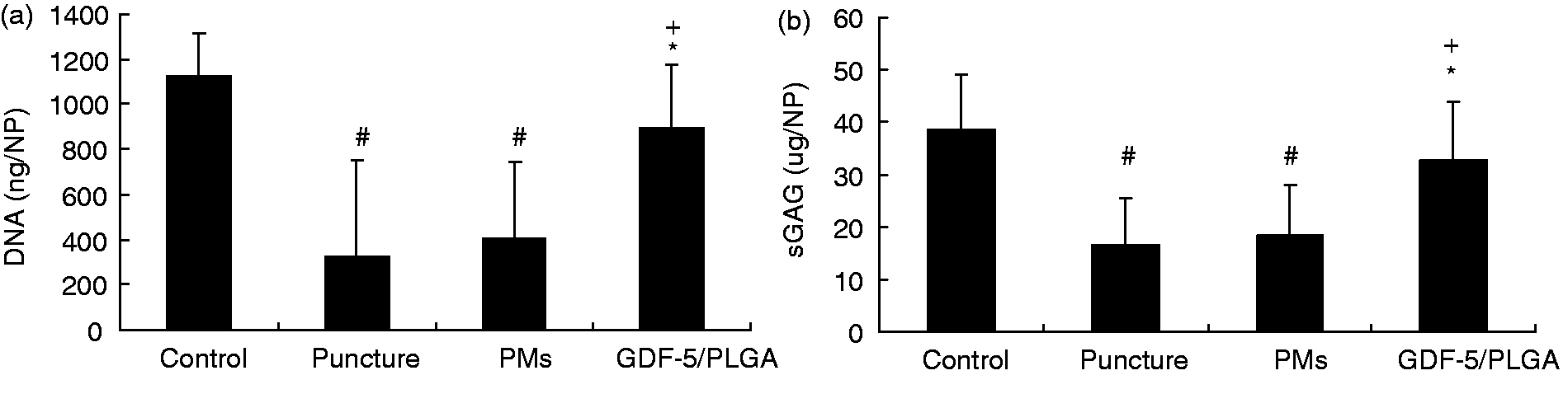
Histology
Representative cuts of the L5/L6, L6/L7, and L7/L8 discs were stained with H&E (Figure 4). Control discs maintained their architecture (Figure 4(a)). The discs by PMs alone injection still showed severely degenerated discs in which of most the NP contents have been lost and collapsed (Figure 4(b)). Further, the AF was irregular and the border between the AF and the NP was not clearly defined in PMs alone treated group (Figure 4(b)). Treatment with GDF5/PMs resulted in an improved cellularity and architecture compared with the puncture group and similar to that of control group (Figure 4(c)). In the treated groups, the invasion of blood vessels or inflammatory cells was not observed within the discs (Figure 4(c)). In addition, no remaining PMs were found in section.
Representative H&E (×4 and ×20) of disc samples obtained at eight weeks for each group after injection. Each group consisted of six discs.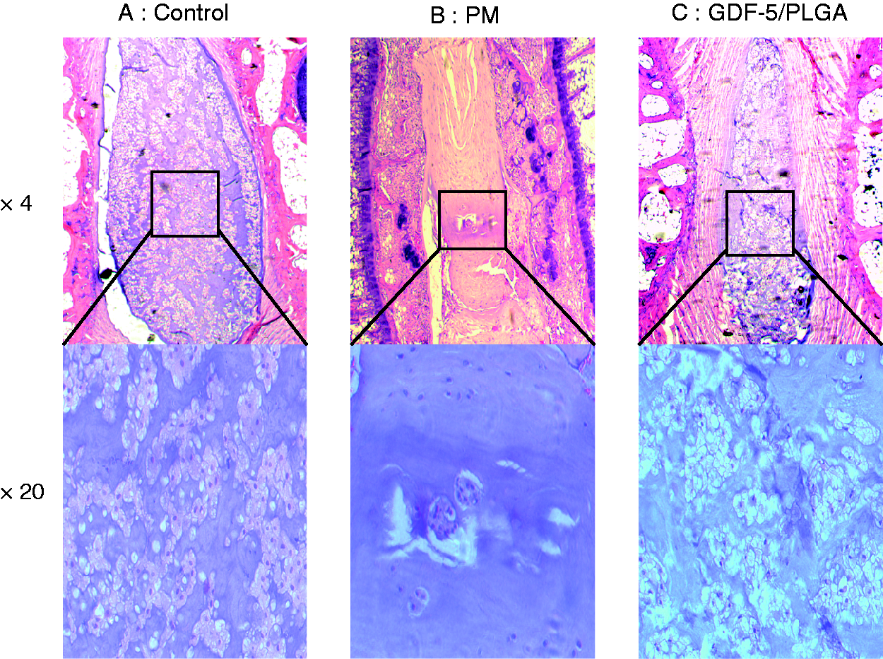
Messenger RNA expression of aggrecan, type I, and type II collagens
Group treated with GDF-5/PMs showed an increase in mRNA expression of aggrecan compared with other three groups although the increase did not reach significance (Figure 5(a)). On the other hand, the mRNA level of type II collagen, which is the phenotypical molecule of chondrogenesis, was significantly increased compared with that of the PMs alone injected group after 8 weeks treatment (Figure 5b, p < 0.01). On the contrary, the mRNA expression of type I collagen that typically reflect dedifferentiation of chondrocytes in the treated with GDF-5/PMs group was significantly decreased compared to that of PMs alone group and puncture group (Figure 5(c), p < 0.05). The differentiation index (the ratio of type II to type I collagen) is typically used to demonstrate the tendency for chondrogenesis. The result showed that the differentiation index of the GDF-5/PMs treated group was significantly higher than other groups (Figure 5d p < 0.01). However, the index in the PMs group and puncture group was significantly decreased when compared with that of the control group (Figure 5d, p < 0.05).
mRNA expression of (a) aggrecan, (b) type II collagen, (c) type I collagen, and (d) differentiation index in the nucleus pulpous of different experimental groups (four discs per group) at eight weeks after injection. The differentiation index is the ratio of type II to type I collagens. *p < 0.05 and **p < 0.01, when compared with puncture injection group. #p < 0.05 and ##p < 0.01, when compared with control group.+p < 0.05 and ++p < 0.01, when compared with PMs alone injection group.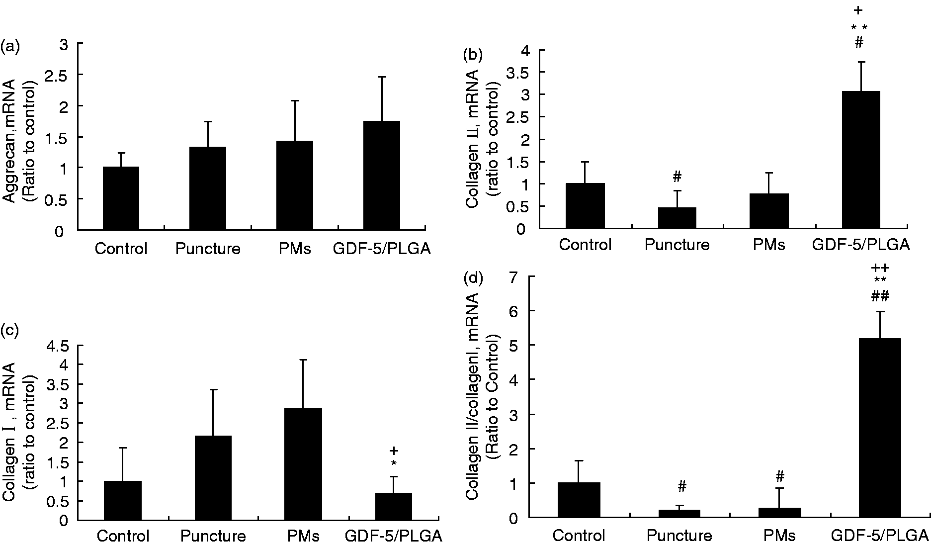
Discussion
Rapid advances of biology-based strategies for disc repair include autologous cell transplantation, growth factor injection, or gene delivery. The delivery of exogenous proteins in the form of growth factors is a promising therapy to address IVD. 5,9,17,20
Several possible effects (such as half-life, solubility of factor, and so on) for the long-term effects of growth factor injection need to further be taken into consideration.
To achieve an ideally controlled release of growth factor during processing or application, the optional carriers can be implemented as growth factor delivery systems to the IVD. PMs were used as ideal protein-encapsulating vehicles.21–26 Incorporation of growth factors within polymeric carriers delays proteolysis and also facilitates the sustained, localized release of growth factors and simultaneously avoided side effects.
In the present experiment, we manufactured rhGDF-5/PMs using the water-oil-water (W/O/W) method. 13 The W/O/W double-emulsion fabrication technique is an effective and well-characterized method for producing PMs. 11 We selected with a high lactic acid ratio to create microspheres as the delivery vehicle for this study. The 75:25 LA/GA copolymer ratio used in this study is less hydrophilic than that of 50:50 ratio, leading to slower degradation. 27
The rate and duration of in vitro growth factor release from the microspheres have showed that the polymer have an initial burst of rhGDF-5 release (30–40%) and continued to release their remaining drug contents up to 42 days. After 42 days, a small amount of rhGDF-5 was still being released from the microspheres in vitro, which achieved a well-controlled release effect. Further, additional protein stabilizers such as bovine serum albumin or additional acid buffers could be used to improve and maintain the bioactivity of growth factor after release.
This study examined the efficacy of the injection of PM-loaded rhGDF-5 into degenerated discs in the well-established SD rat anular needle puncture model. The results of this study demonstrated that the intradiscal injection of rhGDF-5/PMs had an initial burst followed by sustained release rhGDF-5 which significantly improved rat disc regeneration, and it is effective for restoring disc height in this model.
IVD degeneration is mostly characterized by changes in disc morphology and composition of the extracellular matrix as well as loss of disc cells and water content. To achieve optimal disc repair, the ideal therapy should preserve the intact architecture of the disc tissue as much as possible while increasing the synthesis of type II collagen and proteoglycan. Our results show that rhGDF-5/PLGA injected into the NP of rat discs had an effect on the structural restoration of disc. An increase in the number of chondrocyte-like cells in the NP were also found after treatment in histological analysis. Further, the results showed up-regulates in gene expression of markers for chondrocytes including mRNA levels of collagen type II and the differentiation index (the ratio of type II to type I collagen), and down regulation of type I collagen, also significantly increased DNA content, proteoglycan synthesis after treatment.
One major concern in this study is that the degradation PLGA products (lactic and glycolic acids) are at low pH at the delivery site, which may inhibit the synthesis of disc cells. However, initially, 75:25 PL/GA copolymer ratio was used in our study. Thus, it results in extended release and slower accumulation of acidic degradation by PLGA degradation products. Moreover, the tissues of the NP are subjected to continuous fluid flow, and the break down products would rapidly diffuse away from the site. In addition, native environment of disc cells is likely to be more acidic than that of cartilage cells, suggesting that they may be relatively resistant to acidic degradation products. 28 Third, Gorth et al., 29 co-cultured NP constructs with blank microspheres in vitro. There was no significant effect on GAG content, suggesting that the microsphere degradation products did have no effect on production of NP matrix.
There are several limitations to the present study. First, no data of histological and biochemical mechanism changes that the reaction of tissue to therapy factor at defined time intervals. Second, it is no control group of injection of rhGDF/5 alone. However, sustained delivery of growth factor, less dose of rhGDF-5 was used in our study than other reports. One previous study of the injection of rhGDF-5 (100 µg) in the rabbit anular puncture model after 16 weeks, resulted in a restoration of disc height and histologic grading scores. 8 In addition, in our previous pilot test, there is less restoration of disc height in GDF-5/PLGA group (p < 0.05) after 4 weeks injection rhGDF-5 alone in rat degenerated disc. Thus, microencapsulation provides sustained release of rhGDF-5, potentially allowing a reduction dosage of rhGDF-5 therapy. Third, the rat anular puncture model was used in this study; however, there are the disparity between a rat IVD and human IVD. The size of rat disc area is smaller than that of human. Thus, the injury of rat disc is easier to restore for less nutritional requirement than in human. In addition, unlike the human disc, the rat tail disc is not subjected to any mechanical load. Although this model does not truly reflect the course of human disc degeneration, similar histological and biomechanical changes exist between rat and human. So, the result of this research may still provide insight into the therapy strategy for human IVD regeneration.
In summary, this study showed that PLGA-encapsulated rhGDF-5 offers the possibility of controlled and sustained delivery of rhGDF-5 in vivo. The approach allowed the rapid and easy injection of rhGDF-5/PLGA complexes into the site of the defects. On the other hand, it allowed the controlled and sustained delivery of the growth factor in direct contact of the injury site preventing the fast degradation of short half-life growth factors when injected in vivo in a soluble form. No inflammatory cells within the IVD tissue were found, suggesting that rhGDF-5/PLGA had no adverse effects on disc tissues.
Conclusion
In summary, rhGDF-5-loaded PLGA-based microsphere was fabricated by double-emulsion techniques in this study. The rhGDF-5 sustained release from the microsphere may provide a control on the levels of rhGDF-5 over time. The result showed that disc degeneration can be treated successfully. Therefore, growth factor-loaded PM therapies show a promising strategy for IVD regeneration.
Footnotes
Funding
This work was financially supported by the National Science Foundation of China (81271728) and Beijing Natural Science Foundation (3102010).
Conflict of interest
None declared.