
Abstract
The poor retention and efficacy of instilled drops as a means of delivering drugs to the ophthalmic environment is well-recognised. The potential value of contact lenses as a means of ophthalmic drug delivery, and consequent improvement of pre-corneal retention is one obvious route to the development of a more effective ocular delivery system. Furthermore, the increasing availability and clinical use of daily disposable contact lenses provides the platform for the development of viable single-day use drug delivery devices based on existing materials and lenses. In order to provide a basis for the effective design of such devices, a systematic understanding of the factors affecting the interaction of individual drugs with the lens matrix is required. Because a large number of potential structural variables are involved, it is necessary to achieve some rationalisation of the parameters and physicochemical properties (such as molecular weight, charge, partition coefficients) that influence drug interactions. Ophthalmic dyes and structurally related compounds based on the same core structure were used to investigate these various factors and the way in which they can be used in concert to design effective release systems for structurally different drugs. Initial studies of passive diffusional release form a necessary precursor to the investigation of the features of the ocular environment that over-ride this simple behaviour. Commercially available contact lenses of differing structural classifications were used to study factors affecting the uptake of the surrogate actives and their release under ‘passive’ conditions. The interaction between active and lens material shows considerable and complex structure dependence, which is not simply related to equilibrium water content. The structure of the polymer matrix itself was found to have the dominant controlling influence on active uptake; hydrophobic interaction with the ophthalmic dye playing a major role.
Keywords
Introduction
The majority of ocular conditions that affect the anterior eye are treated by topical application of drugs in the form of solutions or suspensions, which together account for approximately 90% of ophthalmic therapeutic usage.1,2 A significant disadvantage of these systems is rapid and extensive pre-corneal drug loss resulting in poor corneal bioavailability, estimated to be only 1–10% of the administered drug.2–7 Loss occurs predominantly by rapid drainage of the fluid through the nasolachrymal duct. Other factors such as extracorneal uptake and binding of the drug to tear proteins also contribute to the reduction of effective drug dosage.2,6,8,9 The nature of the problem is illustrated in the work of Ludwig and Van Ooteghem using fluorescence techniques and Hardenbeger using a radioactive technetium (99mTc) tracer technique.10,11 Figure 1(a) and (b) provide a graphical representation derived from their data.
(a) Effect of dropsize on ocular retention of saline-based formulations.10 (b) Effect of formulation modifiers on ocular drainage times illustrating the effect of viscosity on retention.11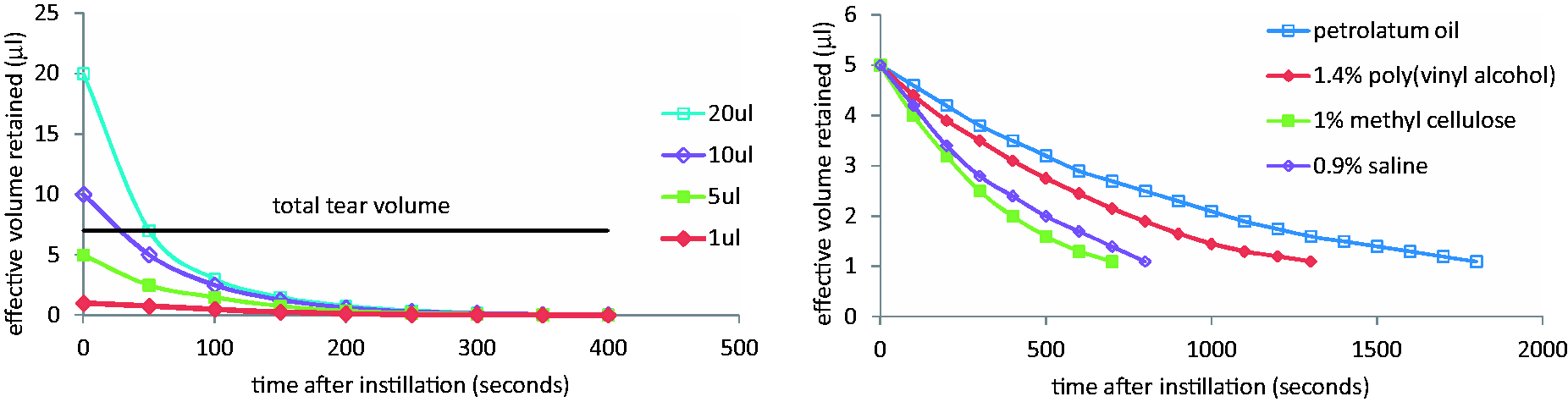
Perhaps surprisingly, mass transport modelling has demonstrated that by reducing the instilled volume from 25 µL to less than 5 µL, the ocular bioavailability of drugs with low corneal permeability should increase fourfold. 12 This is a consequence of prolonged contact time between drug and anterior eye surface, resulting from the fact that normal tear volume is not disturbed. 13 Realistically however, strategies such as reducing administered volume, increasing drug dosage and frequency of application and related strategies, although capable of improving drug effectiveness are difficult to achieve in practice.2,5,7,14,15
Several attempts have been made to harness specific properties of macromolecules (viscosity modification, mucoadhesion and in situ gelling11,16–19) to enhance interaction and retention of the drug with the anterior eye. The marked effect of viscosity increase (0.9% saline < 1% methyl cellulose < 1.4% poly(vinyl alcohol) < petrolatum oil) on the reduction of tear film drainage is illustrated in Figure 1(b). It is clear that neither increased instillation volume nor increased viscosity of the instilled formulation is able to produce effective drug retention over a period of several hours.
Nonetheless, it is apparent that the improvement of pre-corneal retention is key to the development of a more effective ocular delivery system. A system in which the retention of the vehicle itself in the eye is prolonged in order that a drug reservoir can be maintained, as in the case of a contact lens, is therefore a promising potential solution.20–26 Furthermore, because of the changing role of optometrists in prescribing drugs to manage eye diseases,27,28 it is logical to extend optometric technology and practice into the therapeutics field. The increasing availability and clinical use of daily disposable contact lenses provides the platform for the development of viable single-day use drug delivery devices based on existing materials and lenses. This approach is complementary to, but in many ways different from, the many approaches that seek to develop new composite contact lens systems with the aim of achieving release over periods of days and weeks.29,30 In such cases, zero-order release is almost essential in order to achieve predictable delivery over successive day-long periods. For a single-day use device, the need is to replace the erratic administration produced by intermittent administration of liquid drops with day-long release that falls within the upper and lower boundaries defined by toxicity and minimum therapeutic effectiveness.
The concept of using a contact lens as an ocular drug delivery vehicle is, in principle, obvious although as yet no systematic understanding of the design considerations linking specific drug structures to specific polymer matrices has been undertaken. The most commonly used contact lens materials and most obvious target delivery matrices are hydrogels, which are simply water-swollen polymer networks. As with the design of any drug delivery system, an understanding of drug–vehicle interactions and partitioning effects between the vehicle and the administration site are key to the design of successful systems.
Several workers have attempted to harness combinations of existing lenses and existing ophthalmic drugs on an empirical basis by simply soaking the lens in the drug solution and investigating the release by passive diffusion into relatively large volumes of saline.31–35 Although such experiments are extremely useful in providing baseline diffusional studies, they do not address fundamental aspects in sufficient detail. Additionally, the fact that lenses will be supplied in a saline-based packing solution of approximately 1–2 mL volume means that diffusional release will have come to equilibrium in that environment long before the lens is introduced into the anterior eye (placed in contact with the cornea). If we are to understand the subsequent release behaviour in the anterior eye, it is important to address both equilibrium structural interactions and the subsequent triggering effects caused by the change in environment. These triggering effects include inter alia, tear volume and flowrate, the mechanical action of the eyelid (which has an inter blink period of approximately 10 s), the effect of tear components such as lipids, proteins and electrolytes on the extraction process and the effects of pH shift on partition coefficient and thus release characteristics of the drug.
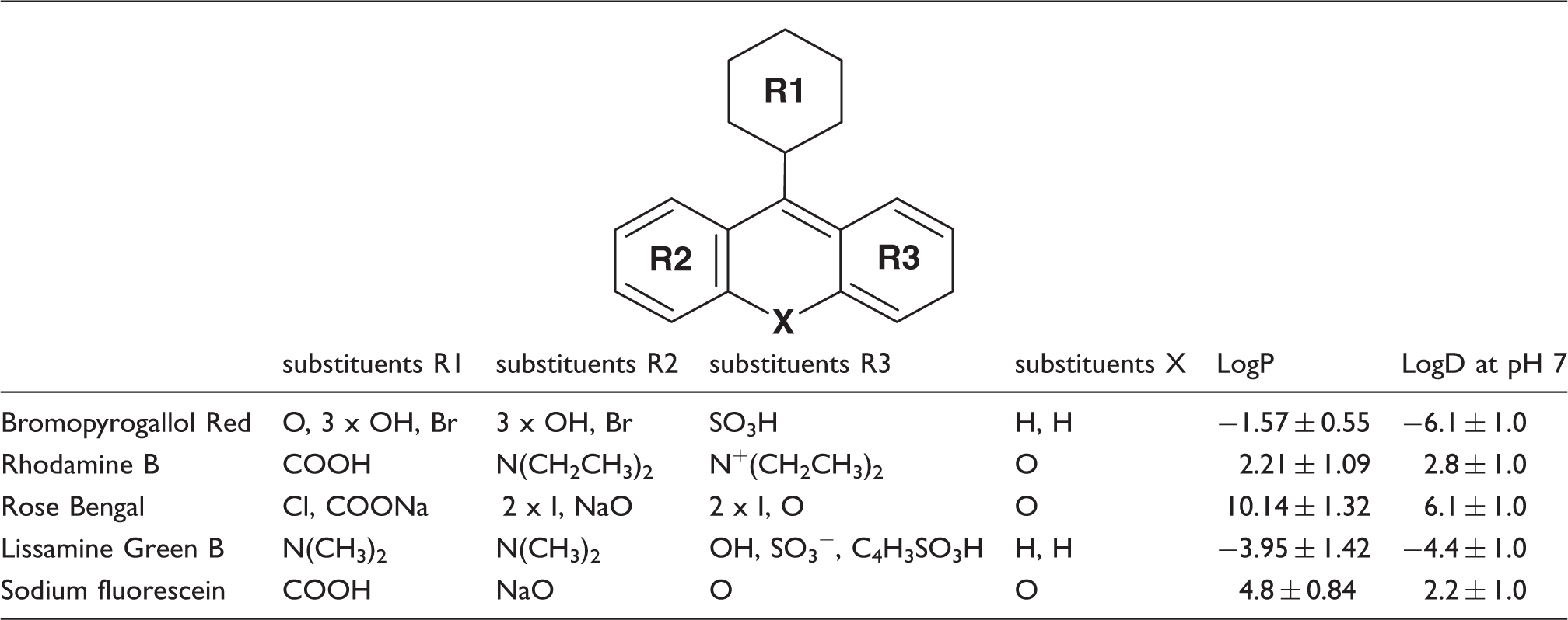
Ring structure common to the chosen ophthalmic and related dyes, indicating variations in the nature of ring substituents and consequent logP (octanol–water partition coefficient) and logD (octanol–water distribution coefficient) values calculated using the ACD/I-Lab service.
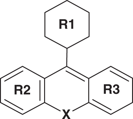
This approach has the advantage of allowing the effects of small structural variations on uptake and release to be readily and directly observed by colorimetric assay. Additionally, it is relatively straightforward to gain ethical approval for in vivo release studies involving commonly used ophthalmic dyes. Importantly, this enables the molecular interactions between the matrix and the active to be assessed, which is a key factor in the design of effective ophthalmic drug delivery vehicles.
Materials and methods
Materials and lens analysis
Materials employed in these studies are commercial hydrogel contact lens materials of known compositions and ophthalmic and related dyes (Table 1). The compiled data for the materials is shown in the results section.
Surface and sub-surface analysis of silicone hydrogel lenses was carried out using XPS. Lenses were dehydrated to a constant weight and transferred to a Thermofisher ESCALAB 250 electron spectrometer equipped with a hemispherical sector energy analyser. A monochromatic Al Kα X-ray source was used for analysis to enhance the resolution. At source an excitation energy of 15 keV and emission current of 6 mA, analyser pass energy of 20 eV with step size of 0.1 eV and dwell time of 50 ms were used throughout the experiments. Sub-surface information was obtained by bombarding the surfaces with 3 keV, 4 mA argon, in order to remove 500 nm of the surface. Elemental (C, N, O and Si) compositions were obtained from the resultant spectra. Combustion C, H, N microanalysis (Medac Ltd, UK) was used for compositional characterisation of conventional hydrogels.
Incorporation of active into the lenses
The contact lenses were loaded with dye by imbibition using conditions specified in the text. Typically, a minimum of three lenses were rinsed to remove any salts from the packing solution and then placed directly into the dye solution (at a ratio of 1 mL of soak solution per soaked lens unless stated otherwise) until equilibrium was reached (approximately 24 h). Loading of fully hydrated hydrogels in this way represents the method most likely to be used in the commercial production of therapeutic contact lenses.
Passive release methodology
Baseline experiments to establish relative structural interactions of active and matrix were carried out by passive release (single lens into 5 mL with gentle agitation); experiments more closely mimicking tear volume, flow rate and pH, and trigger effects of the eyelid are investigated in subsequent studies.
Loaded lenses were blotted on filter paper to remove excess dye, placed in 5 mL of fresh phosphate buffered saline (PBS) release medium at pH 7.4 and stirred constantly (on a shaker at 200 r/min). This regime minimised the formation of a stagnant boundary transfer layer, and maintained optimum sink conditions. At the end of each hour, the lenses were removed and placed in vials containing fresh PBS release media and the process repeated until no further dye was released from the lens.
Release media analysis
The optical density (OD) i.e. absorbance of the release media was measured by UV-Vis spectroscopy using a Molecular Devices SpectraMax M2 spectrophotometer at the maximum absorption wavelength of the released active. The absorbencies were then converted to concentrations using standard calibration curves. Release measurements were carried out in triplicate and averaged.
Analysis of active retained within the lens matrix
Non-destructive measurement of mass of retained active was carried out spectroscopically using a Molecular Devices SpectraMax M2 spectrophotometer at the appropriate maximum absorption wavelength of the active.
Determination of distribution coefficients
Values of logP (octanol–water partition coefficient) and logD (octanol–water distribution coefficient) of both drugs and dyes were determined using the ACD/I-Lab service based on structure. The values for polymeric repeat units were obtained from the literature using the nearest structural equivalence. 36
Statistical analysis
The experimental data are reported for triplicate samples, unless otherwise stated. Figures show averages and standard error bars.
Results and discussion
Material characterisation
Details of contact lenses used in this study, including USAN/ISO nomenclature, constituent monomers, water contents, elemental composition data and estimated octanol–water distribution (logD) coefficients.
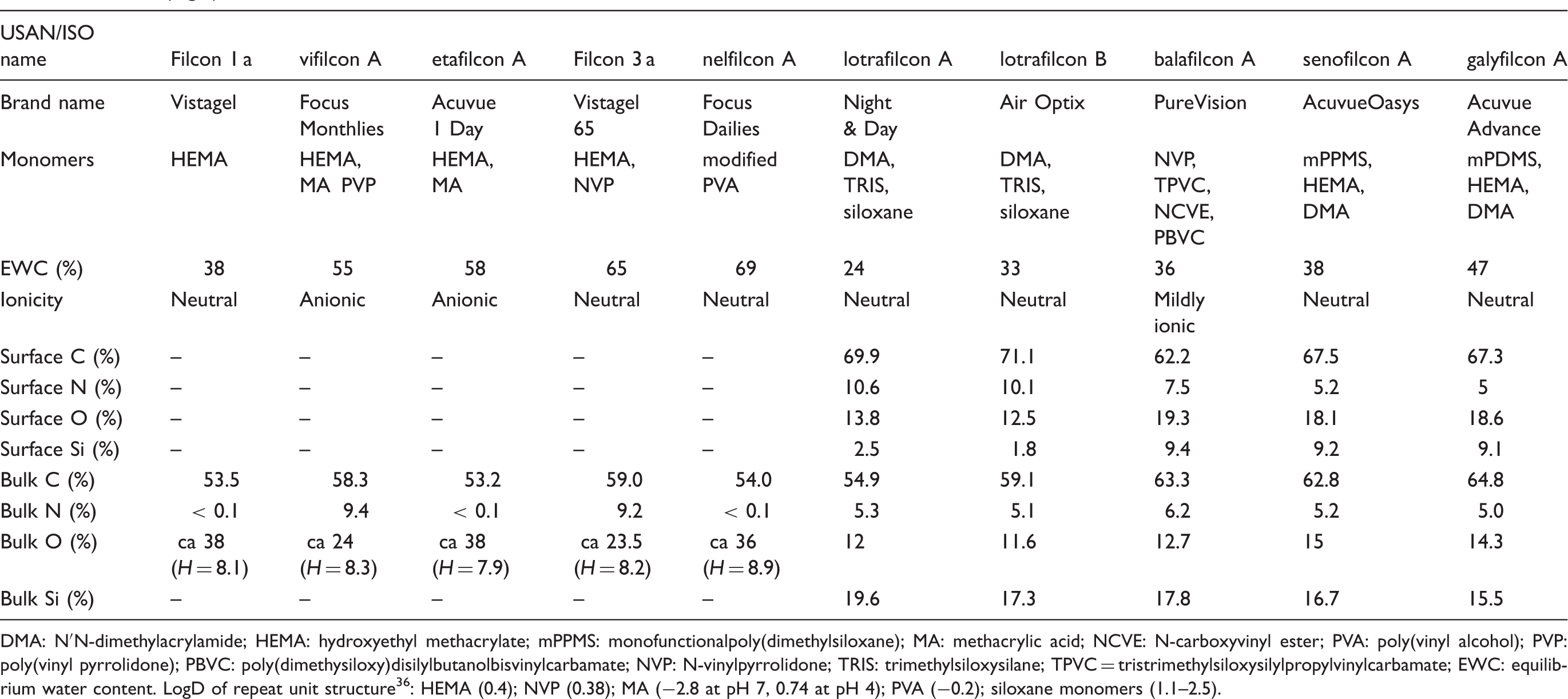
DMA: N′N-dimethylacrylamide; HEMA: hydroxyethyl methacrylate; mPPMS: monofunctionalpoly(dimethylsiloxane); MA: methacrylic acid; NCVE: N-carboxyvinyl ester; PVA: poly(vinyl alcohol); PVP: poly(vinyl pyrrolidone); PBVC: poly(dimethysiloxy)disilylbutanolbisvinylcarbamate; NVP: N-vinylpyrrolidone; TRIS: trimethylsiloxysilane; TPVC = tristrimethylsiloxysilylpropylvinylcarbamate; EWC: equilibrium water content. LogD of repeat unit structure 36 : HEMA (0.4); NVP (0.38); MA (−2.8 at pH 7, 0.74 at pH 4); PVA (−0.2); siloxane monomers (1.1–2.5).
Effect of polymer and dye structure on equilibrium uptake
It is well documented that both water content and matrix composition affect the transport of simple molecules, such as oxygen permeation through hydrogel membranes.37–39 The diffusion of hydrated species (e.g. ophthalmic dyes or drugs) is subject to similar influences, with both size and structural features of the hydrated species together with hydration pathways and molecular structures of the matrix playing a part. It is therefore important to establish the relative extents to which an imbibed species is: (a) held in the aqueous component of the hydrogel and (b) in some way sequestered by the polymer component.
A competitive equilibrium was established between dye, water and polymer by using a fixed volume (2 mL) containing a fixed mass of surrogate active (e.g. 100 µg/mL Rose Bengal) and a hydrogel contact lens (approximately 25 mg). This allowed the distribution of the active between solution and matrix to be determined. This combination of low concentration and limited volume reflects the therapeutic concentration of a typical ophthalmic drug in the volume of a typical packing solution. However, it should be noted that under these conditions the uptake does not necessarily represent the total absorptive capacity of the lens; it simply reflects the differential ability of lens materials to immobilise the dye and thus the partition coefficient between lens and packing solution under these specific conditions. Spectroscopic analysis of the dye concentration in solution enables the mass of dye absorbed by the lens to be calculated; thus the distribution of the dye remaining in solution and in the lens is known.
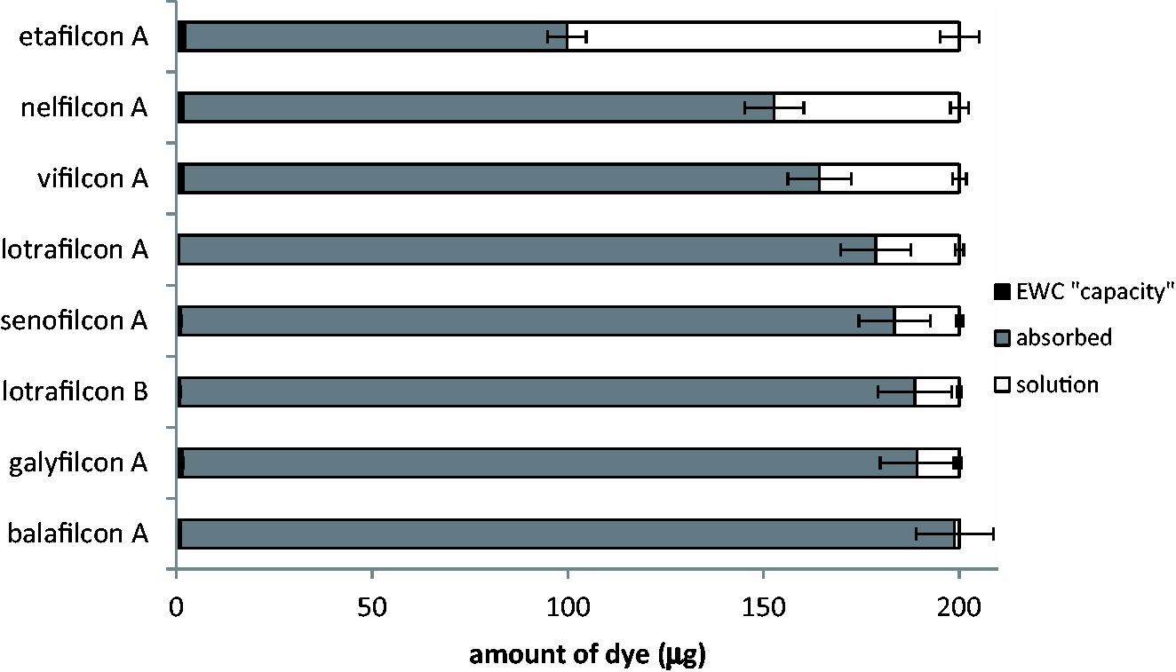
Figures 2 and 3 present data for the interaction of two structurally distinct actives (Rhodamine B and Rose Bengal) with a range of hydrogel matrices. The materials are arranged to show the extremes of behaviour achieved with the range of matrix structures employed.
The mass of dye absorbed by various lens materials and remaining in the PBS solution at equilibrium, with each lens soaked in 2 mL of 100 µg/mL Rhodamine B solution. The mass of dye absorbed by various lens materials and remaining in the PBS solution at equilibrium, with each lens soaked in 2 ml of 100 µg/mL Rose Bengal solution.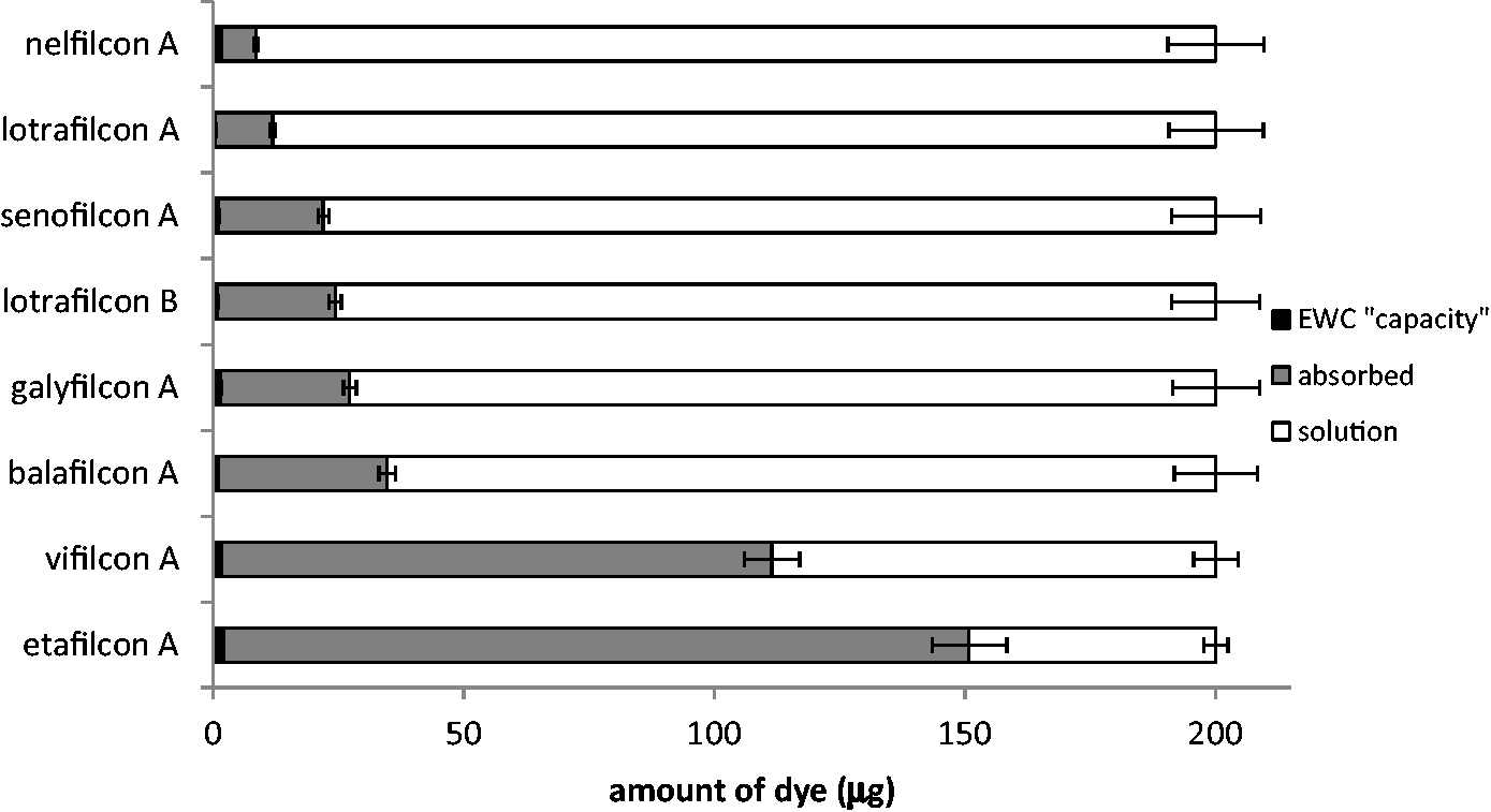
This initial study involved the most hydrophilic (Rhodamine B) and most hydrophobic (Rose Bengal) variants of the intact ring, as reflected by their octanol–water partition coefficient (Table 1). Care must be taken in interpretations of hydrophilicity, however, since association enables relatively hydrophobic molecules to show high aqueous solubility, which enhances interaction with hydrophilic environments. An example of this is the self-association of Rose Bengal, which enables a combination of substantial aqueous solubility and the hydrophobicity reflected in the high positive values of logP (10.1) and logD (6.1 at pH 7). 40 Rhodamine B has also been reported to show aggregation in water but here the two tertiary amine groups in the molecule convey greater hydrophilicity on both unaggregated and aggregated forms as reflected in the less positive values of both logP (2.2) and logD (2.8 at pH 7).41,42 Several ophthalmic drugs (e.g. dexamethasone phosphate, cromolyn sodium and ketorolac tromethamine43–45) are also known to show association behaviour; the use of Rhodamine B and Rose Bengal as models thus provides instructive interpretive information on structural effects in lens–drug interaction.
The ratio of water to polymer as reflected in the equilibrium water content of the hydrogel matrix has a marked effect on the nature of drug–polymer interaction. It may be envisaged that the drug enters the lens matrix in its associated form through the aqueous component of the hydrogel and can then undergo, for example, charge–charge interaction, charge–dipole interaction, hydrogen bonding, weaker polar interactions or hydrophobic interactions with the polymer matrix.
The Rose Bengal results (Figure 3) are relatively easy to understand. The influence of the higher octanol–water partition coefficient now becomes apparent. Rose Bengal, having entered the lens matrix in associated form and consequent enhanced aqueous solubility, then undergoes rearrangement or disaggregation to facilitate hydrophobic interaction with the polymer matrix. Charge interaction plays little part in controlling retention. As indicated in Figure 3, the relatively hydrophilic anionic etafilcon A shows least ability to retain Rose Bengal. On the other hand, silicone hydrogels with their hydrophobic core components show very high competitive absorption behaviour.
It is clear that Rhodamine B behaves differently from Rose Bengal (Figures 2 and 3) and the role of the two amine groups in Rhodamine B is apparent. Not only do we see strong interaction with the anionic etafilcon A and vifilcon A materials but also there is little evidence of the disaggregation leading to enhanced hydrophobic interaction with the lens matrix that was apparent in Rose Bengal interactions. This is most clearly seen in the very low interaction of the five silicone hydrogel materials with Rhodamine B, in contrast to the very strong interaction of Rose Bengal with these polymers. The silicone hydrogels as a family behave relatively similar to each other. The uptake results for silicone hydrogels confirm that it is the bulk composition of the materials (which are comparable, Table 2) rather than their surface compositions (which show marked differences, Table 2) that drive this process. Thus the effect of the surface coating, which reduces the effective surface silicone concentration of lotrafilcon A and B (Table 2) has no observable effect on their uptake behaviour. There is, however, a small but observable effect of water content as evidenced in the fact that the order of uptake is lotrafilcon A < senofilcon A ∼ lotrafilcon B < galyfilcon A. The level of balafilcon A ionicity, although low in comparison to that of etafilcon A and vifilcon A, does produce a noticeable increase in the uptake of the N-containing dyes by balafilcon A in comparison to lotrafilcon B and senofilcon A, which have substantially equivalent water contents.
Small changes in the structure of the dye can therefore have a significant effect on the physicochemical parameters (pKa, logP, logD, Table 1) and consequently on the interaction with the matrix. In order to further understand the dye uptake mechanism, the influence of reduced soak concentration (10 µg/mL) with the same materials on the equilibrium was investigated. The relationship between the concentration of a component in a feed solution and the mass of that compound taken up by a polymer is important in many fields and has been studied for a wide range of absorbed compounds and substrate polymers. The results typically take the form of a conventional type I sorption isotherm with an initial steep slope progressively reducing to a plateau.
46
Comparative data for the two feed concentrations (10 and 100 µg/mL) are shown in Figures 4 and 5, which present both percent uptake from feed solution and actual mass uptake values.
Variation in percentage of Rhodamine B dye uptake at equilibrium, as a function of feed concentration. Each lens material was soaked in 2 mL of 10 µg/mL and 100 µg/mL Rhodamine B. Figures above individual bars indicate mass uptake in micrograms. Variation in percentage of Rose Bengal dye uptake at equilibrium as a function of feed concentration. Each lens material was soaked in 2 mL of 10 µg/mL and 100 µg/mL Rose Bengal. Figures above individual bars indicate mass uptake in micrograms.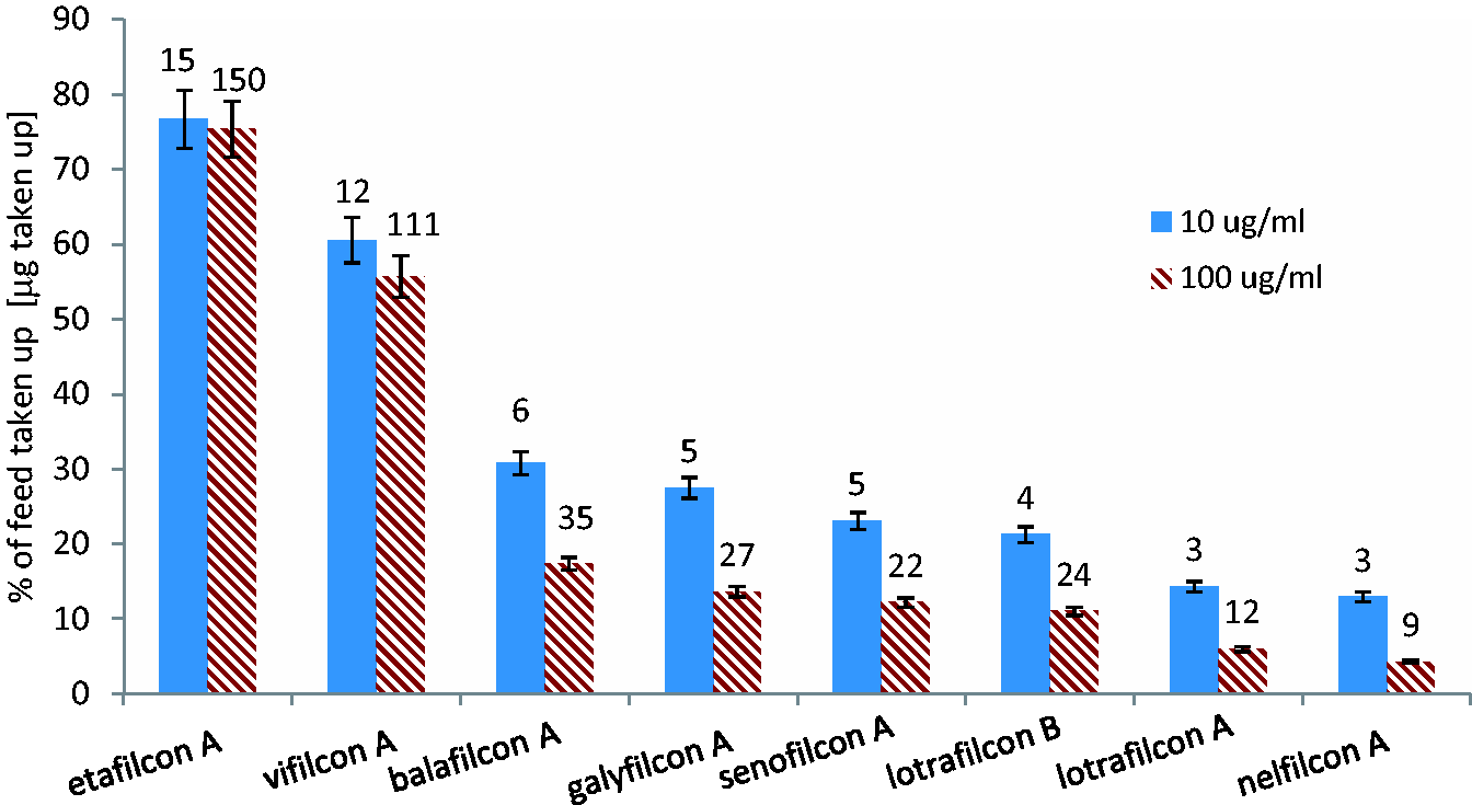
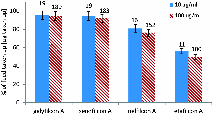
These figures show that in the case of both Rhodamine B and Rose Bengal there is an increase in uptake with increased concentration, but their behaviour differs significantly. The weakness of the hydrophobic interaction between Rhodamine B and the group of substantially neutral polymers, shown in Figure 4, is illustrated by the fact that at a concentration of 10 µg/mL, around 25% of Rhodamine B is taken up by the silicone hydrogels (lotrafilcon B, senofilcon A and galyfilcon A) whereas this falls to around 5% at the higher concentration of 100 µg/mL. In contrast, at these two feed concentrations, approximately 90% of the feed concentration is taken up by the same group of polymers with Rose Bengal (Figure 5). Figure 4 also reflects the charged-based interaction of Rhodamine B with anionic polymers (etafilcon A and vifilcon A). It has been noted (Figures 2 and 3) that the very weakly anionic balafilcon A shows rather more uptake capacity than the completely neutral silicone hydrogels.
Kinetics of release into sink conditions
Having observed the structural effects of both dye and material on uptake behaviour, it is logical to examine the effect of these factors on release, initially passive release into an ‘infinite’ reservoir of saline. It is important to understand passive release behaviour from different matrices as a baseline for further study of trigger release conditions such as agitation, temperature, pH and release medium composition. In order to ensure that an uptake equilibrium ‘plateau’ had been reached, the feed concentration was increased from 0.01% (100 µg/mL) to 1% (10 mg/mL).
The methodology described in ‘Passive release methodology’ was used to study the passive release of a series of ophthalmic and related dyes of different structures from nelfilcon A (Figure 6). The polyvinyl alcohol (PVA)-based nelfilcon A material was chosen because it combines the highest water content and the simplest molecular structure: carbon backbone ‘polyethylene’ chain with pendant hydroxyl groups.
47
PVA is used in several ocular-related applications.
48
Additionally, it is the only strictly neutral commercial lens material (many lenses are based on hydroxyethyl methacrylate (HEMA), which although nominally neutral is known to disproportionate, yielding tracial methacrylic acid residues). In this set of experiments, the range of dye structures has been extended to cover compounds shown in Table 1, in which modification of the same underlying ring structure produces an extended range of logP and logD values.
Cumulative release profiles of ophthalmic and related dyes from nelfilcon A into PBS, following an equilibrium pre-soak of 1 lens per mL of 1% dye solution for 7 days. RB: Rose Bengal; NaFl: sodium fluorescein; BPR: Bromopyrogallol red; RhB: Rhodamine B. The insert shows a more detailed release profile of Rhodamine B.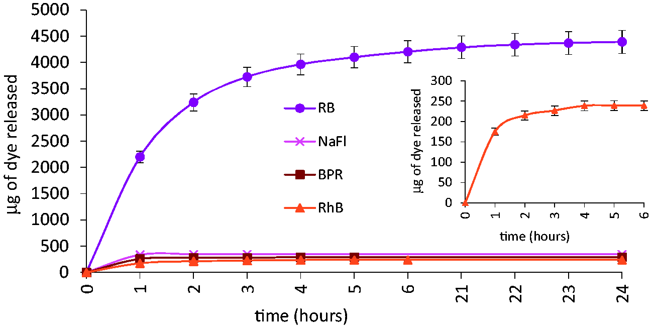
It is clear from Figure 6 that although the detailed release profiles are similar, the mass of dye released is very different. Rhodamine B and Rose Bengal represent the extremes of interaction studied here and both release profiles (Figure 6) bear some apparent resemblance to first-order release kinetics. However, it is apparent that more effective interpretation of the interaction phenomena would be facilitated by direct measurement of retention of surrogate active by the polymer matrix; this is made possible by the use of ophthalmic dyes.
Total cumulative release and optical density (OD) at passive release equilibrium of Rhodamine B and Rose Bengal from nelfilcon A, etafilcon A and Filcon 3 a. The OD was determined at the wavelength of maximum absorbance (λmax) of the dye.
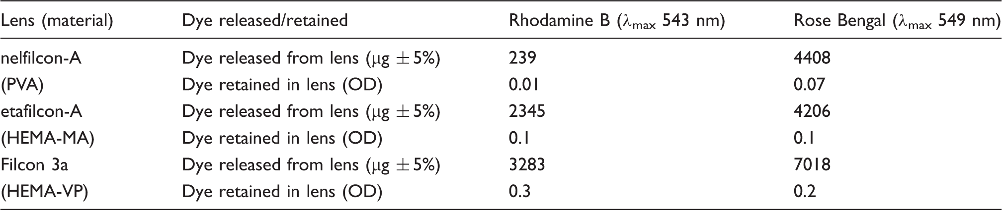
Although effects of ionicity and association complicate dye–polymer interactions, it is apparent that hydrophobic interactions are universal and make a significant underlying contribution in all cases. It is interesting to assess the values of octanol–water distribution coefficients of the polymer repeat units (data, Table 2) to see if they offer any correlation with the rank order referred to above (Table 3). HEMA-NVP (logD approximately 0.4) always shows stronger hydrophobic interaction than PVA (logD approximately −0.2), which accords with expectations. Methacrylic acid-containing polymers are more complicated because the logD value reflects the pH dependence of the carboxyl group, decreasing from 0.74 (non-ionised) to −2.8 (ionised). In consequence, under physiological conditions the behaviour of HEMA-MA in purely hydrophobic terms is intermediate between that of PVA and HEMA-NVP. The question of silicone hydrogels is interesting. The use of the atom fragment contribution method suggests that the partition coefficients of silanols containing a single silicon atom are approximately 0.5 greater than the corresponding alcohols over a wide range of structures. 49 Given the silicon content of silicone hydrogels (Table 2) and the implied siloxy monomer content, the overall logD values would be expected to be little greater than that of HEMA-NVP. This correlates with the results shown in Figure 3 taken together with those in Table 3. It is also consistent with the observations of Kalgard et al who found little difference in uptake and release behaviour of silicone hydrogels in comparison to conventional hydrogels.31,32
Uptake and release of dyes under different soak concentrations
In practice, ophthalmic drugs are formulated at different concentrations according to the required therapeutic need. The simplest way of using the contact lens as a drug delivery vehicle is to soak it in a solution of the ophthalmic drug – for example as a packing solution. It is therefore important to establish the effect of feed solution concentration on the release kinetics of a drug. Nelfilcon A lenses were soaked in 2%, 1%, 0.5% and 0.1% Rose Bengal solution; the cumulative release profile for each soak concentration is presented in Figure 7.
(a) Cumulative release into PBS of Rose Bengal from nelfilcon A contact lenses, each lens pre-soaked for 7 days in 1 mL of solution at a range of feed concentrations. (b) Data from (a) showing release as a percentage of feed (i.e. feed concentration × 1 mL).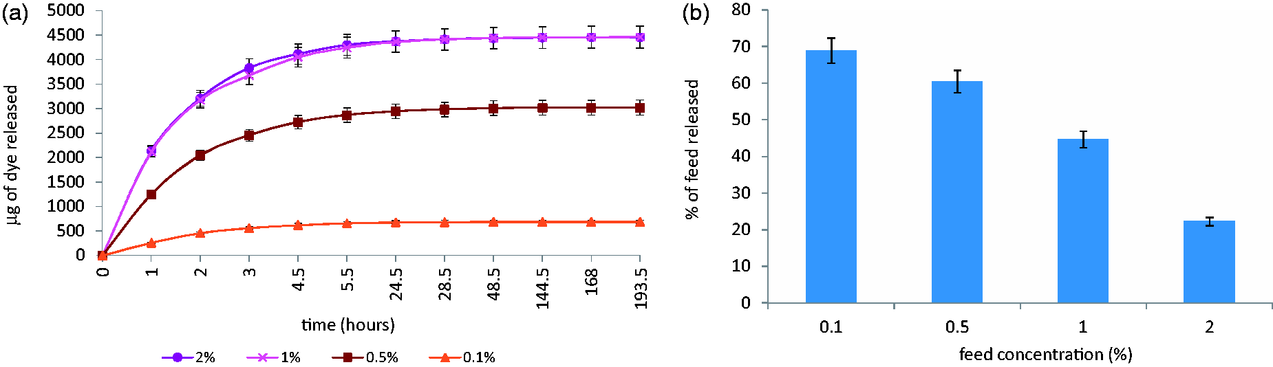
Variation in initial loading concentration did not alter the profile of release from the hydrogel but simply increased the quantity released to a plateau level at 2%. It is of interest to note that the balance of cumulative release and retention is clearly influenced by feed (Figure 7(b)). Data of this type are important in the design of specific lens-drug systems, where the drug is loaded into the lens via a packing solution of given volume (normally 1 mL) that contains the ophthalmic drug at its normal therapeutic concentration.
Structural links between ophthalmic dyes and ophthalmic drugs
Structures, pKa values and octanol–water partition and distribution coefficient data for target ophthalmic drugs.
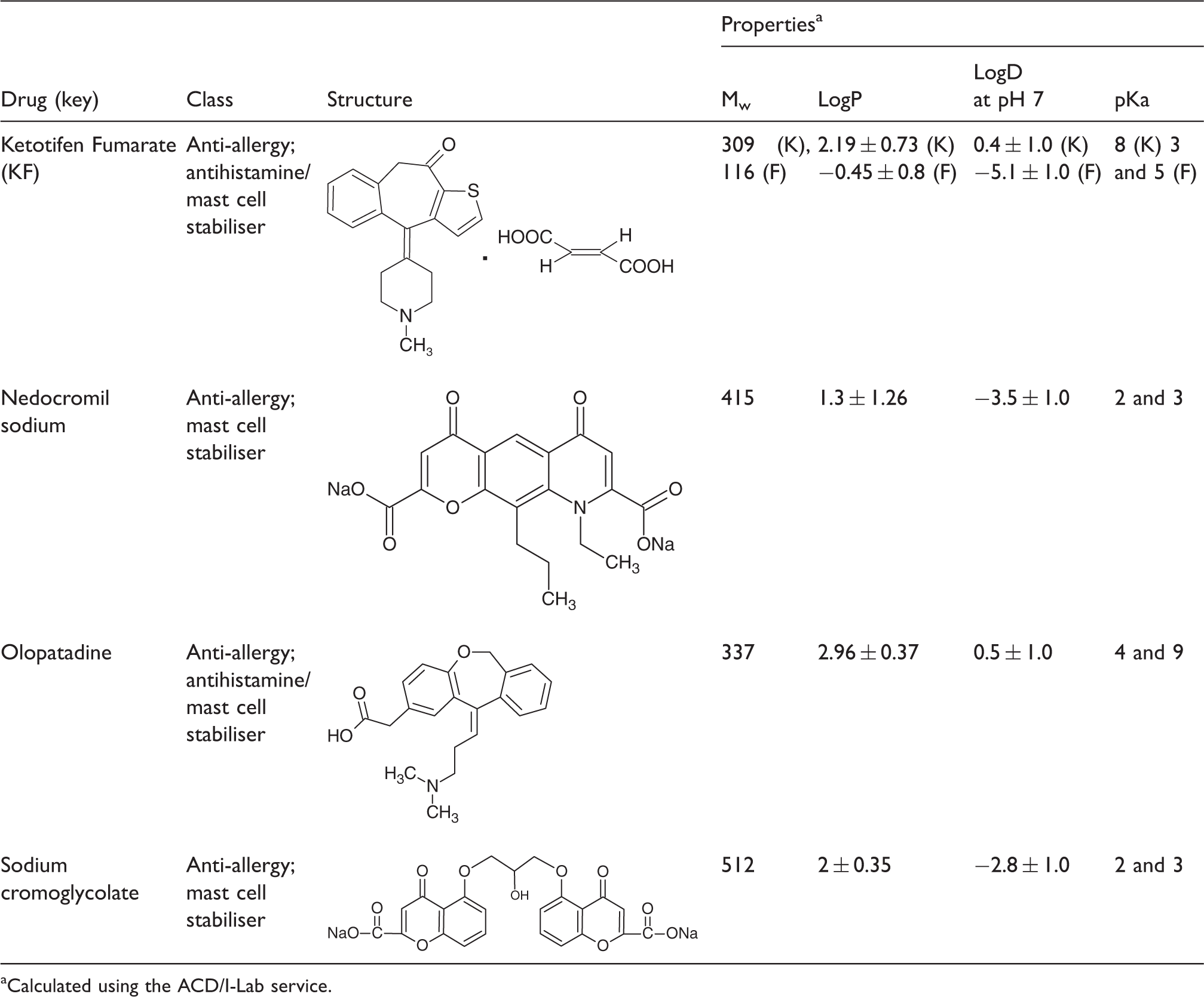
Calculated using the ACD/I-Lab service.
Conclusion
In the design of contact lenses for drug delivery, selection of drug and lens material combinations that will provide the desired therapeutic effect entails knowledge of the interactions and structural factors that influence uptake and release. Even the ingenious approaches (e.g. molecular functionalisation, molecular imprinting, drug encapsulation, use of carriers and incorporation of diffusion barriers35,50–54) that have been currently envisaged as methods of producing enhanced control of drug delivery from the lens ultimately involve diffusion of drug through the lens material. It is therefore essential to understand the factors related to partition and diffusional release in order to design effective systems.
Despite the range of materials and dyes structures studied here, under ‘passive’ conditions the release profiles are very similar in shape and duration. This supports the model for diffusion-controlled release of a hydrated species through the aqueous phase of a polymer matrix. The release profiles shown here are comparable to those observed with ophthalmic drugs in conjunction with commercial lenses and typical monolithic hydrogel devices.31–33,55,56 In general, silicone hydrogels behave very similarly to conventional hydrogels in simple uptake and release experiments. Balafilcon A shows the strongest hydrophobic uptake behaviour, although it may be that the porosity of this and other the silicone hydrogel matrices is an influential factor, rather than innate hydrophobicity alone.
In developing an efficient and effective single-day contact lens-based system for ocular drug delivery, consideration of the key characteristics of the drug, the lens matrix and the anterior eye allow drug/vehicle/delivery site interactions to be anticipated. Modulation of uptake is achieved by altering the concentration of the feed solution; which contributes to the design of a lens matrix-therapeutic agent combination with specific dosage requirements. Although water content enables a water soluble drug to diffuse into the lens, the balance of hydrophobic and ionic effects strongly influences retention and release. These factors also control the distribution of drug between lens and packing solution prior to insertion of the lens. The retained drug is then potentially capable of undergoing ‘triggered’ release stimulated by the transition from packing solution (passive environment) to eye (active environment). This aspect of release behaviour is dealt with in a subsequent paper.
Footnotes
Conflict of interest
None declared.
Funding
This research received no specific grant from any funding agency in the public, commercial, or not-for-profit sectors.