
Abstract
The discovery of new strategies to repair large segmental bone defects is currently an open challenge for worldwide clinicians. In the treatment of critical-sized bone defects, an alternative strategy to traditional bone grafting is always more frequently the use of tailor-made scaffolds modelled on the final size and shape of the implant site. Here, poly-ε-caprolactone-based composite scaffolds including poly-
Introduction
Large bone defects resulting from massive trauma, excision of tumours and congenital malformations require surgical therapy, because spontaneous regeneration is limited to relatively small defects.1,2 In this context, tissue-engineering strategies often involve the use of porous scaffold with or without molecular delivery to develop effective bone graft substitutes.3,4 Indeed, the use of suitable porous scaffolds could be of great advantage for critical defects since it offers the opportunity to fill bone cavity in situ and stabilize the defects while exerting beneficial stimuli, which promoting the cell activity and proliferation.
5
However, to be effective, it is mandatory that employed scaffolds fit into the anatomic defect and possess mechanical properties to bear in vivo loads, enhance tissue in-growth and produce biocompatible degradation.6,7 To date, mechanical and geometric requirements often are not completely satisfied in current tissue-engineering approaches, thus resulting in the main source of failure of many biological implants.8,9 In this context, synthetic scaffolds hold promise due to interesting properties in terms of workability and degradation, which assure an optimal compatibility with local microenvironments, without any infection transmission.10,11 Many synthetic materials as bone substitute are easily available, including metals, natural and synthetic polymers, ceramics and glasses.12–14 Among them, biodegradable synthetic polymers offer a number of advantages over other materials for developing scaffolds in tissue engineering, and they have been used in a number of clinical applications.
15
In particular, they exhibit predictable and reproducible mechanical and physical properties, such as tensile strength, elastic modulus and degradation rate under controlled conditions that can be tailored as a function of the specific application.
16
For instance, the finding of mechanical properties, able to match those of the tissues at the site of implantation, is often in conflict with the need of controlling kinetics of matrix degradation – namely, degradation or resorption rate similar to the rate of tissue formation.17,18 Moreover, a highly interconnected porous structure is mandatory to guide cell in-growth and bone tissue formation by promoting a more homogeneous blood vessels invasion.
19
Besides, scaffolds have to reproduce the complex composition and structural organization of natural bone tissue, which cannot be easily replicated using a unique material due to their limited range of properties.
20
To date, the employment of composite materials may represent a good solution in scaffold design, achieving the ideal balance between strength and toughness due to the improvement of specific properties of the composite compared to its separate components.
18
Recently, Guarino et al.20,21 proposed an interesting approach based on the use of fibre-reinforced porous composites developed by applying the basic theory of continuous fibre-reinforced composites to support the formation of bone segment in long bone treatment applications. Previous in vitro study demonstrates that human marrow stromal cells and trabecular osteoblasts rapidly adhere and proliferate into the 3D scaffold whereas the fibre arrangement into the porous scaffold concurs to an efficient migration of bone cells.
20
In this case, poly(
Materials and methods
Materials
PCL (MW 65 kDa) and PLLA fibres (75 D-tex; Mw 100 kDa) were purchased, respectively, from Sigma–Aldrich and Sofradim Companies. A benzyl ester of HA with 75% of esterification degree (HYAFF11®-75p) was supplied by Fidia Advanced Biopolymers in powder form. Tetrahydrofuran (THF) and dimethylsulfoxide (DMSO) have been purchased from Sigma Aldrich. Sodium chloride crystal (Fluka AT >99.9%) was sieved in specific size range depending on scaffold type (212–300 µm and 300–500 µm). A mixture of Ca2NaK(PO4)2 and monocalcium phosphate monohydrate were supplied by Technical University of Catalunia (UPC) under the name of R-cem. CaCl2, MgCl2·6H2O, NaHCO3 and K2HPO4·3H2O and human bone morphogenetic proteins (hBMPs) have been purchased from Sigma Aldrich (Milano, Italy).
Scaffold preparation
PCL pellets were dissolved in a solvent mixture to form a solution by stirring for 2 h at 40℃ using a PCL/solvent ratio equal to 12/88. Two different solvents were used, respectively, THF and DMSO by 80/20 weight ratio. HYAFF11® and R-Cem were then mixed to the polymer solution imposing a PCL/HYAFF11®/R-Cem weight ratio equal to 80/20/20. NaCl crystals with specific size range (300–500 µm) were employed as templating agents, and the PCL/NaCl volume ratio (32/64 v/v) was selected in order to optimize the fibre/matrix adhesion. Fibre-reinforced composite scaffolds were obtained by combining the phase inversion/salt leaching and filament-winding technology as shown in Figure 1(a). Briefly, PLLA fibres, impregnated through the PCL solution, were wound on a polypropylene tubing-coated stainless steel mandrel by using a specific winding parameters set (winding angle WA = 45°, winding pitch WP = 500 µm). Tubes with 6 mm as outer diameter and 1.5 mm as inner diameter were employed for morphological and mechanical characterization. For the in vivo test, composite tubes have been designed with proper size to match the implant site: 16 mm as outer diameter and 4 mm as inner diameter.
Scheme of the procedure used to prepare and bioactive PLLA fibre-reinforced scaffolds: (a) preparation of composite tube; (b) creation of porous structure; (c) biomineralization; (d) functionalization. PLLA: poly-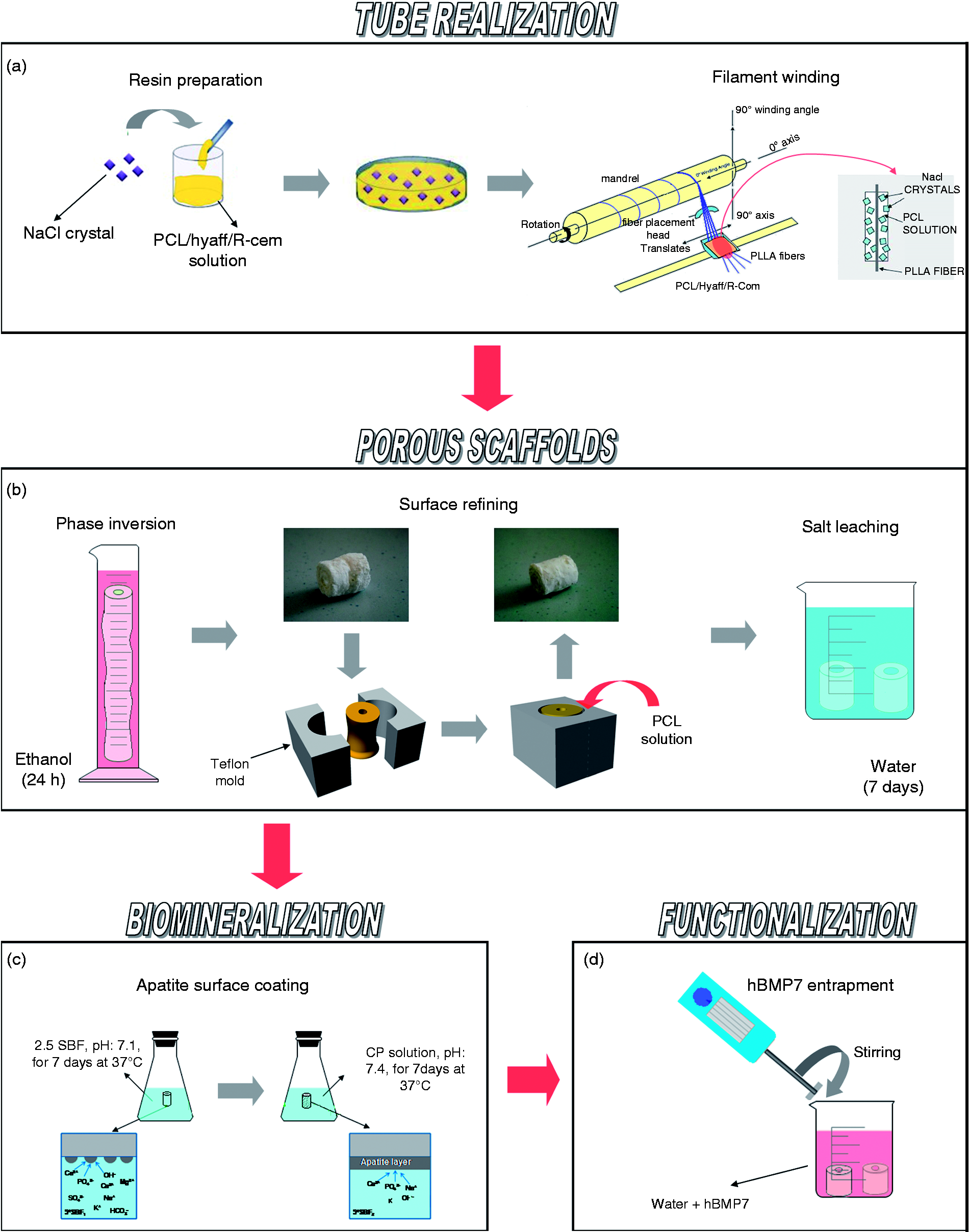
Once the winding process was completed, conventional solvent-casting procedures were employed immerging tubes in ethanol for 24 h. A Teflon round mould was employed to improve the scaffold surface finishing of sample for the in vivo test as shown in Figure 1(b). The porous tube was put into the mould, and it was closed. PCL/NaCl mixture, previously prepared, was used to fill the cavity between the tube and the surface of the mould, in order to create a porous skin around the scaffold with a regular shape. This treatment results in a more homogeneous surfaces that better fit with the in vivo defect. To completely remove the porogen particles, samples were put in distillate water for 7 days at room temperature, and the final porous composite scaffold was obtained.
Fibre-reinforced composite scaffolds (16 mm in diameter) were coated with calcium phosphate using a two-step coating process as reported elsewhere. 33 In the first step, a concentrated SBF solution (SBF 2.5) was prepared by dissolving reagent grade NaCl (40 g), CaCl2 2H2O (1.84 g), MgCl2 6H2O (1.52 g), NaHCO3 (1.76 g) and Na2HPO4·2H2O (0.89 g) salts in 1 L of demineralized water at 37℃. All operations were performed at room temperature by magnetic stirring in order to promote the formation of nuclei. Samples were immersed in the physiological solution (pH 7.1) and left to coat for 7 days under continuous gentle stirring at 37℃. This process was previously shown to result in a formation of thin, amorphous calcium phosphate layer. In the second step, reinforced tubes pre-coated in step 1 were immersed in a calcium phosphate solution (CPS) at physiological pH of 7.4 and temperature of 37℃ for 7 days to deposit a crystallized layer onto previously formed amorphous calcium phosphate layer. CPS was prepared by dissolving NaCl (8 g), CaCl2·2H2O (0.59 g), Na2HPO4·H2O (0.36 g) and Tris (6.05 g) in distilled water, and the pH of the solution was adjusted to 7.4 with 1 M HCl. The coated composite tubes were thoroughly washed in distilled water and dried overnight at 50℃.
Scaffold functionalization by morphogenic signal (BMP-7 Sigma Aldrich, Italy) was performed to guide cell recruitment into the scaffold implanted in vivo. The entrapment of the morphogenetic protein was performed by gently stirring of the sample in water solution with hBMP-7 concentration of 300 ng/mL. hBMP-7 was entrapped into the scaffold by gently stirring in spinner flask for 30 min. The amount of 1.6 µg of BMP was used for scaffold gram. The BMP-7 release in phosphate-buffered solution was determined using a commercial ELISA kit (R&D Systems Inc, Minneapolis) according to the manufacturer's instructions. Concerning the encapsulation efficiency, composite scaffolds – 10 mg in weight – were rinsed with acetonitrile three times. After centrifuging (16,000 rounds min−1) for 10 min, the precipitation was dissolved by isotonic phosphate-buffered saline (PBS).
Release was determined by incubating composite scaffold in 4 mL PBS (pH 7.4) with 0.02% sodium azide at 37℃. The tubes were centrifuged at 5 and 12 h and every other day starting on day 1 and ending on day 6. The collected 0.8 mL supernatant was assayed by ELISA for BMP-7 concentration, and 0.8 mL of additional PBS was added to the remaining one for the next incubation.
Morphological investigation
Scaffold morphology was investigated via scanning electron microscopy using a Quanta FEG200 (FEI, the Netherlands) scanning electron microscopy. Briefly, specimens were cut using a razor blade along preferential directions, parallel and perpendicular to the surface, respectively, after they were gold-coated by automatic sputter coater (EMscope SC500, UK) up to reach a conductive layer thickness of around 30 nm. The resulting transverse and longitudinal sections were gold-coated under vacuum by using an automatic coating sputter set at 15 mA for about 20 min. The porosity was assessed in terms of pore size, shape and spatial distribution by images at different magnifications. Furthermore, X-ray energy-dispersive spectroscopy (EDS – Inca Energy 200, Oxford Instruments, UK) was used for a qualitative estimation of Ca/P ratio.
Thermal analysis
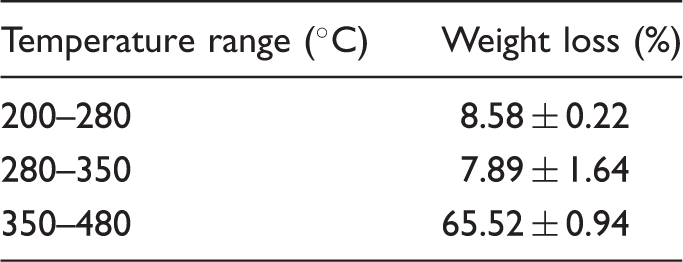
Weight losses and decomposition temperatures for the composites sample.
Dynamic mechanical analyses
Mechanical tests under compression were done using a dynamometric machine (Instron 4204) equipped with a load cell of 1 kN and requiring a crosshead speed of 1 mm/min. Porous cylindrical specimens characterized by a height of 12 mm and a diameter of 6 mm were fabricated according to the ASTM 695/2a standard. Stress and modulus versus strain were recorded. The elastic modulus E′ has been calculated as the slope of the tangent to the initial linear portion of the deformation curve. This test is preparatory to estimate basic information to be used in the dynamic mechanical tests. Indeed, the definition of the elastic range allowed to establish the average load and amplitude to apply for the cyclic stimulation. The dynamic mechanical analysis (DMA) results are presented in terms of storage (E′) and loss (E″) moduli and damping ratio (tan δ).
DMA was performed on the porous three-dimensional scaffolds under compression by using a dynamic system (Bose Electroforce Biodynamic system). Tests were performed under compression from 0.1 to 3.2 Hz at a constant dynamic load of 2 N. Porous scaffolds have been tested both in dry and wet condition. The dry and wet-state measurements of the porous scaffolds were carried out at a physiological temperature of 37℃.
In vivo test
All in vivo experiments were performed in accordance with the European and Italian Law on animal experimentation. The research protocol on animals was approved by the Ethical Committee of Rizzoli Orthopaedic Institute and then by the Italian Ministry of Health. Eleven crossbred (Bergamasca) female adult sheep, 55 ± 10 kg b.w. (Pancaldi Raffele, Budrio, Bologna, Italy), were housed in standard and controlled conditions (T: 22℃ ± 1℃, H.R.: 55% ± 10% and 10 air/changes/h ventilation) and fed with maintenance diet (Mucedola, Settimo Milanese), clover and water ad libitum. After a quarantine period, the animals were submitted to surgery. Sheep were premedicated with an i.m. injection of ketamine (10 mg/kg, Imalgene 1000 Merial, Italia s.p.a.) and xylazine (0.3 mg/kg, Rompun, Bayer, Milano) and subcutaneous atropine sulphate 0.125 mg/kg, general anaesthesia was induced by intravenous (e.v.) thiopenthone sodium in 2.5% solution (6 mg/kg, Thiopental Inresa Freiburg) and maintained with 50%/50% O2 7 L min−1 and 2% isoflurane (Aerrane, Baxter SpA, Roma). All surgeries were carried out in aseptic conditions. Each animal was placed on its left side, and the right hind-limb was shaved and disinfected for the surgical procedure. A medial approach to the metatarsus shaft was performed directly above the bone, and the medial aspect of the metatarsus was exposed. A 3.5-mm broad titanium dynamic compression plate with eight holes was contoured to the shaft; a high-speed perforator was used to drill the holes. The plate was fixed to the bone using 3.5 mm screws and distributing three screw holes distally and three proximally to the planned defect. Tapping and screw insertion were done manually. Thereafter, the defect was marked on the bone ensuring a standardized 2 cm defect between the fourth and fifth screw hole. The screws and plate were removed. With an oscillating saw, the defect was cut under constant irrigation and preservation of soft tissues using two retractors inserted on both sides of the defect. The 2-cm segment was carefully removed; once the implant was applied, the screws were placed again in the proper site, and the soft tissues were closed.
The created gap was treated according to the following groups: scaffold without treatments (CS group) (3 sheep), scaffold with PRP and BMSCs (CS-PRP-BMSC group) (3 sheep) and scaffolds with human BMP-7 (Sigma, St Louis, Missuri, USA) (CS-BMP group) (5 sheep). In the animals treated with CS-PRP-BMSC, PRP-containing BMSCs was applied with a 20-mL syringe around the scaffold mixed immediately before transplantation. In the animals treated with CS-BMP, BMP-7 was added to the scaffold: in sterile conditions under laminar flow, 4.8 mL of PBS and 200 µL of BMP-7 solution were added to the scaffold at a final concentration of BMP-7 of 0.4 µL mL−1. After wound dressing and while the animals were still anaesthetized, anterior–posterior X-ray were made by means of a portable X-ray machine (Nessey HF30-Raffaello-ACEM SpA, Bologna, Italy). Successively, a Scothcast casting-type (3M, St. Paul, MN) immobilization, that includes the limb up to the hock joints, was applied. During the postoperative period, antibiotic (cephalosporin 1 g per die for 5 days, Cefamezin Pfizer Italia Srl, Latina, Italy) and analgesic (metamizole sodium 50 mg kg−1 die for 3 days; Farmolisina Ceva Vetem, S.p.A, Agrate Brianza, Milan, Italy) therapy was administered i.m., and sheep were housed in single boxes under the same environmental conditions.
The animals' general conditions and casts were checked daily. The cast was removed under pharmacological sedation after 1 and 2 months. After radiographic examination, another cast was applied at 1 and 2 months. At selected experimental times, a fluorochrome intramuscular injection was given to the sheep: tetracycline (2 and 4 weeks before euthanasia) (30 mg kg−1; Terramicina 100, Pfizer Italiana, LT, Italy), xylenol orange (6 and 8 weeks before euthanasia) (90 mg kg−1; Sigma, St Louis, MO) and alizarin red (10 and 12 weeks before euthanasia) (25 mg kg−1; Sigma, St Louis, MO). Three months after surgery, the animals were pharmacologically euthanized with the i.v. administration of 10 mL m-butamide, mebenzonium iodine and tetracaine chloride (Tanax, Hoechst, Frankfurt am Main, Germany), under general anaesthesia. For each animal, the right metatarsus was excised and stripped of soft tissues; the presence of haematomas, oedema and inflammatory tissue reactions was macroscopically evaluated. Then, bone segments were antero-posterior and medio-lateral radiographed and fixed in 4% buffered paraformaldehyde for 48 h and processed for histological investigations.
Preparation of PRP
PRP was prepared according to the method described in 2004 by Weibrich et al. 34 as follows: before scaffold implantation (within about 2 h), approximately 20 mL of peripheral venous blood was collected into siliconized tubes containing 3.8% sodium citrate at a blood/citrate ratio of 9:1. 34 PRP was obtained by two sequential centrifugations: at 200 g for 5 min and at 1000 g for 15 min. The platelet button was resuspended in 5 mL of platelet poor plasma. For platelet activation, a sterile 10% solution of CaCl2 (Sigma-Aldrich S.r.l. Milan, Italy) in the proportion 50 µL mL−1 was added to the PRP immediately before transplant. The number of platelets (PLTs) was determined on whole blood and on PRP under a microscope with a haemocytometer chamber after 1:100 dilution with 1% ammonium oxalate. The % yield was calculated in the following way: number of PLTs in PRP/number of PLTs in the whole blood × 100. Platelet number (mean ± SD) on whole blood was 276 ± 45 × 103 mm−3; on PRP was 1714 ± 334 × 103 mm−3, with a mean % yield of 619% ± 24%.
Preparation BMSCs
Before scaffold implantation, a 16-gauge bone-marrow needle was inserted into the posterior iliac crest. A 10-mL syringe was attached to the needle, and a total bone marrow volume of 6–7 mL was collected in sterile vials containing 0.5 mL of 1:1 × 10³ heparin to prevent clotting and was immediately processed by isopyknic centrifugation to concentrate the relatively few stem cells, thus improving bone marrow osteogenic efficiency. Briefly, an equal volume of PBS was added to the bone marrow, and the mixture was then layered over undiluted Ficoll Paque (Sigma-Aldrich, Italy), which had a density of 1.077 g mL−1 and was centrifuged for 20 min at 400 × gravity. The band of light-density cells was separated, washed, counted (1 × 10⁶ cells mL−1) and re-suspended in 200 µL of PBS for immediate implant. The whole procedure was performed under sterile conditions.
Histology
Un-decalcified bone specimens were fixed in 4% paraformaldehyde, dehydrated in graded series alcohols and embedded in polymethyl methacrylate (Merck, Schuchardt, Hohenbrunn, Germany). Metatarsi were sectioned longitudinally and along a plane parallel to the long axis of the plate, respectively, using the same cutting–grinding system (EXAKT 400CS Micro Grinding System, EXAKT Apparatus GmbH, Norderstedt, Germany). Two sections for each sample were then automatically thinned to 40 ± 10 µm and subsequently polished to a thickness of 20 ± 5 µm. Next, the sections were stained with Toluidine Blue, Acid Fuchsin, Fast Green and processed for routine histological analyses by using a transmission and polarized light AxioSkop Microscope (Carl Zeiss GmbH, Germany).
Results
Scaffold optimization
SEM images show an ordered structural organization with PLLA fibres well integrated into the PCL matrix without any significant alteration of pore morphology (Figure 2). Fibre-reinforced samples showed a bimodal porosity characterized by two different pore size: a microporosity with small pore sizes ranging 1–10 µm and a macro-porosity with 100–400-µm pore size have been distinguished. PLLA fibres were well integrated into the PCL matrix without any significant alteration of pore morphology. More specifically, the arrangement of single pore between the fibres was guaranteed by the adequate selection of the fibre step during the winding process, which must be larger than the NaCl crystal size, in order to maintain the needful spaces for developing an interconnected pore network. The presence of single pores, interspersed among the fibres and larger than the NaCl crystals, was ensured by adjustment of the winding pitch during the winding process. In Figure 2, significant differences can be detected in the surface morphology before and after the refining treatment. Before treatment, samples show smooth surfaces covered by micropores with pores of 5–10 µm in size, created by phase inversion. After the refining treatment, the surfaces show larger macropores due to the removal of NaCl particles. The refining treatment allowed improving the surface properties creating the optimal conditions for cell seeding and attachment onto the bone/scaffold interface. The morphology and composition of the mineral layer on the surface of PCL scaffolds in this study were investigated by SEM and EDS. The SEM photographs of the apatite coating soaked in SBF solution for 14 days were presented in Figure 3. After immersion in SBF for 7 days, isolated spheroid particles have grown on the surface. The quantity and size of these precipitates increase as the immersion time increases. After 14 days immersion, the surface is covered by an inhomogeneous layer of calcium phosphate. The size and the length of the apatite particles are about 100 and 300 µm, respectively.
SEM analysis of fibre-reinforced composite scaffolds at different magnification: (a) pore morphology (magnification 120 × scale bar 1 mm), (b) organization of PLLA fibres inside porous PCL matrix (magnification 300×, scale bar represent 400 µm) and (c) and (d) show different surface morphologies before and after the refining treatment (magnification 2000× (c) 500× (d), scale bar 50 µm). PCL: poly(ε-caprolactone); PLLA: poly- SEM micrographs of scaffold surface after 2 weeks of biomimetic treatment at different magnification: (a) 70 × (scale bar 1 mm), (b) 500 × (scale bar 200 µm) and (c) 2000 × (scale bar 50 µm). EDS analysis demonstrates that the mineral nucleated is primarily composed of calcium and phosphorous with a Ca/P ratio similar to bone apatite. EDS: energy-dispersive spectroscopy; SEM: scanning electron microscope.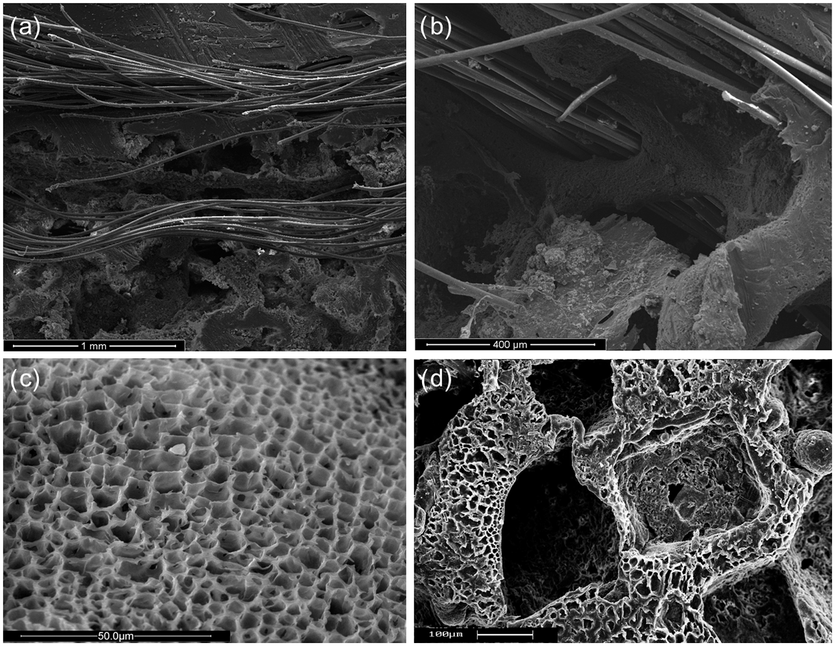
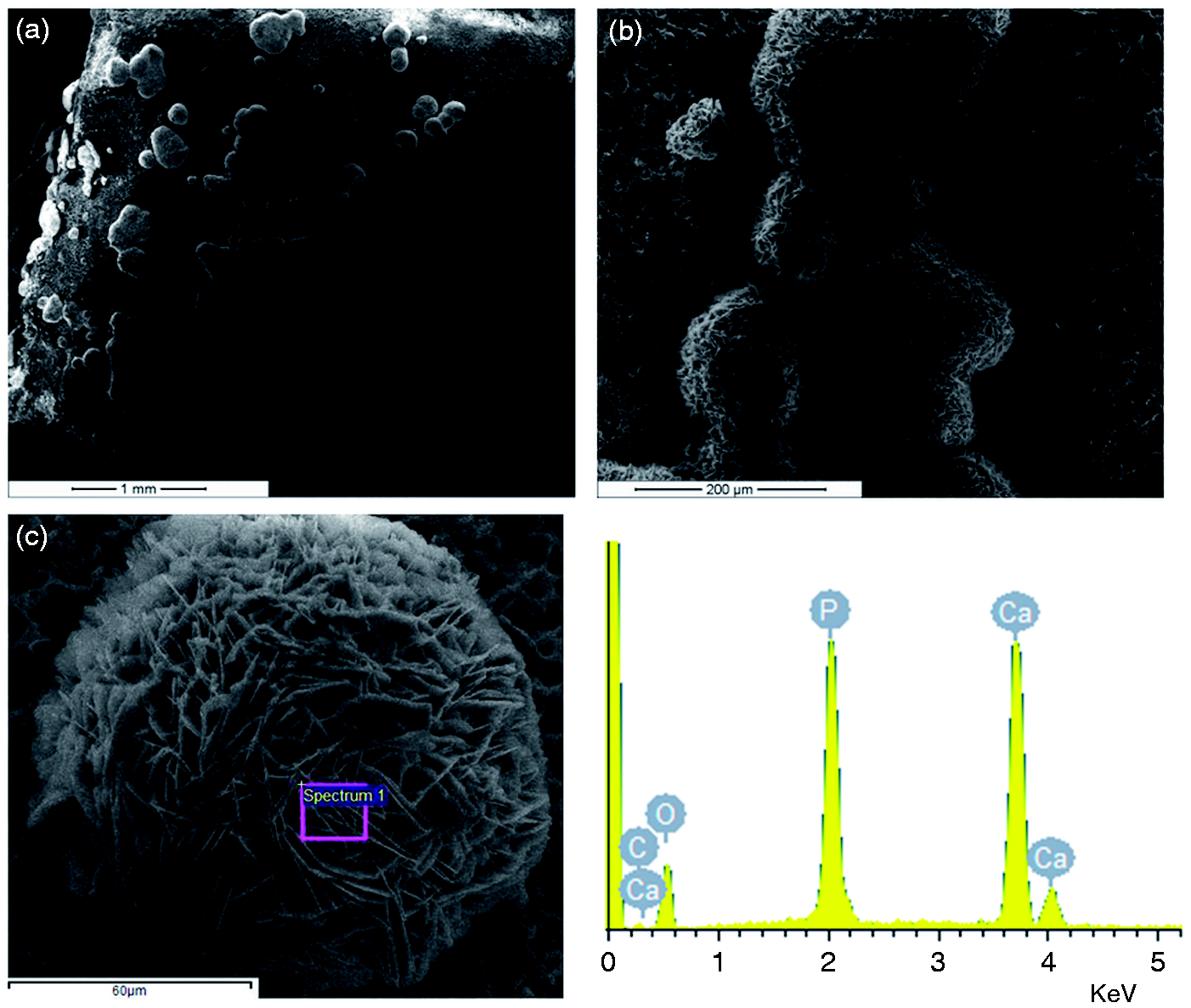
Higher magnification of SEM micrograph of mineral nucleated on PCL scaffolds displayed primarily a spherulitic morphology on the micrometer scale and a plate-like morphology on the nanometric scale. Therefore, smaller spheres represent crystals that have recently nucleated while the larger ones represent crystals that have grown for longer periods of time. EDS result shown in Figure 3 identifies these precipitates as calcium and phosphorus-rich phases, which contain carbon and oxygen as well. The mineral nucleated on composite scaffolds had a composition consistent with natural bone apatite.
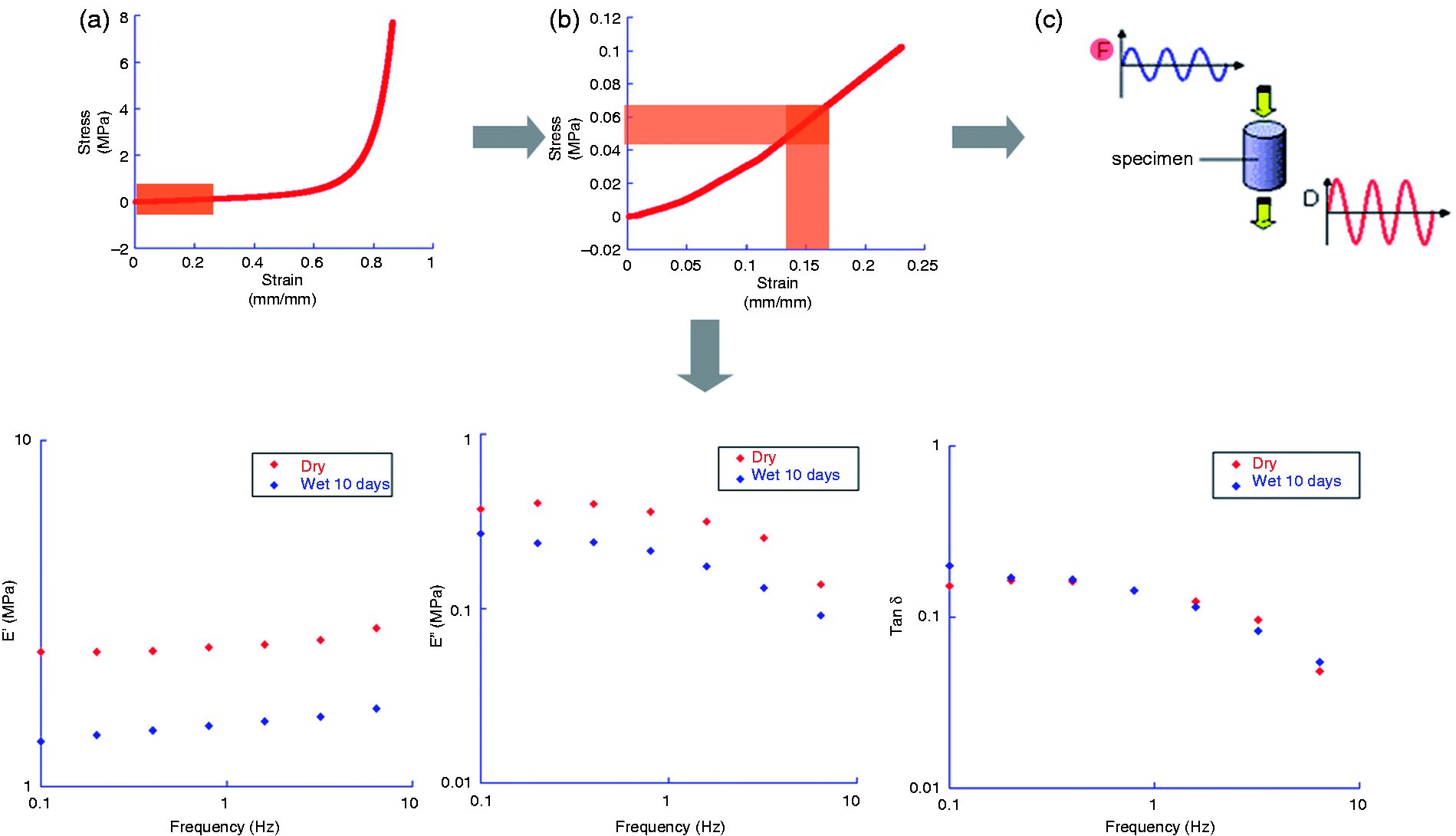
The thermal stability and composite composition were checked by TGA analyses. TGA curves of the composite and the pure components are reported in Figure 4(a) and (b). Comparing the degradation of the composite with the analysis of the pure components, three stages of degradation can be distinguished as reported in Table 1. First, thermal degradation took place in a range of temperature between 200 and 280℃, and it was related to the HYAFF11® decomposition. The second stage of thermal degradation observed in the temperature range of 280–350℃ can be attributed to the decomposition of PLA, and it partially covers the next step of degradation due to the PCL phase between 350 and 480℃. An initial weight loss can be detected between 50 and 70℃. It is well known that HYAFF11® is highly hygroscopic, and it is able to bind large quantities of water molecules. Probably, small quantities of moisture water bonded to the HYAFF11® phase vaporize as the temperature starts to approach to 100℃ causing a weight loss of 1.5–2%. Over 700℃, a certain mass retain, of about 20 wt%, can be attributed to the presence of calcium phosphate stable down to 700℃ and to the residual of HYAFF11® degradation over 700℃. TGA analysis also shows quantitative information about the sample composition in terms of fibre/matrix ratio equal to 10. The presence of HYAFF11® phases is crucial to control the mechanisms of BMP-7 entrapment and release and biomechanical functions. In particular, high-encapsulation efficiency (over 90%) has been estimated in agreement with requirements for bone therapy to reach a sustained therapeutic dose into the implant. The study of the cumulative release confirms the absorption of BMP-7 in the hydrogel-like phases, showing a slowly sustained release – nearly the 5% of the total amount after 6 days. Furthermore, the static response under compression of composite scaffolds has been reported in terms of stress–strain curve (Figure 5). It shows a linear elasticity at low stresses followed by a long collapse plateau truncated by a regime of densification in which the stress rises steeply. This evaluation was mandatory to identify the optimal load to impose during the dynamic test where the variation of elastic moduli was investigated as a function of frequency ranging from 0.1 to 3.2 Hz. A comparative analysis of the storage modulus E′ (elastic component) equivalent to the energy stored through deformation, loss modulus E″ equivalent to the energy dissipated through the cycled stimulation and tan δ that is an indicator of how efficiently the material loses energy to molecular rearrangements and internal friction was performed. Figure 5 shows the plot of the storage, loss modulus and damping values against the frequency. The increase of E′ as the frequency increases confirms the viscoelastic behaviour of the samples. In particular, it is possible to distinguish two different zones: at the low-frequency range, a viscous behaviour is predominant. As the frequency increases, the elastic behaviour progressively dominates, and the scaffolds begin to work in a totally elastic way. The estimation of E″/E′ ratio (tan δ) allowed to directly visualize the material ability to dissipate applied mechanical energy into heat at different frequencies. Mechanical tests on wet samples underline the initial degradation of PLA fibres and the hydrophilic behaviour of HYAFF11® phase; in fact, after 10 days of immersion, a partial decay in mechanical properties is recognized as confirmed by the reduction of the elastic modulus (E′) from 2.5 to 1.3 MPa (at 0.1 Hz). This study is mandatory to evaluate the ability of the scaffold to transfer the stress to cells, thus reaching relevant indication about the mechanical behaviour of samples for in vivo studies.
Thermogravimetric curve of PCL/PLLA composite scaffold. PCL: poly(ε-caprolactone); PLLA: poly- Scheme of different steps to analyse dynamic mechanical properties of fibre-reinforced scaffolds.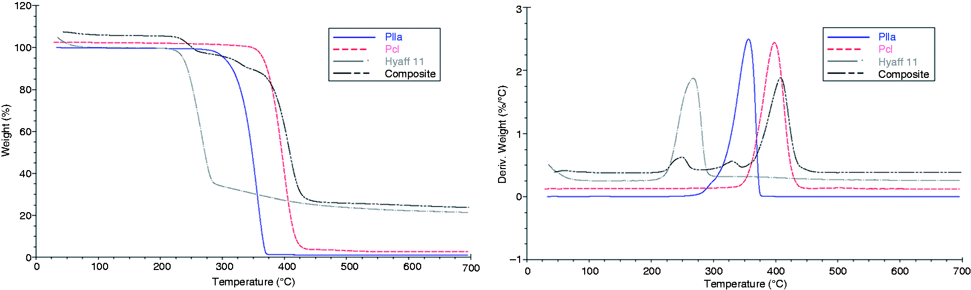
In vivo test
All animals recovered rapidly after surgery and well tolerated the cast. One animal treated with scaffold alone died in the postoperative period for causes unrelated to the study and was not included in the evaluation. Two sheep treated with CS-BMP scaffold had some osteolysis occurred due to the infection of pin insertion. In the last 4 weeks, when the cast was removed, in two sheep treated with CS-BMP scaffold, a non-complete alignment of the proximal part of the limb was observed without any evident lameness. Evaluation of the osseous implantation at the bone biomaterial interface by light microscopy showed the biocompatible behaviour of implants that did not evoke tissue inflammation or foreign body reaction in the surrounding bony tissue at the examined experimental time. The formation of new bone in the CS scaffold alone seems to be restricted to the edge of the defects while the largest part of the scaffold remained free of bone up to 12 weeks (Figure 6(a)). Additionally, the scaffold was still present into the bone gap after 12 weeks but there was absence of fibrous tissue all around it. In comparison to CS, CS-PRP-BMSC showed a better response with more newly formed trabecular bone observed at the bone-scaffold interface and on the external surface of bone and scaffold (Figure 6(b)). The best results were obtained in CS-BMP scaffolds where the remodelling processes were coupled with a good amount of new bone apposition, and a fair amount of newly formed bone within its structure was evident (Figure 6(c)). The presence of new bone was evident in one case even in the most internal part of the scaffold as clearly evidenced by Figure 6(d); xylenol orange and alizarin red labelling revealed an intense bone formation from 8 weeks after surgery (Figure 6(e–f)).
Histological evaluation at 12 weeks: CS (a), CS-PRP-BMSC (b), and CS-BMP (c–d). Fluorochrome labeling with alizarin red (e) and xilenol orange labelling (f). BMP: bone morphogenetic protein; BMSC: bone marrow stromal cells; C: cortical bone; NB: newly formed bone; PRP: platelet-rich plasma; S: scaffold.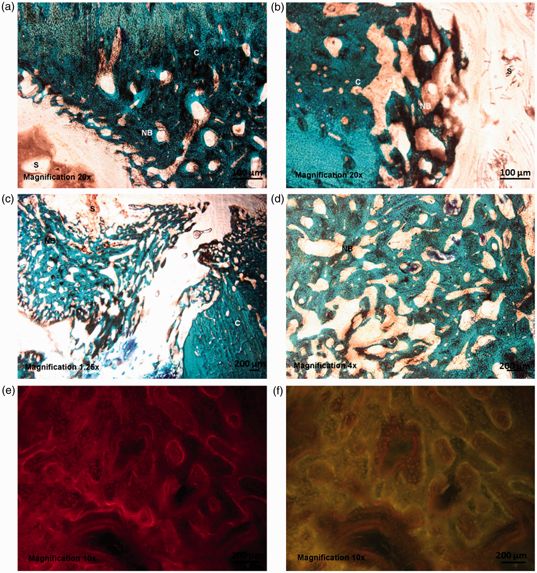
Discussion
The current challenge in scaffold design is to fabricate reproducible bioactive and bioresorbable 3D scaffolds with tailored porosity and pore morphology, capable of maintaining structure integrity for a predictable period, even under load-bearing conditions. In this work, we proposed to study a composite scaffold obtained by the integration into the PCL matrix of different material phases: HYAFF11 as a hydrophilic cue, R-cem particles as bioactive signals and PLA fibres as mechanical reinforcement. The morphological features, the mechanical properties and the cellular response of composite scaffolds have been investigated in order to match anatomy and functionalities of natural tissue during the in vivo implant. First of all, the proposed prototype shows a bimodal porosity, characterized by macropores arising from salt crystal dissolution, and micropores formed by the controlled removal of solvent via non-solvent exchange, which both concur to create a fully interconnection pore network able to support cell invasion and molecular transport. Moreover, the inclusion of material phases with different chemical and physical aspect, in turn, allows to particularize peculiar functions of the PCL matrix, adding new functionalities to the scaffold.
The incorporation of PLLA fibres into the PCL polymer matrix strongly improved the mechanical properties leaving unaltered the pore morphology. The peculiar spatial distribution of fibres confers to the scaffold a peculiar mechanical response with an increase of one order of magnitude in elastic modulus if compared with non reinforced PCL scaffolds. 18 Young modulus was evaluated from compressive static test and results to be over 2.5 MPa in dry condition. This value was similar, in order of magnitude, with the elastic modulus of cancellous bone as reported in literature.35,36 Moreover, dynamic analysis confirms the results obtained from the static characterization and highlights the viscoelastic behaviour of the samples showing that E′ increases with the frequency while E″ seems to be not really affected as frequency varies. HYAFF11 is highly hydrated, and soluble polymer that resembles native hyarulonic acid, basic constituent of the native extracellular matrix and efficiently concurs to the sustained release of molecular factors from the scaffolds with any relevant toxic response in the biological microenvironment. 37 Indeed, while HYAFF11 begins to degrade, via hydrolysis, growth factors are able to freely move out and elute from the matrix. The peculiar chemistry of polymer may provide to influence the molecular retention, from hours to months, dependent upon the degradation of the polymer, although limited research has been conducted in the field. 38
Meanwhile, the growth of apatite-like mineral phase promotes an efficient biomimesis at the interface with the defect site. In this context, we proposed to adapt the process technology to create tailor-made prototypes, which model the geometry of the animal defect, in order to prevent all possible failures due to scaffold/implant interface mismatches. So, a pilot in vivo study in diaphyseal defects in a sheep model has been performed on the composite scaffold in three different configurations: scaffold alone (CS), scaffold with BMSCs and PRP (CS-PRP-BMSC) and scaffold enriched with BMP7 (CS-BMP). Even if this in vivo study has several limitations such as the low number of evaluated animals and the lack of histomorphometric and microtomographic evaluations that would allow bone microarchitecture to be quantified, this is a preliminary study in which we evaluated histologically the bone response and enhancement of bone regeneration. After 12 weeks, scaffolds did not evoke tissue inflammation or foreign body reaction in the surrounding bony tissue. Our research demonstrated that CS-PRP-BMSC showed an enhanced response with more newly formed trabecular bone observed at the bone-scaffold interface and on the external surface of bone and scaffold in comparison to CS. PRP is a blood-derived product, which shows promise in enhancing tissue regeneration due to the growth factors released from the platelet alpha granules. 39 BMSCs have receptors for the growth factors contained in PRP, and in vitro studies have shown that the addition of 10% PRP is enough to promote a marked BMSC proliferation. 40 Additionally, BMSCs are easily isolated from patient tissues and have the potential to develop either in vitro or in vivo into distinct mesenchymal tissues and to release healing signals in the microenvironment, becoming an attractive cell source for tissue-engineering approaches. 41 Therefore, the addition of autologous BMSCs and activated PRP to the scaffold provides an immediate source of viable osteogenic precursor cells and growth factors at the implantation site and provides a direct biological contribution to osteogenesis. Despite this direct contribution to osteogenesis provided by the association of BMSC and PRP, in this study, the best results were obtained when the scaffold was used in combination with BMP-7. BMPs induce mesenchymal stem cells to differentiate into osteoblasts and produce new bone tissue. 42 In particular, BMP-7 has been shown to be able to accelerate bone remodelling, thanks to its osteoconductive and osteoinductive characteristics but the literature emphasised the need for scaffolding to contain the cytokines and to control the shape of the bone regeneration.43–50
Conclusions
The efficient encapsulation and sustained release in the implant site led to the positive findings documented by this study on a large bone defect model. The currently available clinical treatments for bone-related deficiencies are not ideal for the healing of large bone defects. Scaffold-aided tissue-engineering approach is promising to tackle this issue, providing a wide range of functions, including physical robustness, as well as promoting de novo bone formation. Here, composite scaffolds offer promising physical characteristics and osteoinductive capacity due to HYAFF11 inclusions and biomineralization. Moreover, the ability to entrap within polymeric scaffolds growth factors such as hBMP-7 allows to improve the osteogenic properties of the scaffolds to promote bone formation in a clinical environment. All these preliminary findings suggest that the proposed composite scaffold promises to be interesting for further preclinical evaluations towards the treatment of critical-sized bone defects.
Footnotes
Acknowledgements
Scanning electron microscopy was supported by the Transmission and Scanning Electron Microscopy Labs (LAMEST) on the National Research Council.
Declaration of conflicting interests
None declared.
Funding
This study was financially supported by the IP STEPS EC NM FP6-IP, NMP3-CT-2005-500465P3-CT-2005-500465.