
Abstract
The objective of this study was to determine biocompatibility of zirconia-based coatings obtained by the sol–gel method. Two matrices, ZrO2 and SiO2/ZrO2, were created and applied on stainless steel type 316L with dip-coating technique. The morphology and topography of biomaterials’ surface were characterized using energy-dispersive X-ray spectroscopy and atomic force microscopy, while chemical composition was analyzed by Raman spectroscopy. Additionally, wettability and surface free energy were characterized. Biocompatibility of obtained biomaterials was evaluated using an in vitro model employing mesenchymal stem cells (MSCs) of adipose and bone marrow origin. Biological analysis included determination of proliferation activity and morphology of MSCs in cultures on synthesized biomaterials. Osteoinductive properties of biomaterials were determined both in non-osteogenic, as well as osteogenic conditions. The results showed that investigated biomaterials exerted different impact on MSCs. Biomaterial with ZrO2 layer was more biocompatible for adipose-derived MSCs, while SiO2/ZrO2 layer promoted proliferation of bone marrow derived MSCs. Moreover, hybrid coating exhibited greater osteoinductive properties than ZrO2 coating, both on cultures with adipose-derived stromal (stem) cells and bone marrow stromal cells. Observed biological effects may result not only from different chemical composition, but also from diverse wettability. The ZrO2 coating was characterized as hydrophobic layer, while SiO2/ZrO2 exhibited hydrophilic properties. The results obtained suggest that behavior of MSCs in response to the biomaterial may vary depending on their origin, therefore we postulate, that screening analysis of implants’ biocompatibility, should incorporate model applying both adipose- and bone marrow derived MSCs.
Keywords
Introduction
Stainless steel, in particular alloy SS316L, remains the most frequently used metallic substrate, both in orthopedics and dentistry. Compared to other metallic materials such as cobalt–chromium or titanium alloys, stainless steel has a low resistance to corrosion. 1 Biomedical devices used both in orthopedics as well as in dentistry should possess high osteogenic properties that would promote proper integration of the implant with the bone tissue. 2 Coating of biomaterials with bioactive layers is currently a common procedure used to improve prosthetic devices. The sol–gel route is one of the most effective methods to synthesize materials for this purpose, allowing not only to enhance corrosion resistance, but also to provide layers of high bioactivity.3,4 Mixing of the reagents in the liquid phase allows to control the composition at the molecular level. Furthermore, this process can be applied to produce single- or multi-component oxide coatings (SiO2, ZrO2, SiO2/ZrO2, etc.). In addition, this method provides various options of coating the materials, for example using spin-coating, meniscus-coating, or dip-coating techniques, which in turn, allows to modify implants of various shapes and sizes.5,6 Therefore, the amorphous coating may be more beneficial for early bone ingrowth than a coating with high crystallinity. 7,8 Physical and chemical properties of the biomaterial are key parameters affecting adhesion, morphology and growth pattern of cells, and in consequence, determining other aspects of cell life such as proliferation and differentiation. The sol–gel matrices are mainly composed of silica (SiO2), which in addition to anti-corrosion properties, is a highly active agent stimulating the proliferation of bone cells (osteoblast) and inducing the formation of hydroxyapatite.9,10 Other sol–gel films frequently applied for medical purposes are composed of zirconium dioxide (ZrO2),11,12 which also exerts an osteoinductive effect. Moreover, studies on biological behavior have demonstrated that zirconium and silica create favorable non-toxic layers with good biocompatibility. 13 Additionally, ZrO2 exhibits high mechanical strength, high fracture toughness, and good corrosion resistance, making it a suitable protective material.3,14–17
The interaction of cells with the biomaterial is modulated by the characteristics of the material surface. Alterations in surface chemistry, composition, roughness, and wettability can influence the cellular response, including adhesion, proliferation, and differentiation.18,19 Wettability and roughness, in addition to the chemical composition of the sol–gel-derived coatings, are other crucial parameters that affect the biological properties of implants. Numerous studies emphasize the importance of the surface nature (hydrophobic or hydrophilic) in the modulation of cell’s attachment.20–22
The assessment of bio-improvement of the material in vitro is often carried out using committed osteoblast-like cell lines. However, advanced biological models increasingly incorporate mesenchymal stem cell (MSCs) for these purposes. 23 The combination of MSCs and biofunctional implants is a new strategy in regenerative medicine that focuses on enhancing tissue regeneration. 24 Two populations of MSCs are predominantly used in regenerative medicine, i.e. adipose-derived stromal (stem) cells (ASCs) and bone marrow stromal cells (BMSCs). ASCs and BMSCs have been proposed to be applied therapeutically simultaneously due to the high similarity of their morphology and surface antigens. Both ASCs and BMSCs have high plasticity, manifested by the ability to differentiate into bone precursor cells (multipotency) and the potential of self-renewal (proliferation). 25 These properties make MSCs a kind of “litmus paper”—an ideal model for testing the biocompatibility of implants in vitro.26,27
Given above, we postulate that the analysis of the biocompatibility of biomaterial in the context of therapies involving ASCs and/or BMSCs should include in vitro models with the population of MSCs of interest. Thus, the aim of this study was to analyze the biocompatibility of ZrO2 and SiO2/ZrO2 sol–gel-derived biomaterials using both ASCs and BMSCs. Further, to evaluate the osteogenic potential of the surfaces investigated both populations of MSCs were propagated in non-osteogenic and osteogenic condition. The assessment of the cytophysiology of MSCs, in response to the surfaces synthesized, may provide essential information concerning not only the toxicity of biomaterials but also its biofunctionality. The results were also evaluated in the context of wettability and nano-roughness of the surfaces.
Materials and methods
Synthesis of biomaterials
Substrates
Discs (15 × 2 mm) made of stainless steel AISI 316L (ITALINOX) were selected as coating substrate, because this material is commonly used in biomedical applications. The chemical composition of the steel was as follows: C—0.03% max, Mn—2% max, Si—1% max, P—0.045% max, S—0.03% max, Ni—10–14%, Cr—16–18%, Mo—2–3%, F—balance. In order to promote adhesion between the substrate and the coating layer, steel substrates were successively cleaned by ultrasonic cleaner (Polsonic, SONIC3) with acetone, ethanol, and distilled water prior to coating.
Sol–gel synthesis
The following substrates were used for the synthesis of zirconia (ZrO2) and silica/zirconia (SiO2/ZrO2) sol–gel-derived coatings: (a) zirconia and silica precursors, zirconium(IV) isopropoxide (ZrOP, Sigma Aldrich) and tetraethyl orthosilicate (TEOS, Sigma Aldrich), respectively, (b) n-propanol (n-PrOH, POCh S.A) was used as a solvent, (c) hydrochloric acid (35–38% HClaq, POCh S.A.) was used as an acid catalyst, and (d) diethanolamine (DEA, Sigma Aldrich) as a chelating agent of zirconia precursor. The ZrO2 sol was prepared from ZrOP, n-PrOH, and DEA, in the following molar ratio: 1:16.8:3.3, while SiO2/ZrO2 sol was obtained from the mixture of TEOS, ZrOP, and n-PrOH in a molar ratio of 3.3:1:27.9. The sol–gel route was performed in an acidic medium by addition of 0.011 M hydrochloric acid to both sols. The resulting mixtures were homogenized on magnetic stirrer for 2.5 h at a speed of 500 r/min at room temperature. Sols obtained were allowed to age for 4 h in a closed container before coating.
The hydrolyzates were deposited on 316L stainless steel substrates by a dip-coating method with controlled process parameters such as the dipping and pulling speed (v = 34.26 mm/min) and a residence time of the substrate in the hydrolyzate (1st layer: 60 s, 2nd layer: 30 s, 3rd layer 15 s). This process was conducted three times in order to obtain a three-layer coating. Each individual layer was applied when the previous layer became dry. Afterwards, the samples (substrates + coating) were dried at room temperature on air and stabilized for 12 h at 250℃ with a controlled temperature gradient (1.5℃/min).
Surface morphology and topography
The morphology and topography of the coating surfaces were investigated using a scanning electron microscope (SEM, Evo LS 15 Zeiss) and atomic force microscope (AFM, XE-100 Park Systems). The quantification of the elements and determination of their distribution were performed at 500x magnification using a Bruker detector in SEM microscope. Energy-dispersive X-ray spectroscopy (EDX) was performed as described in the previous work. 28 AFM XE-100 Park Systems was used in the contact mode to obtain quantitative topographical images of the surface of the coatings. Microfabricated tip NCS36/Al BS from MikroMasch was applied. The cantilevers had a force constant of 1 N/m and a radius of curvature of 100 Å. The areas of sampling were 45 × 45 µm.
The parameters used to characterize the roughness of coatings obtained were: Ra—arithmetic mean roughness and Rq—root mean square roughness. According to ISO 4287 standard, parameters of the surface roughness were calculated based on the standard formula integrated in the software.
Raman spectroscopy
The chemical structure of synthesized coatings was determined using Raman spectroscopy. Raman spectra were measured by a dispersive Raman Spectrometer LabRAM HR800 Horiba JobinYvon. The incident laser excitation was provided by a water-cooled argon laser source operating at 514.5 nm. Spectra were recorded in the region 4000–50 cm−1 with a spectral resolution of 2.5 cm−1. The resulting zirconia and silica/zirconia coatings were examined as thin films deposited on metallic substrate. In the presented results, Raman spectra are given in two ranges, i.e. 150–1500 cm−1 and 1950–3500 cm−1, for zirconia and silica/zirconia coatings, respectively, due to the presence of characteristic bands in the selected ranges.
Measurements of wettability and surface free energy (SFE)
The contact angles were measured by the sessile drop method using an automatic drop shape analysis system, DSA 10 Mk2 (Kruss, Germany). Droplets (0.2 µL) of UHQ-water and diiodomethane (Aldrich, Germany) were placed on the surface of each sample and the contact angles were obtained by averaging the results of 12 measurements for each liquid. SFE was calculated from the contact angles using the Owens-Wendt approach, which allows to obtain dispersive (γd) and polar (γp) components of free energy of the total surface.
Biocompatibility evaluation
The study was approved by the Second Local Ethical Committee, localized at the Wroclaw University of Environmental and Life Sciences (Chełmońskiego 38C, 51-630, Wroclaw, Poland. The decision no. 84/2012).
Isolation and propagation of MSCs
MSCs were isolated according to the principles and methods accurately described before.29,30 Cultures were passaged twice to obtain sufficient number of cells for the biocompatibility test of the materials synthesized. Passages were performed when 78–80% cell confluence was reached. The immunofluorescence staining was performed routinely to confirm the presence of CD29, CD44, and CD105 mesenchymal lineage markers and the absence of CD34.
In vitro tests
Cultures of MSCs were propagated in 24-well plates, pre-coated with biomaterials. The initial cell inoculum was equal to 3.5 × 10⁴ of MSCs per well. The proliferation activity of cells and their morphology was evaluated after 24, 48, 120, and 168 h of culture. The results obtained for cultures on biomaterials were analyzed relative to the cultures propagated on a pure surface of 316L steel, which were considered as controls.
Analysis of cell viability
Cytophysiological activity of cells was determined using resazurin-based assay (Sigma Aldrich), performed according to the manufactures’ instruction. The results allowed to define an arbitrary unit of the proliferation factor (PF) and the population doubling time factor (PDT-F). Both normative values reflected the activity of MSCs in cultures with biomaterials tested in relation to the cultures on a pure surface of 316L steel, giving an arbitrary value equal to 1. The rate of proliferation higher than 1 (PF >1), in cultures propagated on biomaterials, expressed an increase in cellular activity, whereas values lower than 1 (PF <1), expressed a reduction of the proliferation rate. The acceleration of cell growth in the case of PDT factor was expressed as values lower than 1. The PDT of MSCs was determined using an algorithm established previously by Jin et al., 31 supported by an online application calculating PDT. 32
Cell morphology
The analysis was performed using an inverted fluorescent microscope (Zeiss, Axio Observer A.1). Cytological evaluation of MSCs included visualization of nuclei stained with diamidino-2-phenylindole (DAPI) after fixation with 4% ice cold paraformaldehyde and permeabilization of cell membranes with 0.1% Triton X-100. The procedure was performed according to the manufacturer’s instructions, as described in detail previously.27,33
Osteogenesis
Osteogenic differentiation of MSCs in culture with biomaterials was performed using StemPro® Osteogenesis Differentiation Kit (Life Technologies). Stimulation of both populations of MSCs lasted 21 days and was performed according to the manufacturer’s instruction. Cultures were maintained in 24-well dishes coated with biomaterials as described in the previous section (“In vitro tests”). The only difference was that the cells were inoculated at a concentration of 2 × 10⁴ cells per well. The osteogenic medium was changed every three days. Cultures expanded in standard growth medium served as controls, allowing determining the effectiveness of differentiation.
Analysis of gene expression—RT qPCR
Sequences of primers used in qPCR.
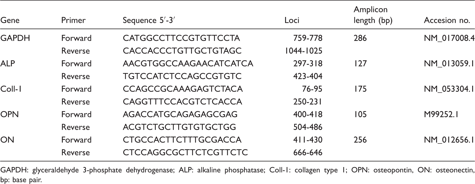
GAPDH: glyceraldehyde 3-phosphate dehydrogenase; ALP: alkaline phosphatase; Coll-1: collagen type 1; OPN: osteopontin, ON: osteonectin, bp: base pair.
Detection of osteopontin and osteocalcin with enzyme-linked immunosorbent assay (ELISA)
Extracellular forms of osteopontin (OPN) and osteocalcin (OCL) were detected in supernatants collected after 21 days of cell culture. The presence of OPN was determined using a Mouse/Rat Osteopontin Quantikine ELISA Kit (R&D Systems, sensitivity 8.5 pg/mL; intra- and inter assay %CV <8), whereas OCL a Rat Gla-Osteocalcin High Sensitive EIA Kit (Takara, sensitivity 0.25 ng/mL; intra- and inter assay %CV <8). Procedures of OPN and OCL detection were performed according to manufacturers’ instructions. The amount of proteins secreted was expressed in ng per mL of culture medium collected.
Quantification of matrix mineralization
The elemental composition of the extracellular matrix (ECM) of cultures was measured using SEM–EDX. SEM imaging (Zeiss Evo LS 15) was performed on post-fixed cells, rinsed with distilled water and dehydrated by graded series of ethanol (from 50% to 100%, at 10% interval).
Quantax detector (Brüker) was used for analysis and the following settings were applied: 10 kV of filament tension and 11 mm of working distance (WD) at a magnification of 200x. The values were presented as weight percentage (wt%).
Statistical analysis
The normality of the population data was determined using Shapiro-Wilk test, while equality of variances was assessed by Levene’s test. Differences between groups were analyzed using parametric (Student’s t test) or non-parametric (Mann-Whitney U test) methods. Statistical analysis was performed with STATISTICA 10.0 software (StatSoft, Inc., Statistica for Windows, Tulsa, OK). Differences with a probability of p < 0.05 were considered significant.
Results
Surface morphology and topography
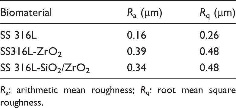
Both zirconia (Figure 1c) and silica/zirconia (Figure 1g) coatings were transparent, homogeneous, smooth and uniformly covered the substrate, steel 316L (Figure 1a). AFM demonstrated similar nano-roughness of the coatings (Figure 1d, f; Table 2) applied to the steel 316L substrate. The parameter of root mean square roughness for zirconia and silica/zirconia coatings had the same value (Rq = 0.48 µm), while the arithmetic means of roughness parameter (Ra) were slightly different in ZrO2 (Ra = 0.39 µm) and SiO2/ZrO2 (Ra = 0.34 µm) coatings. In addition, the application of sol–gel-derived coatings on a 316L substrate (Ra = 0.16 µm) significantly increased the surface roughness. Summary of the roughness parameters is presented in Table 2.
Scanning electron microscope images (a,c,e) at 500x magnification (scale bar: 10 µm) and the images of surface morphology in atomic force microscopy (b,d,f) of zirconia (c,d) and silica/zirconia coatings (e,f), deposited on AISI 316L annealed at 250℃ (a,b). Characteristics of the surface roughness parameters for all materials measured by AFM. Ra: arithmetic mean roughness; Rq: root mean square roughness.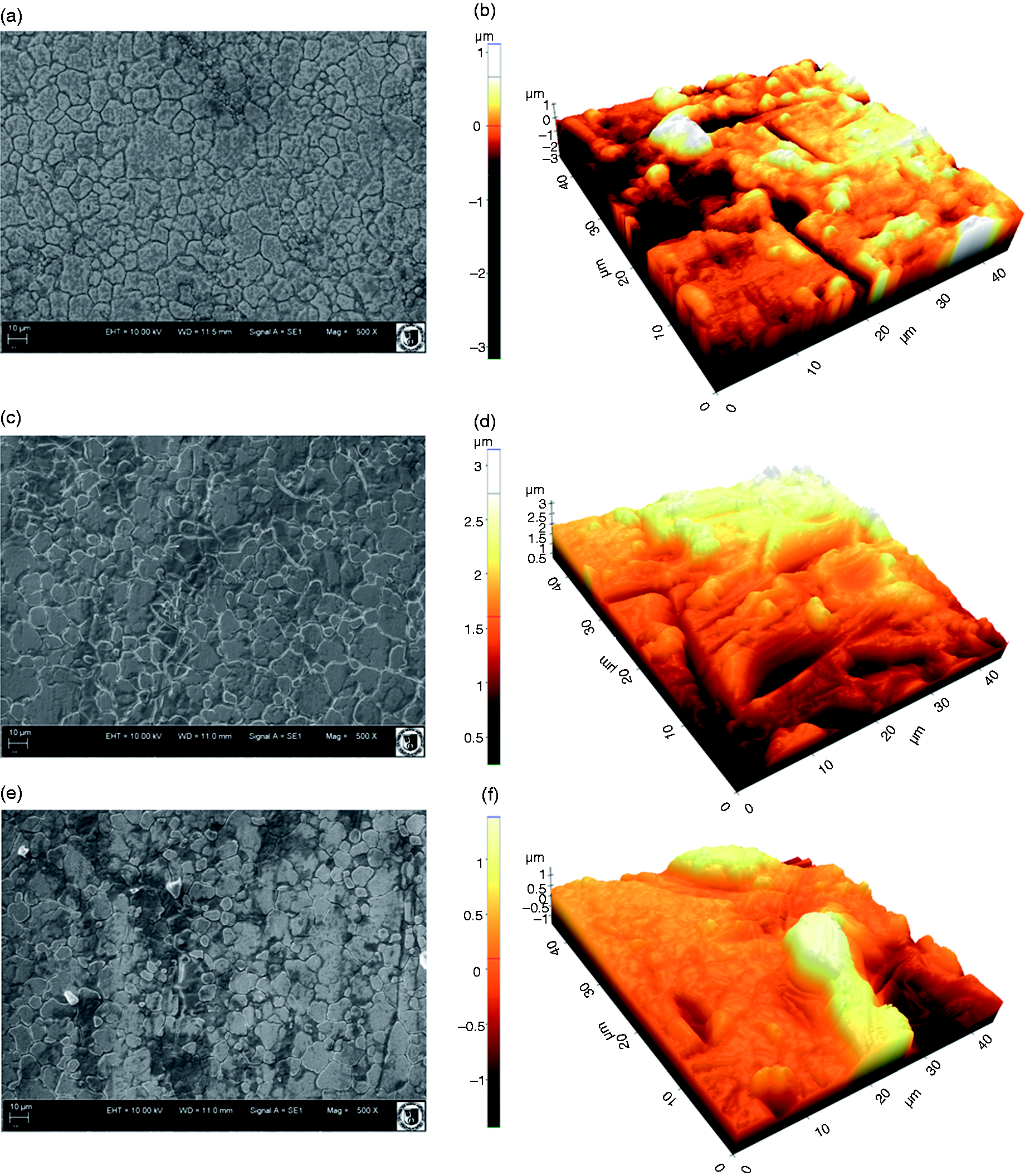
Raman spectroscopy and SEM-EDX
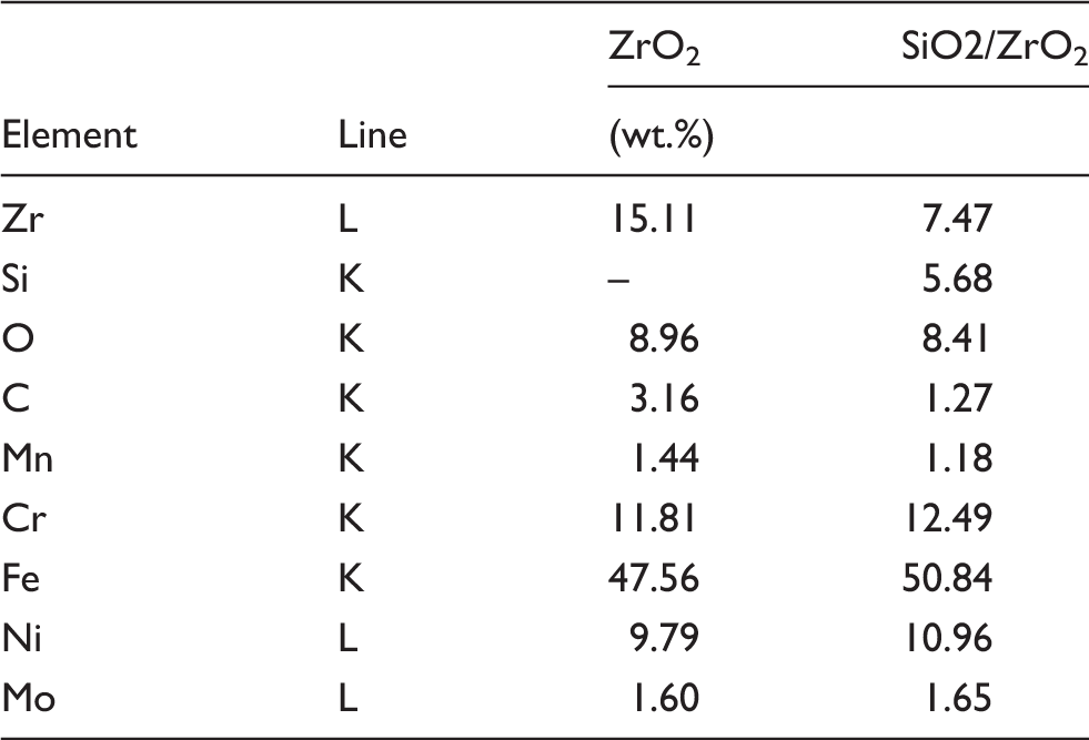
The combination of SEM-EDX analysis and Raman spectroscopy allowed to quantify the elemental content and qualitatively evaluate the distribution of chemical moieties on the test surfaces. Uniform distribution of elemental components of coatings obtained (Zr, Si, O) was observed in both investigated materials (Figure 2b, d). Quantitative data of SEM-EDX analysis are presented in Table 3.
Raman spectra (a,c) and SEM-EDX mapping images of elemental distribution (b,d) in zirconia (a,b) and silica/zirconia coatings (c,d) deposited on AISI 316L, annealed at 250℃. Quantitative data of SEM-EDX analysis of the sol–gel-derived coatings.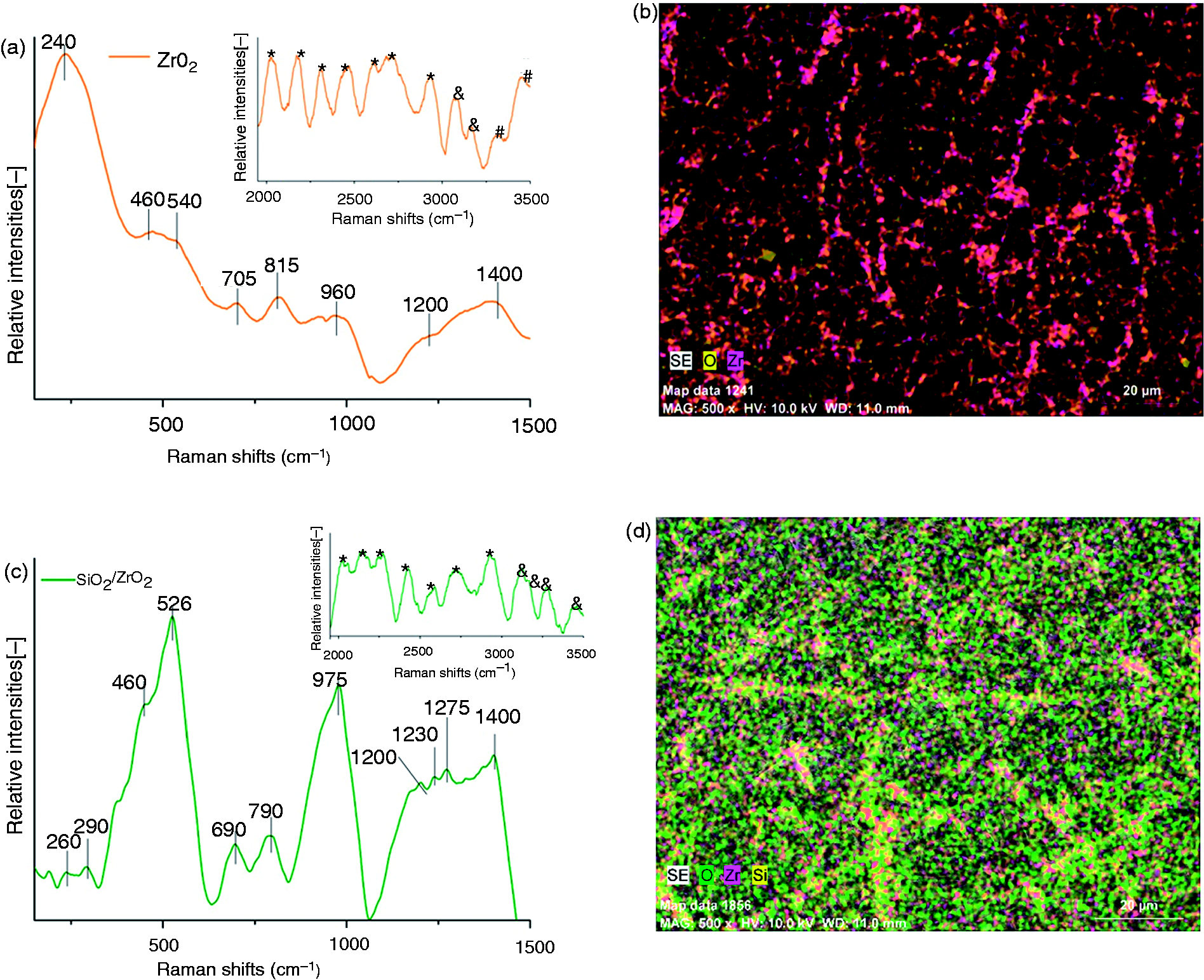
The Raman spectra of ZrO2 and SiO2/ZrO2 coatings are shown in Figure 2a and c in two ranges: 150–1500 cm−1 (larger graph) and 1950–3500 cm−1 (smaller graph in top right corner), due to the presence of characteristic bands in selected ranges. Three bands at 260 and 290 cm−1 in SiO2/ZrO2 (Figure 2a) and at 240 cm−1 in ZrO2 (Figure 2c) correspond to the torsional vibrations of ethyl groups. 36 The Raman spectra of both coatings showed a characteristic broad band around 500 cm−1, consisting of two components centered at 460 and 540 cm−1 in ZrO2 (Figure 2a) and at 460 and 526 cm−1 in SiO2/ZrO2 (Figure 2c) coating. These bands are characteristic of amorphous phase of zirconia. 37 In addition, the overlap of multiple vibrations is represented as a broad band in the spectral range of 1200–1400 cm−1, making it difficult to evaluate the contribution of particular vibrations to the total intensity of the band. This spectral range can be attributed to the in-plane OH deformation vibration of alcohol molecules (Figure 2a and c). 36
The Raman spectra of ZrO2 coating (Figure 2a) showed much broader bands, and most intense of them were located at 705 (C–H), 815 (CH2), and 960 (n-PrOH), while the Raman spectra of SiO2/ZrO2 coating (Figure 2c) had three well-defined bands, and the most intense of them were located at 690, 790, and 975 cm−1. These vibrations correspond to the stretching mode of Si(OCH3)2(OH)2 (∼690 cm−1), symmetric stretching mode of Si–O–Si network (∼790 cm−1), and Si–OH and Si–O–Zr bonds (∼975 cm−1). 38 In the region of 1950–3500 cm−1, bands occurred, which were mainly derived from (i) organic compounds assigned to CH2, CH3 (marked with ‘*’), (ii) OH bonds (marked with ‘&’) and (iii) N–H bonds from DEA (marked with ‘#’) used for the synthesis of zirconia coating. 36
Contact angle and SFE
The surface properties of the materials obtained.
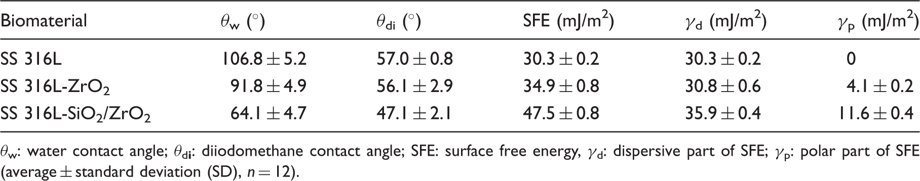
θw: water contact angle; θd
Analysis of proliferation activity of MSCs in cultures with synthesized biomaterials
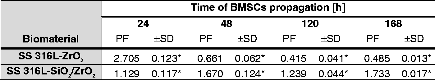
The analysis of cytophysiological activity of cells showed that proliferation activity of ASCs in culture with 316l-ZrO2 biomaterial was not significantly accelerated in comparison with the culture on a pure metallic substrate. The highest PF was recorded after 48 h, however, it decreased significantly after 120 h. The proliferative activity of ASCs cultured on SiO2/ZrO2 layer was significantly increased after 24 h of propagation as compared with the culture on pure steel and steel coated with a layer of ZrO2. The proliferation of ASCs in culture on SiO2/ZrO2 layer was stabilized after 48 h of propagation. The PF values obtained afterwards reached a level comparable to the control culture (Figure 3). Although, the activity of BMSCs was significantly increased after the first 24 h of culture on 316L-ZrO2 biomaterial, the proliferation of BMSCs was significantly reduced in the later course of experiment. The proliferation of BMSCs in culture on SiO2/ZrO2 layer was significantly elevated during the experiment (Figure 4).
Correlation of cytophysiological activity of ASCs in cultures with biomaterials. Correlation of cytophysiological activity of BMSCs in cultures with biomaterials. PF: proliferation factor expressed by arbitrary unit, determined based on the activity of cells on stainless steel 316L without the sol–gel-derived layer; mean ± standard deviations (SD) are based on three independent measurements. *Differences statistically significant at p < 0.0001.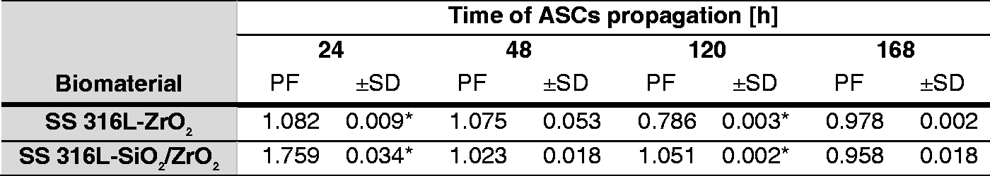
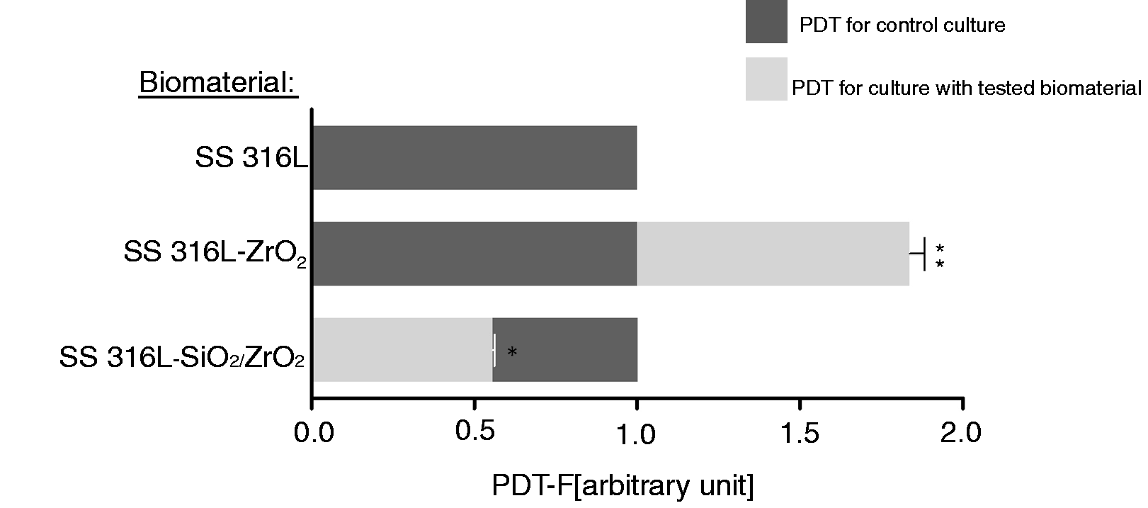
The estimation of PDT of ASCs in cultures on biomaterials showed that cultures propagated on SS 316L-ZrO2 had PDT-F similar to the control culture, while PDT of ASCs cultured on SS 316L-SiO2/ZrO2 was significantly elongated in comparison to cultures on stainless steel (Figure 5). The rate of expansion of BMSCs propagated on stainless steel 316L with ZrO2 layer was significantly extended. However, the time needed to double the population of BMSCs cultured on SS 316L-SiO2/ZrO2 was considerably reduced (Figure 6).
Population doubling time factor (PDT-F) calculated for ASCs. Mean ± standard deviations (error bars) are based on three measurements. *Difference significant at p < 0.0091. Population doubling time factor (PDT-F) calculated for BMSCs. Mean ± standard deviations (error bars) are based on three measurements. Asterisks mark differences significant at *p < 0.0001; **p < 0.0008.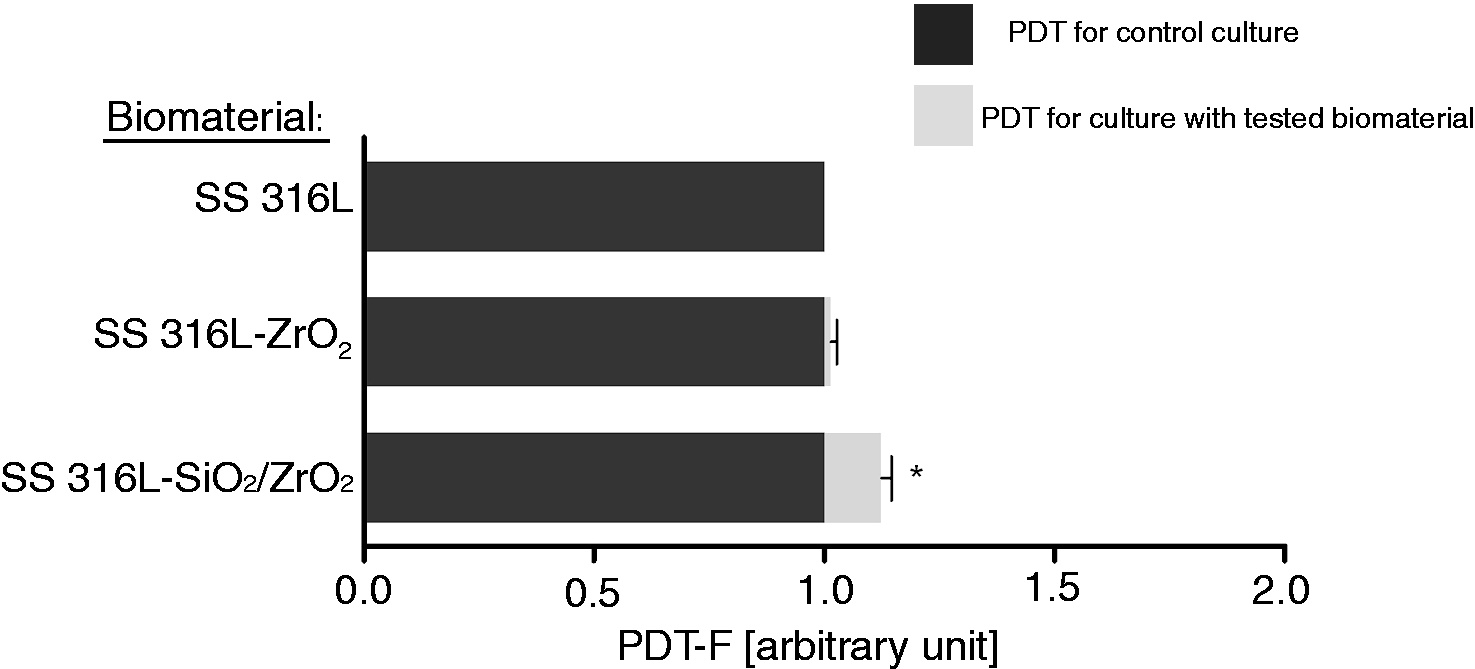
Morphology of MSCs in cultures on synthesized biomaterials
The morphology of MSCs as well as changes in the pattern of the culture growth were monitored during the whole course of the experiment. Significant alterations in the morphology and in the growth architecture of MSCs are shown in the images obtained after 24 and 168 h of propagation in Figure 7. Both ASCs and BMSCs shared similar mesenchymal-like shape and flat polygonal morphology. However, the dipolar shape of cells was dominant in the ASC cultures, while BMSCs were multipolar. In contrast to BMSCs, ASC cultures showed a dense network of intercellular connections, clearly visible after 168 h of culture. ASCs on stainless steel 316L had a uniform distribution, whereas ASCs propagated on synthesized biomaterials showed a tendency to aggregate. Cell aggregates reached a significant size after 24 h in cultures on SS 316L-ZrO2 biomaterial, and on SS 316L-SiO2/ZrO2 after 168 h of propagation. The characteristic aggregates of ASCs on the 316L-SiO2/ZrO2 formed spot-like structures.
Analysis of the morphology of MSCs and growth pattern of the culture in response to the synthesized biomaterials. Blue fluorescence indicates nuclei of cells (DAPI–DNA staining). Magnification 100x, scale bar: 200 µm.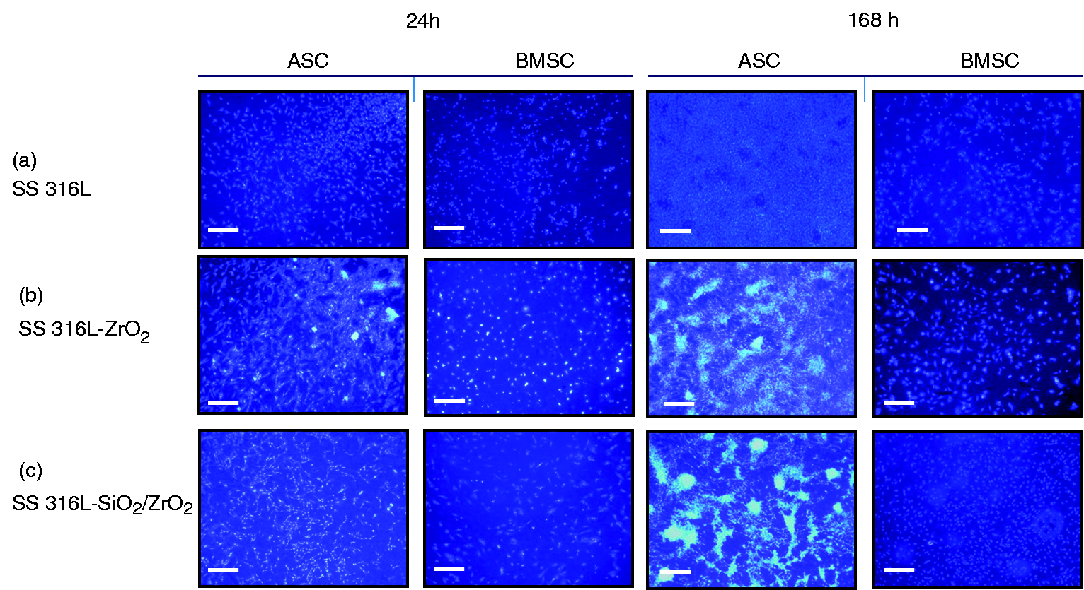
The cultures of BMSCs, in contrast to ASCs, did not reveal a tendency to aggregation but adopted a regular specific distribution. When compared to the control culture, the most evident development of active monolayer was obtained in cultures on 316L-SiO2/ZrO2 biomaterial. After 168 h of expansion on this biomaterial cells developed a dense network of intercellular connections, while BMSC culture on SS 316L-ZrO2 showed signs of apoptosis.
Evaluation of osteogenesis in cultures with synthesized biomaterials
Analysis of gene expression in the ASC cultures showed that the level of alkaline phosphatase (ALP) mRNA increased significantly after osteogenic stimulation, whereas the induction of osteogenesis in the BMSC cultures caused a decrease in the level of ALP transcript. BMSCs in non-stimulated cultures expressed the highest level of ALP mRNA in cultures on SS 316L-SiO2/ZrO2 biomaterial (Figure 8a,b). Analysis of expression of collagen type I (Coll-1) showed that both ASCs and BMSCs expressed a higher level of this transcript in cultures that did not undergo osteogenic stimulation. The type of surface had no effect on the expression of gene encoding collagen type I (Figure 8c,d). The level of OPN in ASCs was elevated in osteogenic cultures. This effect was also present in BMSC cultures, even though a deviation was observed in cells propagated on SiO2/ZrO2 layer, where osteogenic stimulation did not affect the expression of OPN. Culture of BMSCs propagated without osteogenic medium reached the highest level of OPN mRNA on SS 316L-SiO2/ZrO2 biomaterial. The surface used had no effect on OPN expression in ASCs. Osteonectin (ON) in ASC cultures was expressed in a constitutive manner in response to standard as well as an osteogenic medium and was not affected by the type of biomaterial. The mRNA level of ON in BMSC cultures was higher in non-osteogenic conditions and was also significantly increased in BMSC cultures on SS 316L-SiO2/ZrO2 biomaterial.
Gene expression in MSCs cultured on synthesized biomaterials in standard and osteogenesis-promoting conditions. The values are expressed as arbitrary units calculated in relation to the mRNA level of housekeeping gene GAPDH. Data include average standard deviation (error bars) of three experiments. Significant difference at *p < 0.0001; **p < 0.0008, and ***p < 0.005; #insignificant difference at p > 0.05. Unstimulated cells are indicated as groups: a (culture on SS 316L), b (culture on SS 316L-ZrO2), and c (culture on SS 316L-SiO2/ZrO2).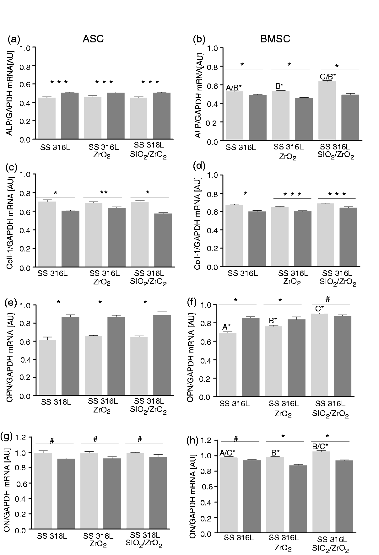
ELISA test performed on the supernatants of ASC cultures on a substrate of stainless steel 316L did not detect the presence of OCL in cultures without osteogenic stimulation. The induction of osteogenesis increased the level of OCL in cultures on SS 316L-ZrO2 biomaterial, while the secretion of OCL in cultures on SS 316L-SiO2/ZrO2 was elevated under non-osteogenic conditions. Significantly increased level of OCL was measured in osteogenic culture of BMSCs propagated on SS 316L-ZrO2 biomaterial. The concentration of secreted OCL was comparable in the cultures on stainless steel and SS 316L-SiO2/ZrO2 (Figure 9).
The concentration of OCL (ng/mL) in the supernatants on day 21 of ASC (a) and BMSC (b) cultures. Mean ± standard deviation (error bars) was derived from three measurements. Significant difference at *p < 0.0001; #no significant difference at p > 0.05.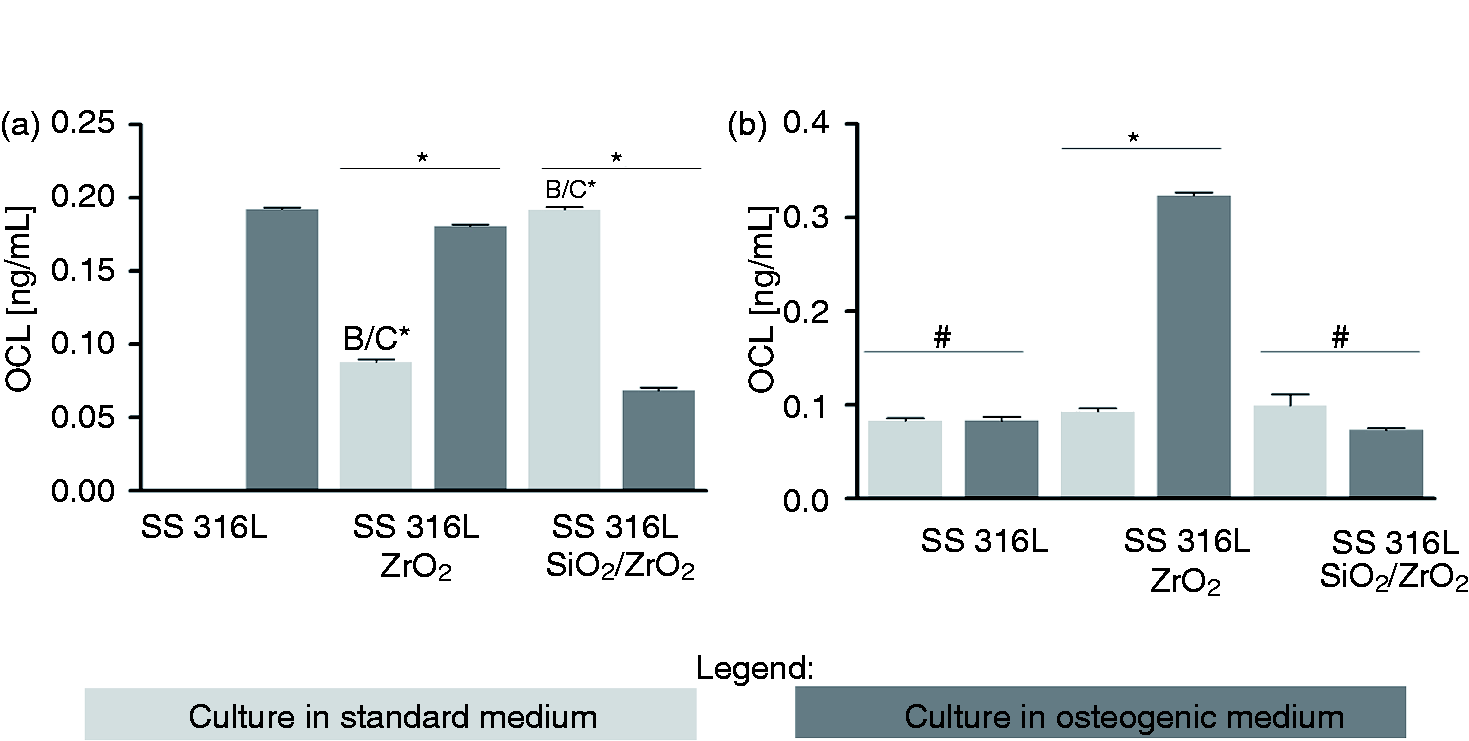
The level of OPN protein in the supernatants of ASCs from cultures on stainless steel substrate and SS 316L-SiO2/ZrO2 biomaterials was increased in osteogenic conditions. The approximate concentration of OPN in ASC cultures on SS 316L-ZrO2 was tested in standard and in osteogenic medium. The highest secretion of OPN was detected in osteogenic culture on SS 316L-SiO2/ZrO2 and on SS 316L with ZrO2 layer in non-osteogenic conditions (Figure 10).
The concentration of OPN (ng/mL) in the supernatants on day 21 in ASC (a) and BMSC (b) cultures. Mean ± standard deviation (error bars) was derived from three measurements. Significant difference at *p < 0.0001; #insignificant difference at p > 0.05.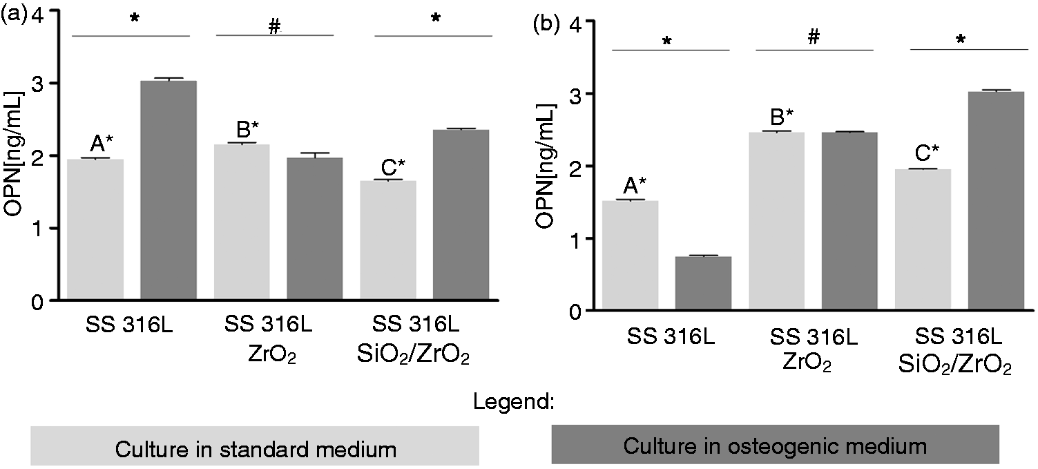
The analysis of mineral deposition in the ECM produced by ASCs showed that the stimulation with osteogenic medium caused an increase in calcium accumulation, nevertheless a significantly higher ratio of this element in ECM was found only in culture with SS 316L-SiO2/ZrO2 biomaterial. The same result was noted in BMSC cultures. The deposition of phosphorus in the ASC matrix was not altered in cultures on pure stainless steel and ZrO2 coating. However, ASCs propagated on SS 316L-SiO2/ZrO2 showed a lower concentration of phosphorous after osteogenic stimulation. Consistent observations were made in BMSC cultures. Cells propagated on SS 316L-SiO2/ZrO2 deposited had lowered concentration of phosphorus in osteogenic conditions, while cultures on pure SS 316L and SS 316L coated with ZrO2 had significantly higher phosphorous content after osteogenic stimulation. The results of SEM-EDX are presented in Figure 11.
Analysis of the composition of mineral matrix in ASC (a,b) and BMSC (c,d) cultures on investigated biomaterials. Mean ± standard deviation (error bars) was derived from three measurements. Significant difference at *p < 0.0001.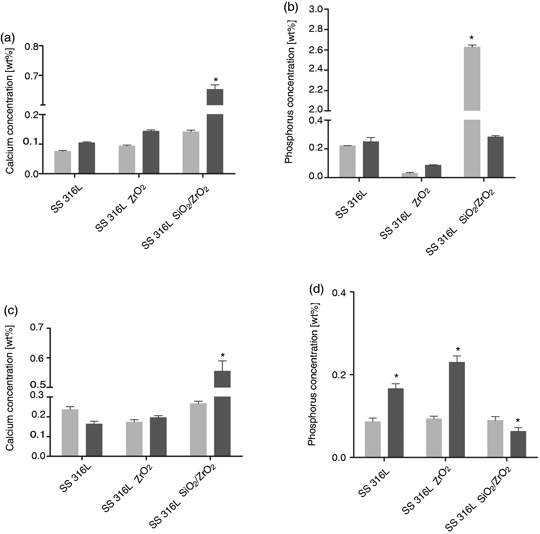
Discussion
Zirconium precursors are often modified due to the extreme sensitivity to hydrolysis. A general method of controlling the reactivity is the stabilization of the alkoxide ligands by chelating organic compound. Acetic acid, acetylacetone, and DEA are the most frequently used stabilizing agents for zirconium, titanium, and other metal alkoxides in the sol–gel studies.39–42 For our purposes and synthesis process we decided to use DEA, which can produce stable solutions for the oxides by controlling the rate of hydrolysis of the precursors used. A continuous and homogeneous morphology of the resulting zirconia and silica/zirconia coatings shows that the thin film might be constructed by an amorphous network. These results are in good agreement with the outcomes of Raman spectroscopy that demonstrated amorphous phase of ZrO2 in both coatings obtained. It is well known that zirconia can adopt an amorphous phase and three crystalline phases. A large number of studies have been performed to investigate the annealing effect on the structural and physicochemical properties of ZrO2 films.43–45 Ehrhart et al. 37 described the sol–gel method for the preparation of zirconia films, using the same precursor as we did, i.e. n-propoxide. Films remained in the amorphous phase to the annealing temperature of 400℃. Our study also showed an amorphous structure of the obtained coatings annealed at 250℃.
Based on the literature, it can be concluded that the hydrophilicity is primarily governed by polar interactions at the solid surface, which in turn arise from the inorganic component of the coating. Masking effects of alkyl chains are primarily responsible for the wettability parameter in the sol–gel derived coatings. An increasing amount of alkyl chains would conjointly decrease the polar and dispersive components of the solid surface energy, which in turn would decrease water wettability of the final material.46–48 Additionally, inhibition of water wettability is more efficient in long alkyl chains. 46 It is also known that the –OH groups are responsible for the hydrophilic behavior of the material. We observed the hydrophilic character of SiO2/ZrO2 coating that may result from a greater number of –OH groups (from the hydrolysis of TEOS). These results are consistent with the Raman spectra that demonstrated a higher intensity of absorption band and a greater number of –OH groups (from the hydrolysis of TEOS) in the case of sol–gel SiO2/ZrO2 coating. There is significant evidence that the surface roughness plays an important role in determining the successful osseointegration of metallic implants. 49 AFM measurements allow to characterize nanoscopic surface features and surface roughness. Ra is a widespread and often used parameter to describe the roughness of the implant surface. The study of Lim et al. found very different static contact angles on variously treated Ti surfaces, which had highly similar Ra values. 50 Therefore, they concluded that apart from the roughness parameter, other surface properties should also be considered important in the biological response. It should also be noted that the surfaces with considerably different topographies may possess similar Ra values and that the height descriptors alone do not adequately describe the surface roughness.51–53 Our study is in good agreement with these statements as our coatings had very similar roughness but different wettability. In our opinion, other properties of the surface should also be considered relevant in the biologic response and may actually constitute more important parameters of biocompatibility than the surface roughness analyzed separately. Our study clearly shows the feasibility of dip-coating sol–gel method in the production of smooth, ultra-thin ZrO2 and SiO2/ZrO2 films with controllable wettability and retained roughness. Biomaterial roughness and wettability together play a key role in the cell attachment and adhesion in the initial stage of interaction of the implant and cells. Early adhesion affects the cell growth, differentiation, viability, and spreading, i.e. the biocompatibility of the implant. We have considered the contribution of wettability to the cellular response because obtained biomaterials had similar surface roughness. The influence of wettability on the proliferation of cells remains unsettled. In general, anchorage-dependent cells are characterized by a lower proliferation on hydrophobic surfaces than on hydrophilic ones. 54 Our observations confirm this dependency, particularly in the context of BMSC cultures. The proliferation of BMSCs was accelerated in cultures on a hydrophilic SiO2/ZrO2 layer, and the time needed for doubling the population was significantly reduced. Proliferation activity of ASCs was also considerably increased on hydrophilic SiO2/ZrO2 surfaces, but only after 24 h and 120 h of propagation. Furthermore, increased proliferative activity did not translate into the acceleration of the PDT in ASCs. The assessment of adhesion parameter is also a crucial indicator of implant biocompatibility, next to the proliferative activity of cells. Knowledge about the potential role of surface wettability in the context of cell adhesion is still incomplete and often contradictory. Majority of studies support the hypothesis that hydrophobic surfaces promote cell attachment, and therefore, have a more pronounced effect on cell behavior than hydrophilic materials. However, Das et al. 20 have reported that human osteoblasts colonized willingly hydrophilic surface, whereas Chang et al. 54 observed an increase in the apoptosis of osteoblasts on hydrophobic materials. These data are consistent with our observations. Both ASCs and BMSCs propagated on SiO2/ZrO2 surface adhered to the surface while maintaining proper mesenchymal-like morphology. However, after 168 h of culture, ASCs started to form large cellular aggregates. The focal localization of ASCs was also detected in cultures on SS316L-ZrO2 biomaterial, but at the same time a high ratio of apoptotic bodies was observed. Similarly, BMSCs cultured on ZrO2 layer displayed signs of apoptosis, which could be caused by delayed proliferation and low cell adhesion. The study of Das et al. 20 highlighted the fact that the hydrophilic biomaterials adsorb attachment proteins, and thus can provide more adhesion sites for the precursors of bone cells and enable osseointegration by minimizing the generation of non-functional tissue or even aseptic loosening. Although this reasoning has been confirmed by other studies,55,56 we do not exclude the influence of chemical composition of the layers obtained on cell behavior and morphology of MSCs. Both silica and zirconia precursors are highly osteogenic and can affect the focal localization of cells. Liu et al. 17 showed that ZrO2 thin films are bioactive and cytocompatible, however Randeniya et al. 57 observed adverse impact on osteoblasts resulting in specific coalescence of cells. Randeniya’s observation concerning morphological changes of ostoeoblast cells are in agreement with our observations. Cells cultured on ZrO2 layers were less spread out in comparison with cultures on hybrid coatings. While Randeniya et al. related improper cell adhesion with the presence of nanocrystalline ZrO2, our results may suggest that chemistry of zirconia is one of essential attributes affecting cell adhesion, as coatings obtained had amorphous character.
The ASC aggregates formed in 168-h culture on hybrid layer resembled the spot-like growth pattern characteristic of osteogenic cultures, 58 while BMSCs exhibited more round and oval shape.
We have analyzed the relative transcript levels of main markers of osteogenesis using real-time PCR to ascertain if surfaces obtained promote and/or enhance the process of differentiation of MSCs toward osteoblasts. The analysis included an evaluation of mRNA expression of ALP, collagen type I (Coll-1), OPN, and ON. The level of gene expression was determined in non-osteogenic and osteogenic culture conditions. The results showed that in ASC cultures the type of surface did not have an influence on osteogenic markers expression, either before or after osteogenic stimulation. Differentiation of ASCs toward osteoblasts increased the level of ALP and OPN transcripts, while the expression of collagen type I decreased after osteogenic stimulation and ON expression was constitutive. BMSC cultures were more sensitive to surface-derived stimuli as well as signals from the culture medium. The activity of ALP was enhanced irrespective of the osteogenic stimulation. BMSCs cultured on a hydrophilic SiO2/ZrO2 surface in non-osteogenic culture conditions expressed the highest levels of ALP and OPN.
A decrease in the expression of collagen type I under osteogenic conditions has also been described by Kyllönen et al. 59 This phenomenon may be affected by the concentration of dexamethasone, which is a necessary component of the osteogenic medium and enables efficient osteoinduction of MSCs. Osteogenic differentiation was also evaluated by immunoenzyme assay that allows detection of OCL and OPN. The highest concentration of OCL was found in the supernatants of ASCs and BMSCs after osteogenic stimulation on ZrO2 surface. The concentration of OCL was also increased in non-osteogenic conditions, in the supernatants of cultures on SiO2/ZrO2 coating, although significantly higher OCL level was detected in the medium collected from ASC cultures. The data suggested that the secretion of OCL was accelerated spontaneously on hybrid coatings, while the culture on ZrO2 enhanced osteogenic differentiation of MSCs.
The concentration of OPN measured in the supernatants of both populations of MSCs was significantly increased in osteogenic cultures on SS316L-SiO2/ZrO2. The highest accumulation of OPN in non-osteogenic conditions was observed in cultures on ZrO2 layer. High OPN level was also noted in ASC cultures on a pure stainless steel substrate. It should be emphasized that OPN is a pleiotropic protein and likely promotes not only osteogenic differentiation, but also other cytophysiological processes such as cell spreading and proliferation. The osteogenic properties of SiO2/ZrO2 layer have been eventually confirmed by the analysis of elemental composition of the ECM. ASCs and BMSCs in culture on SS316L-SiO2/ZrO2 biomaterial produced ECM deposits of calcium and phosphorous. Interestingly, after stimulation the concentration of calcium was elevated, while the amount of phosphorous decreased. Comparison of ECM produced by ASCs and BMSCs showed that the activity of the latter resulted in more effective mineralization. The morphological analysis and the evaluation of osteogenesis confirmed that ASCs had a greater potential to create nodular aggregates compared to BMSCs. However, the overall osteogenic potential of ASCs is inferior in comparison with BMSCs.60,61 Stainless steel represents a biocompatible material itself, but our results indicate that the application of synthesized coatings enhances the functional differentiation of MSCs into osteoblasts, thereby promoting early bone healing and implant stabilization.
Conclusion
We expect that a comprehensive characterization of zirconia surfaces and an extensive analysis of its biocompatibility will provide valuable information concerning the development of implants that can be applied to the in vivo model. Furthermore, the biomaterial-dependent commitment of MSC populations is vital in the context of cell-based therapy. There is a high demand for implants with osteoinductive properties, and therefore, they should be continuously developed and improved. We do hope that promoting positive cellular responses of MSCs and/or osteoblast will contribute to their success in clinical practice.
Footnotes
Acknowledgments
The authors would like to thank to Professor Elżbieta Pamuła from Department of Biomaterials at AGH University of Science and Technology in Krakow for the opportunity of the measurements of wettability and surface free energy presented in this paper. The authors would like to thank Aneta Skaradzińska, PhD and Adam Dobrowolski, PhD from Department of Biotechnology and Food Microbiology at Wroclaw University of Environmental And Life Sciences for the opportunity of the total RNA isolation and evaluation.
Declaration of conflicting interests
The authors declared no potential conflicts of interest with respect to the research, authorship, and/or publication of this article.
Funding
The research was supported by Wroclaw Research Centre EIT + under the project “Biotechnologies and advanced medical technologies”—BioMed (POIG.01.01.02-02-003/08) financed from the European Regional Development Fund (Operational Program Innovative Economy, 1.1.2).