
Abstract
Three types of commercially available ultra-high molecular weight polyethylene (UHMWPE) acetabular cups currently used in total hip arthroplasty have been studied by means of Raman micro-spectroscopy to unfold the microstructural modification induced by the oxidative degradation after accelerated aging with and without lipid absorption. The three investigated materials were produced by three different manufacturing procedures, as follows: irradiation followed by remelting, one-step irradiation followed by annealing, 3-step irradiation and annealing. Clear microstructural differences were observed in terms of phase contents (i.e. amorphous, crystalline and intermediate phase fraction). The three-step annealed material showed the highest crystallinity fraction in the bulk, while the remelted polyethylene is clearly characterized by the lowest content of crystalline phase and the highest content of amorphous phase. After accelerated aging either with or without lipids, the amount of amorphous phase decreased in all the samples as a consequence of the oxidation-induced recrystallization. The most remarkable variations of phase contents were detected in the remelted and in the single-step annealed materials. The presence of lipids triggered oxidative degradation especially in the remelted polyethylene. Such experimental evidence might be explained by the highest amount of amorphous phase in which lipids can be absorbed prior to accelerated aging. The results of these spectroscopic characterizations help to rationalize the complex effect of different irradiation and post-irradiation treatments on the UHMWPE microstructure and gives useful information on how significantly any single step of the manufacturing procedures might affect the oxidative degradation of the polymer.
Introduction
Acetabular cups of modern hip implants are manufactured from ultra-high-molecular-weight polyethylene (UHMWPE), a material with extreme durability in its non-oxidized state. 1 However, polymer engineers and scientists have long studied methods to concurrently improve wear resistance, deformation/fracture resistance and oxidative stability of UHMWPE by varying manufacturing methods, consolidation techniques, resin recipes, bearing surface treatment (e.g. laser-assisted ion implantation or coating with cross-linked polyethylene glycol (PEG) to improve wear resistance), or reinforcing the polyethylene matrix with carbon fibers. Studies dating back to the 1990s clearly proved that the use of ionizing radiation to crosslink the polymeric chains effectively could reduce adhesive/abrasive wear of UHMWPE bearings.2–4 However, more recent studies have shown that the performance of conventional gamma-sterilized UHMWPE is excellent during the first decade of implantation of the bearing component, while wear and oxidation might become an issue for the mechanico-chemical stability of the material starting from the beginning of the second decade. In fact, the irradiation causes hemolytic cleavage of C-C and C-H bonds of the polyethylene molecular chains. The resulting alky radicals are not stable and enter a complex system of reactions, which not only lead to crosslinking in the amorphous phase, but also to chain scissions, oxidation, formation of C=C double bonds and transformations to more stable allyl and peroxy radicals. The formation of C=C double bonds is not assumed to have a significant effect on the performance of the polymer, but all the other reactions influence wear and mechanical properties adversely.5,6 Chain scission leads to lower molecular weight polyethylene, whose wear and mechanical performances are inferior as compared to the original polymer. In the effort of counteract such a degradation of the material, combining irradiation process with a post-irradiation annealing to eliminate residual free radicals has been identified as the solution to improve wear and oxidation resistance.7–11 In a recent publication, Oral et al. reported an interesting study in which oxidative degradation of irradiated and melted UHMWPE components was observed as a consequence of exposition to body fluids during shelf-aging. 12 The absorption of lipids, which are present in the synovial fluid and are in contact with the polyethylene component during implantation, might trigger accelerated oxidation and degradation of the mechanical properties of these materials. The oxidation rate is dependent on lipid concentration, oxygen concentration, and temperature. Diffusion of lipids in the semicrystalline polymer induces plasticization, especially on the surface where the concentration of absorbed species reaches its highest value. In turn, plasticized polymers show deterioration of wear resistance, whose extent depends on the nature and the amount of the lipids.13,14 Although Greenbaum et al. 15 did not notice any reduction of wear resistance in their in vitro experiments on polyethylene with absorption of lipids, they confirmed the deterioration of some mechanical properties (significant reduction of compressive elastic modulus and compressive yield strength). The debate regarding these important issues, recently brought to the consideration of the scientific community, made clear that a comprehensive characterization of the response to oxidation of the new generation UHMWPEs is still lacking. In other words, the idea of removing the free radicals generated during processing might not be sufficient to guarantee a bearing component immune to long-term oxidation if the microstructure of the material is prone to absorb a conspicuous amount of lipids and if the initial density of residual free radical is high.
The purpose of this study was to investigate the microstructural modifications induced by accelerated aging in three different (commercially available) highly crosslinked UHMWPEs which where in vitro oxidized with and without the presence of absorbed lipids in their microstructure. Two materials belonged to an early generation single-step irradiated polyethylene (Crossfire™, Stryker Orthopaedics, Inc., Mahwah, NJ and Longevity™, Zimmer, Inc., Warsaw, IN, USA), while the third investigated material belonged to a successive generation of sequentially irradiated polyethylene (X3™, Stryker Orthopaedics, Inc., Mahwah, NJ, USA).
Materials and methods
UHMWPE materials
Three commercially available liners were investigated in the present study. The main characteristics and peculiarities of the three materials, including the steps of the processing, can be summarized as follow:
Longevity™, hereafter referred as Liner A, is manufactured by Zimmer, Inc. (Warsaw, IN, USA) and it is a first-generation remelted liner clinically introduced in the Trilogy acetabular cup design since 1999. Molded sheets, consolidated from GUR 1050 resin (5.5–6 million g/mol), are radiation crosslinked by electron beam with a total dose of 100 kGy and then remelted (>135℃) to quench residual free radicals.
Crossfire™, hereafter referred as Liner B, is manufactured by Stryker Orthopedics, Inc. (Mahwah, NJ, USA). This brand also belongs to the first-generation of annealed liners. This liner was clinically introduced in 1998. The manufacturing procedure of Crossfire™ also starts from GUR 1050 resin but the resin has the morphology of extruded rods. The rods are gamma-irradiated with a nominal dose of 75 kGy and subsequently annealed at 130℃. After being machined into liner shape and then barrier packaged, Crossfire™ is exposed again to gamma irradiation for sterilization purpose with the nominal dose of 30 kGy in nitrogen atmosphere.
X3™, hereafter referred as Liner C, is manufactured by Stryker Orthopedics, Inc. (Mahwah, NJ, USA). It belongs to a second-generation annealed liner and was clinically introduced in the Trident and Tritanium acetabular cup design in 2005. GUR 1020 (3.5 million g/mol) compression molded sheets are gamma irradiated at the nominal dose of 30 kGy and then annealed at 130℃. The same procedure is sequentially repeated three times (i.e. the cumulative radiation dose being 90 kGy).
The polyethylene liners as received by the manufactures were cut through their thickness to obtain rectangular prisms from the area of the alleged main wear zone (contact area between hip cup and femoral counter surface). In every prism-shaped specimen, one face preserved to the original surface of the liner, as shown in Figure 1(a). For each sample, this area was investigated using confocal Raman microspectroscopy after in vitro oxidation. Three cups for each of the three materials were cut and two specimens were obtained from each cup: one specimen underwent accelerated aging while the second one was immerse in lipid solution (squalene) before accelerated aging. The procedure of in vitro oxidation and absorption of lipids are described in the next section.
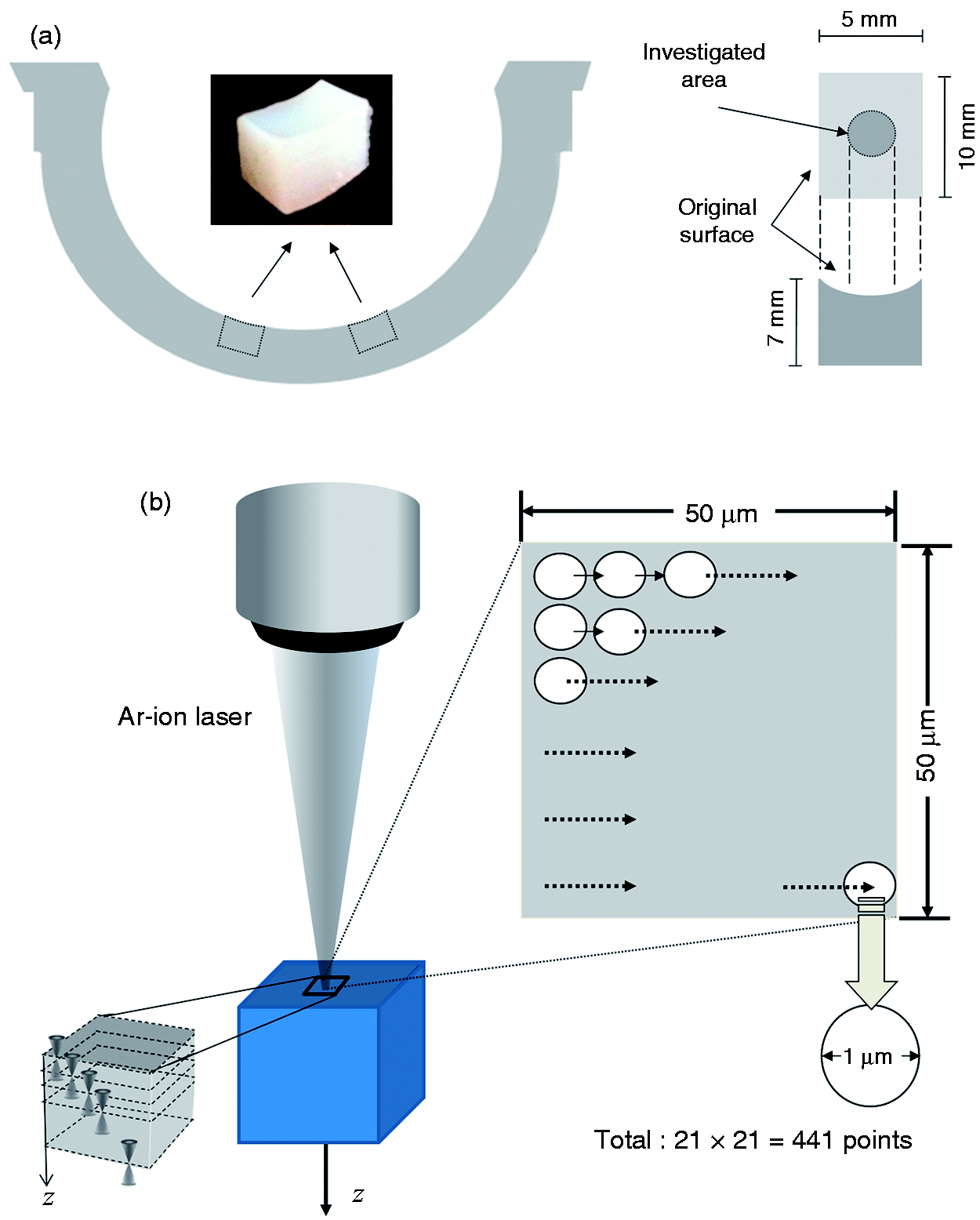
(a) Schematics showing the geometry of the samples for accelerated aging and the locations of the liner from which they were cut. (b) Schematics of the spectroscopic measurement protocol.
Accelerated aging with and without lipid absorption
The accelerated aging of the samples was performed according to the standards ASTM F2003-02 (reapproved in 2008) 16 with the intent to investigate the oxidative stability of Liners A, B, and C. Following the standard procedures, the three materials were oxidized under equal conditions prior to characterization of their microstructures by Raman spectroscopy. Unlike infrared spectroscopy, Raman spectroscopy allows non-destructive characterization without further sample manipulation. A set of samples obtained from new cups of the three materials were immersed for 4 h in squalene (95% solution, Wako Pure Chemicals Industries, Ltd., Osaka, Japan) at elevated temperature (100℃ in a convection oven) to induce diffusion of lipids in the microstructure before accelerated aging. As a lipid to be diffused through the specimens, we chose squalene because it is a precursor in cholesterol synthesis and it is present in the synovial fluid with unsaturated bonds (thus inducing generation of free radicals in UHMWPE during in vivo implantation). At the completion of the procedure of lipid diffusion, all specimens were cooled down to room temperature in their respective lipid solution and remnants of the solution were carefully removed from the surface of the samples. Subsequently, all samples (with and without diffused lipids) underwent accelerated aging, whose preliminary conditioning step consisted of irradiating the specimens with γ-ray (25 kGy dose) and maintaining them at 23℃ for 28 days. An oxygen bomb was used to age the specimens at 70℃ in pure oxygen (5 atm) for 14 days. All the specimens were analyzed by micro-Raman spectroscopy within 10 days after the completion of the test.
Confocal Raman spectroscopy
All the spectroscopic assessments in this study were made by means of a Raman microprobe
spectrometer (T-64000, Horiba/Jobin-Yvon, Kyoto, Japan) in back-scattering geometry. The
excitation source was a 488.0 nm Ar-ion laser (Stabilite 2017-Spectra Physics, Mountain
View, CA) yielding a power of approximately 35 mW on the UHMWPE liner surface. The
confocal configuration of the probe adopted throughout the present experiments
corresponded to a 100 × objective; numerical aperture, focal length, and pinhole diameter
were fixed as: NA = 0.9, f = 11 mm, and Φ = 100 μm,
respectively. Individual spectra were typically collected in 10 s in both polarized and
non-polarized measurements. The recorded spectra were averaged over three successive
measurements at each selected location. A spectral resolution better than
0.15 cm−1 was achieved by means of an 1800 grating l/mm. Spectral mapping was
non-destructively performed in order to characterize crystallinity from the surface down
to 100 μm inside of UHMWPE acetabular liners. A map 50 × 50 μm2 in dimension
was collected at eight different depths depth (z
lab
= 0, 5, 10, 20, 35, 50, 75 and 100 μm) with an
in-plane sampling of 2.5 μm step (for a total of 441 spectra per each map). A schematic of
the spectroscopic measurement protocol is given in Figure 1(b). The relationship between the observed
Raman bands and the vibrational modes of the polyethylene molecular structure has been
amply documented in the literature.17–23
Figure 2 shows a typical Raman
spectrum of polyethylene with its related individual bands after spectral deconvolution.
Spectral deconvolution has been performed by means of an automatic fitting algorithm
enclosed in a commercially available computational package (Labspec 3, Horiba/Jobin-Yvon,
Kyoto, Japan), using mixed Gaussian/Lorentzian curves. The Raman spectrum of polyethylene
(shown here in the range between 950 and 1600 cm−1) can be divided into three
main regions: region I, dominated by the C–C stretching
vibrational mode in the interval 1000–1150 cm−1; region
II, mainly represented by the –CH2– twisting vibration at around
1300 cm−1; and region III, which is
characteristic of –CH2– bending between 1350 and 1500 cm−1. The
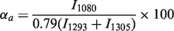
broad band at 1080 cm−1 is associated with the presence of an amorphous
phase.
21
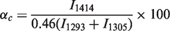
The
peak at 1414 cm−1 is characteristic of an orthorhombic phase, which represents
the almost totality of the crystalline phase, and the peak at 1293 cm−1 can be
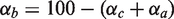
used to approximate the degree of crystallinity of the overall UWMWPE structure.19–23 The percentages of crystalline (α
c
) and amorphous
(α
a
) phases can
be calculated from the Raman spectrum of polyethylene, according to the following
equations
21
Typical Raman spectrum of polyethylene with its related individual bands after
spectral deconvolution. The bands used for the calculation of phase fractions are
also labeled.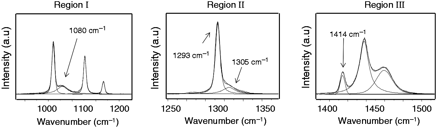
Statistical analysis
Specification of the groups (material and treatment), the dependent variables (studied parameters), and the number of measurements used for the statistical analysis.
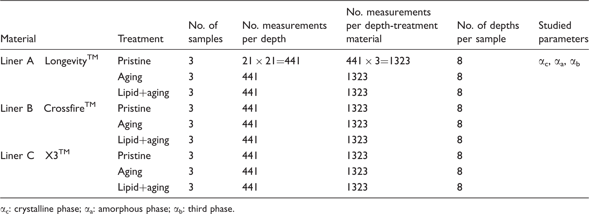
αc: crystalline phase; αa: amorphous phase; αb: third phase.
Results and discussion
Two-dimensional Raman maps collected at different focal depths, as explained in the
Confocal Raman spectroscopy section, enabled to visualize microstructural patterns along the
material subsurface. In order to give a statistically meaningful picture of the
microstructural conformation of the three materials, we decided to analyze three different
samples for each material and specific condition of in vitro
oxidation. The average values of crystalline, amorphous and third phase as calculated from
the three maps were considered in the discussion as the parameters characterizing each liner
at each oxidative condition. Although the practice of accelerated aging might not simulate
the real degradation occurring in hip components during implantation (e.g. the influence of
cycling loading on the resistance to oxidation is not considered), as clearly stated in the
ASTM standards themselves, these experimental procedures are extremely substantial and
meaningful to judge and discuss the structural modifications induced by the oxidation of
different UHMWPE materials as a function of starting resin, consolidation process,
sterilization method and post-irradiation treatment. Figure 3 shows in-depth profiles of crystallinity
fraction, α
c
, as
calculated from the three liners at different aging conditions. Specifically, Figure 3(a) to (c) shows the trends of
crystalline phase for the three materials in their non-oxidized state (hereafter referred as
Case 1), after accelerated aging (hereafter referred as Case 2) and after lipid absorption
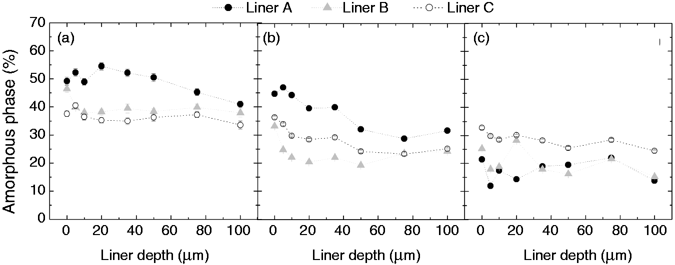
followed by accelerated aging (hereafter referred as Case 3), respectively. Similarly, Figures 4(a) to (c) and 5(a) to (c) report the trends of
amorphous phase fraction, α
a
, and third phase fraction, α
t
, respectively. The scattering bars represent the standard
variations of the calculated means, which were low and reliable in all the cases because of
the large number of measurements for each map. For each profile, one-way ANOVA test was
performed to confirm the differences among the average values of phase fraction calculated
at different depths. In all the profiles showed in Figures (3) to (5), the results of the test confirmed that the population means are different
(p < 0.05). As far as the crystallinity is concerned, all
the three materials revealed gradients of crystallinity along the first microns of the
subsurface in all the three cases, with the average lowest values on the surface. The
crystalline phase fraction linearly increases along the first microns of the subsurface and
it reaches a plateau at around 35–50 μm, but in Cases 2 and 3, such a gradient became
steeper. This evidence might prove that in the sub-surface, there is the most favorable
trade-off between high concentration of free radicals and oxygen. In Case 1 (Figure 3(a)), the differences among the
retrieved profiles can be interpreted as the direct consequence of the selected processing
(namely, reduced crystallinity by remelting (Liner A) vs. substantial preservation of the
as-irradiated structure upon annealing (Liner B and Liner C)). Moreover, the higher
crystallinity in Liner C (62.3%) as compared to Liner B (58.1%) can mainly be explained by
considering the lower molecular weight of the resin used in X3™ material (GUR1020 vs.
GUR1050). In Cases 2 and 3 (Figure 3(b) and
(c)), all the three materials after accelerated aging clearly showed increases of
crystallinity underneath the surface (especially deeper than 20 μm), as compared to their
respective non-oxidized states (Case 1). On the surface of Liners B and C, the increase of
crystallinity is not substantial as in Liner A. In fact, if on one hand, in Liner B the
modification induced by oxidation on the surface leads to a robust increase of third phase
rather than crystalline phase (see Figure
5), on the other hand, on the surface of Liner C the lowest content of oxygen-rich
amorphous phase in the pristine (see Figure
4(a)) and, presumably, the lowest concentration of free radicals might have
preserved the microstructure from oxidation. One of the most detrimental effects of
oxidation in UHMWPE is the reduction of molecular weight induced by chain scission
(triggered by the formation of oxidized products, such as ketones and carboxyl acids). In
our in vitro degradation experiments, recrystallization is
univocally correlated to the shortening of molecular chains, which acquire higher mobility
to reorganize in crystalline domains at lower and more stable energy.
23
Moreover, this
microstructural modification is promoted and accelerated by the temperature (70℃ in our
case) and, possibly, by the presence of chemical species such as lipids, which might form
new free radicals. In other words, no further external variables had been added to the
system under investigation and any observed increase of crystallinity (and third phase
content) at the expensive of the amorphous phase should be correlated to the oxidative
degradation. It has already been demonstrated in several studies that oxidation leads to
deterioration of the mechanical properties, such as loss of ductility (embrittlement) and
lower wear resistance.24–30 Therefore, the variations of phase fractions as calculated by Raman
spectroscopy and presented in this paper are directly correlated to the worsening of wear
and mechanical properties in UHMWPE. Keeping in mind these considerations, comparison
between Case 2 and 3 showed that the presence of lipids promoted recrystallization, namely
higher oxidation, after aging in all the investigated materials at their very surface
(z = 0). Conversely, Case 3 for Liner B and C did not show
any remarkable difference in the sub-surface as compared to Case 2, while in Liner A the
diffusion of lipids inhibited the formation of crystallinity. In order to understand and
discuss the latter experimental evidence, we must take into consideration also the variation
of the other two phases in the material. In fact, Figures 4(b) and (c) and 5(b) and (c) show that in Liner A the incorporation of
lipids induced an abrupt decrease of amorphous phase followed by and increase of third
phase. This phase is considered to be an intermediate phase forming an interfacial layer
between crystalline and amorphous material. The molecular chains contained in this layer are
stretched, conferring an anisotropic character to this phase and properties that are
intermediate between the orthorhombic crystallites and the amorphous material. Strobl and
Hagedorn described the third phase as consisting of crystalline-like structures which have
lost their lateral order but which cannot simply be identified as another crystal
modification.
19
As
a matter of fact, lipids diffuse through the amorphous phase,
5
where they can promote the formation of
oxidized species. In liner A, the amount of amorphous phase in the pristine is higher than
the other two materials (cf. Figure
4(a)), which means that it might have absorbed the highest concentration of lipids.
On the other hand, Liner B and C might have incorporated fewer lipids, which could explain
the moderate decrease of amorphous phase (especially in Liner C) as compared to Liner A. In
addition, oxidation-induced recrystallization in the amorphous phase of Liner A might have
been impeded by the higher concentration of lipids, which, in turn, led to the formation of
new third phase rather than a perfectly ordered orthorhombic crystallographic structure. In
Figure 6(a) to (c), Cases 2 and 3
are compared in terms of variation from the non-oxidized state (Case 1) of crystallinity,
amorphous phase, and third phase, respectively, as retrieved from the bulk of the three
materials. We considered as representative of the bulk the average calculated from the maps
collected at 50, 75, and 100 μm in depth. One-way ANOVA tests and post hoc Tukey tests
confirmed that the population means plotted in Figures 3–5 are statistically different (p < 0.05). In-depth profiles of crystallinity fractions (% volume) as collected in the three
materials at different oxidative conditions: (a) non-oxidized material (Case 1), (b)
after accelerated aging (Case 2), and (c) after absorption of lipids and accelerated
aging (Case 3). In-depth profiles of amorphous phase fractions (% volume) as collected in the three
materials at different oxidative conditions: (a) non-oxidized material (Case 1), (b)
after accelerated aging (Case 2), and (c) after absorption of lipids and accelerated
aging (Case 3). In depth profiles of third phase fractions (% volume) as collected in the three
materials at different oxidative conditions: (a) non-oxidized material (Case 1), (b)
after accelerated aging (Case 2), and (c) after absorption of lipids and accelerated
aging (Case 3). (a) Variation of bulk crystallinity for the three liners under investigation after
accelerated aging (Case 2) and after aging with lipid absorption (Case 3). (b)
Variation of bulk amorphous phase for the three liners under investigation after
accelerated aging (Case 2) and after aging with lipid absorption (Case3). (c)
Variation of bulk third phase for the three liners under investigation after
accelerated aging (Case 2) and after aging with lipid absorption (Case 3).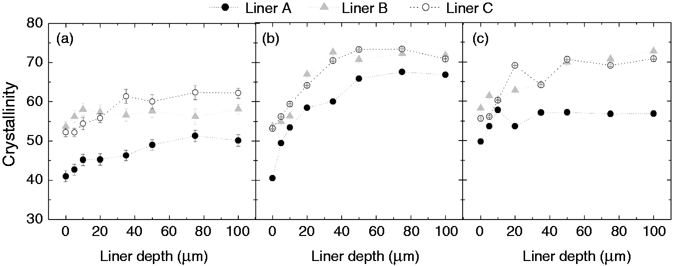
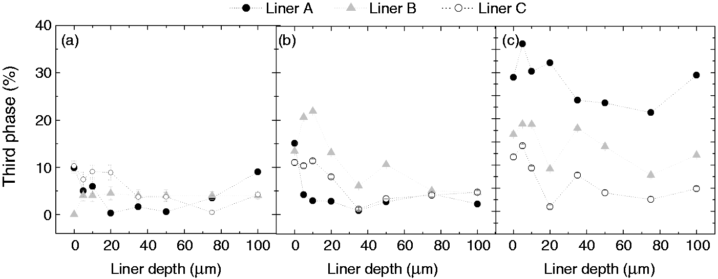
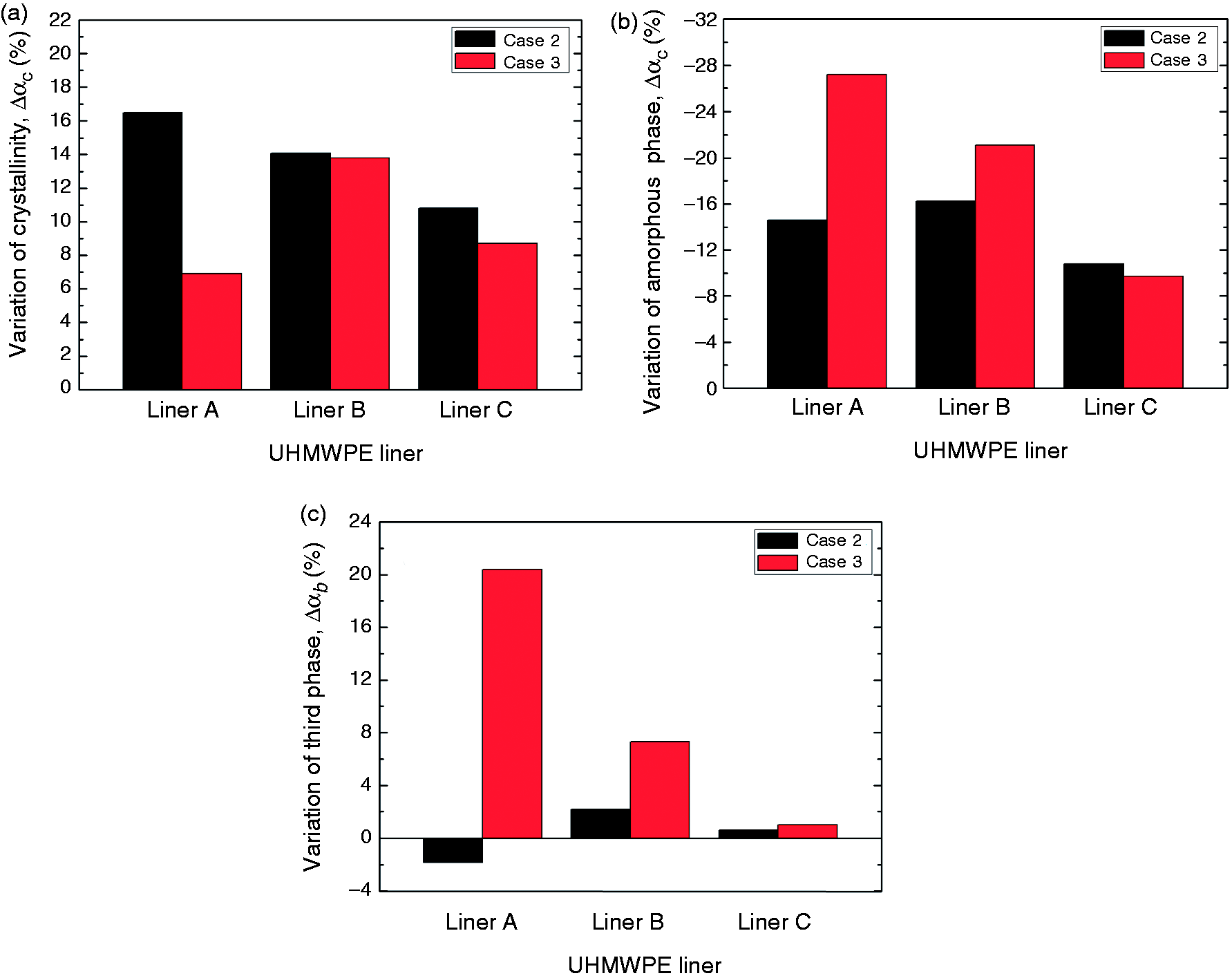
In Case 2, namely the samples that underwent accelerated oxidation without lipids, Liner A experienced the highest variation of crystallinity, while Liner C the lowest one. Concurrently, Liner C showed lowest variation of amorphous and third phases. Liner B showed a similar trend to Liner A in Case 2, although an interesting difference is worthy to remark: in both materials the reduction of amorphous phase is similar, but in Line A also part of the third phase transformed to crystalline phase (cf. Figure 6(c), showing third-phase decrease in Liner A). We might hypothesize that free radicals were trapped also in the intermediate phase of the initial microstructure of Liner A leading to oxidation and subsequent transformation to crystalline phase. In Case 3, as previously discussed comparing the in-depth profiles, the most striking evidence resided in the lowest variation of crystallinity of Liner A (cf. Figure 6(a)), but, conversely, the most remarkable reduction of amorphous phase and increase of third phase as compared to Liners B and C (cf. Figure 6(b) and (c)). In presence of lipids, Liner B showed a higher reduction of amorphous phase and, similarly to Liner A, formation of third phase was promoted. Liner C showed the lowest variation of the three phases also in Case 3 runs. Comparing Cases 2 and 3 for Liner C, it is clear that the presence of lipids did not affect the oxidative resistance of the polymer, presumably due to the combined effect of low starting concentration of free radicals and the small fraction of amorphous phase in which lipids can be absorbed.
Conclusions
In accelerated oxidation without lipids, the three-step annealed highly crosslinked UHMWPE experienced the lowest variation of crystalline, amorphous and third phases. The two materials belonging to the first generation of highly crosslinked UHMWPE showed similar trends for phase-fraction variation induced by in vitro oxidative degradation: the total decrease of amorphous phase in which oxidized species are formed is slightly superior for the single-step annealed material as compared to the remelted one, presumably due to the lower concentrations of free radicals in the remelted material. Also in lipid-assisted accelerated oxidation, the new generation of highly crosslinked polyethylene showed the lowest (total) variation of the three phases: the presence of lipids did not affect the oxidative resistance of the polymer, presumably due to the combined effect of its low starting concentration of free radicals and the low amorphous phase in which lipids could be absorbed. The latter hypothesis seems to be confirmed by the observation that, in the remelted material, the oxidative stability given by the initial low concentration of free radicals seems to be offset by the highest amount of amorphous phase in which lipid absorption is favored. The microstructure of the new generation 3-step annealed material was thus found to be the most stable as compared to previous generation remelted and single-step annealed polyethylenes.
Footnotes
Declaration of conflicting interests
None declared.
Funding
This research received no specific grant from any funding agency in the public, commercial, or not-for-profit sectors.