
Abstract
Novel antibacterial nanomaterials have been developed for biomedical applications. The present study involves the preparation and properties of antibacterial nanofibers from chitosan/polyethylene oxide electrospun nanofibers incorporated with silver nanoparticles. Silver nanoparticles were efficiently synthesized in situ after ultra violet (UV) with AgNO3 as precursor and chitosan/polyethylene oxide as reducing agent and protecting agent, respectively. Then the resultant solutions were electrospun into nanofibers. The formation of silver nanoparticles was confirmed with ultraviolet visible (UV-vis) and transmission electron microscopy (TEM), and the electrospun nanofibers were characterized by scanning electron microscopy and energy dispersive X-ray. The resultant fibers exhibited uniform morphology with silver nanoparticles distributed throughout the fiber. Also, the fibers showed certain tensile strength and excellent antibacterial activity against Gram-positive (Staphylococcus aureus) and Gram-negative (Escherichia coli) bacteria. Sustained release of silver nanoparticles from fibers could last for over 72 h. The silver-containing chitosan/polyethylene oxide nanofibers showed excellent cytocompatibility.
Introduction
In recent years, electrospinning has been extensively studied as the most cost-effective and versatile method to prepare nanofibers with the diameter ranging from several micrometer down to tens of nanometer. 1 In the conventional electrospinning processing the spinning fluid is driven into the orifice of single nozzle connected with high-voltage power supply, forming micro-nanofibers which are finally received on the grounded receiving screen. The single needle/nozzle electrospinning device can effectively prepare a variety of uniform nanofibers, but the efficiency of its preparation is relatively low. In order to improve the yield of electrospinning, it is desirable to develop highly efficient multineedle/multinozzle and needle-free electrospinning device. For example, the concentric nozzle, 2 juxtaposition nozzles, 3 multinozzles, 4 and so on were used to produce the nanofibers of hollow type, sheath core type, or multiporous channel type instead of a single nozzle. The needle-free electrospinning, for example, in the bubble electrospinning process, 5 a pump was used to inject a lot of gas to polymer solution or melt, resulting in numerous bubbles in the polymer solution. Once the bubbles ruptured on the free surface of the polymer solution, numerous Taylor cones were likely generated from the solution, which is similar to multineedle electrospinning improving the efficiency of nanofiber production.
To date, hundreds of synthetic and natural polymers have been successfully electrospun, such as polylactide, poly(ɛ-caprolactone), collagen, silk fibroin, etc. 6 Owing to the high specific surface area and three-dimensional interconnected nano-porous structure, electrospun nanofibers provide a structure similar to native extracellular matrix.7–9 In addition, electrospun nanofibrous mats have great absorbance and high gas permeability. They can provide moist environment and protect the wound from exogenous infection. 10
Chitosan has many excellent biological properties, and it is a very important and beneficial natural polysaccharide. It is nontoxic, biodegradable, biocompatible, antibacterial, and hemostatic. However, due to limited solubility of chitosan and the polyelectrolyte nature, the repulsive interaction between chitosan molecules prevents the necessary chain entanglement for fiber formation, thus electrospinning of pure chitosan is still a tricky problem. 6 To date, 1,1,1,3,3,3-hexafluoro-2-propanol (HFIP), 11 tri-fluoro acetic acid (TFA), 12 and 90% aqueous acetic acid solution 13 have been studied as the solvents to prepare pure chitosan nanofibers. Compared with HFIP and TFA, both volatile, toxic, and expensive solvents, aqueous acetic acid solution is a more environment-friendly and cost-effective solvent to fabricate chitosan electrospun nanofibers.
Polyethylene oxide (PEO)14,15 and polyvinyl alcohol6,16 are often added to the solution as additives to facilitate the electrospinning of chitosan.17–20 In the blends, the fiber forming ability is improved by adjusting the interaction between polymer molecules. PEO is a water soluble polymer with good biocompatibility, low toxicity, and it is quite feasible and versatile for electrospinning. PEO fibers exhibit great interest for biomedical devices contacting with living organisms. 20
In order to enhance the antibacterial properties of chitosan-based nanofibers, antimicrobial agents have been introduced into the fibers. For example, Dilamian et al. 15 prepared chitosan/PEO nanofiber incorporating poly(hexamethylene biguanide) hydrochloride. Besides, as a natural antimicrobial agent, silver is emerging as a viable treatment choice for infections caused by burns, chronic wounds. 21 Silver-embedded polymer matrix can release silver continuously to provide a lasting and effective antibacterial activity, 22 which has wide applications in biomedical materials. As an efficient antibacterial material, silver nanoparticles (Ag-NPs) embedded polymer nanofibers are traditionally prepared by two-step process. 23 However, in this method, toxic chemical reagents are often used as the reduce agent, and it is also a challenge to disperse nanoparticle in the viscose electrospinning solution. 24
In this work, Ag-NPs were prepared by ultra violet (UV) irradiation with chitosan/PEO acting as stabilizer and protector and then Ag-NPs incorporated chitosan/PEO solution was electrospun into nanofibers. The resultant fibers and nanoparticles were characterized and tensile strength, antibacterial property, and silver release property of the nanofiber mats were investigated.
Experimental
Materials
Chitosan with degree of deacetylation of 95.28% and viscosity of 235 mPa s was provided by Zhejiang Golden-Shell Biochemical Co., Ltd, China. PEO with a molecular weight of 900 kDa was purchased from J & K Chemical. Acetic acid and silver nitrate (AgNO3) were supplied by Shanghai Ling-Feng Chemical Reagents Ltd, China. The Gram-positive bacteria Staphylococcus aureus (ATCC 25923) and Gram-negative bacterial (Escherichia coli) were used for the bacterial assay. All chemical reagents were used as received without any further purification.
Preparation of Ag-chitosan/PEO electrospinning solution
In a typical procedure, 90% concentrated aqueous acetic acid solution was used as the solvent. AgNO3 aqueous (1 mol/l) solution was added into the 90% acetic acid solution to prepare the AgNO3 solution with 0, 2, 4, 8 wt%, AgNO3 to polymer. PEO and chitosan powders were then dissolved in the solutions. The total concentration of polymers in the solutions was 4 wt%, and chitosan to PEO weight ratio was 75/25. The Ag-chitosan/PEO solution was stirred overnight in brown bottle. The obtained solution was irradiated by UV light to get Ag-NPs incorporated electrospinning solution.
Electrospinning of Ag-chitosan/PEO electrospinning solution
The UV irradiated Ag-chitosan/PEO solutions were loaded into 5 ml syringe with a blunt 20-gauge stainless capillary. The distance between needle tip and collector was 20 cm, and the flow speed was 0.5 ml/h. A positive voltage of 10 kV was applied. The resultant nanofibers were collected on an aluminum foil. For comparison, Ag-chitosan/PEO solution and chitosan/PEO solution were electrospun under the same condition. All the fibrous membranes were dried to remove residual solvent.
Characterizations and measurements
The formation of Ag-NPs was confirmed by ultraviolet visible (UV–vis) spectrum (TU-1901, China). The size and distribution of Ag-NPs in the nanofibers were characterized by transmission electron microscopy (TEM, JEOL JEM-2100, Japan). The morphology of nanofibers was observed by scanning electron microscopy (Hitachi TM-3000, Japan) following sputter coating with gold. The average diameter and diameter distribution of fibers were analyzed by Adobe Acrobat Pro software. The presence of silver element in the nanofiber was proved by energy dispersive X-ray (EDX, Oxford AZtec X-Max 20, Britain). Fourier transform infrared (FTIR, Nicolet 6700, Thermo Fisher) spectra were recorded in the transmittance mode. X-ray diffraction (XRD, Rigaku D/max-2550 PC, Japan) was recorded with 2θ in the range of 10–60°.
Tensile strength
The nanofiber membranes (n = 5 for each group) were cut into 5 mm × 25 mm rectangular stripes, and the thickness was measured and averaged from five different points. The mechanical property of the stripes was tested on an electron tensile machine (LLY-006, China) with loading speed of 10 mm/min, distance of 10 mm, break limit of 50% at ambient temperature of 20℃, and humidity of 65%. The tenacity was divided by the average thickness and the width to obtain break strength.
Antibacterial evaluation
The antibacterial activity of the silver-embedded nanofiber membrane was examined by inhibition zone method. Chitosan/PEO nanofiber membranes without Ag-NPs were tested as a control. Gram-positive bacteria S. aureus (ATCC 25923) and Gram-negative bacteria E. coli (ATCC 25922) were used as model strains. Sterilized Luria–Bertani (LB) agar nutrition medium was used for bacteria growth. To evaluate the antibacterial activity, the S. aureus and E. coli bacteria culture with concentration of 10–8–10–7 colony forming unit per ml (CFU/ml) was sprayed on LB agar plate separately, following by nanofiber membranes (circular disk, 10 mm in diameter) placed on it. The diameter of the clear inhibition zone was measured after incubation at 37℃ for 24 h.
In vitro silver release experiments
Chitosan/PEO nanofiber membrane containing 8 wt% AgNO3 was used to study the release property. The amount of silver released from the nanofiber membrane was examined by inductively coupled plasma mass spectrometry (Prodigy, Leeman, USA). The specimen with a weight of 30 mg was immersed into 8 ml phosphate-buffered saline (PBS, 0.01 M, pH = 7.2–7.4) and was then shaken in an incubator for 3 days at 37℃. To evaluate the accumulative release of silver ions, the solution (1 ml) was extracted at different time intervals (Figure 7), and fresh PBS (1 ml) was added into the release solution for continuing incubation.
In vitro cell culture
The cytotoxicity of the electrospun nanofiber mats was evaluated based on a procedure adapted from the ISO 10993-5 standard test method. Pig iliac endothelial cells (PIEC) (Department of Biology, Donghua University, China) were cultured in Dulbecco’s modified Eagle medium (Gibco, USA) supplemented with 10% fetal bovine serum (Gibco, USA). Culture was maintained at 37℃ in a wet atmosphere containing 5% CO2. When the cells reached 80% confluence, they were trypsinized with 0.25% trypsin and 0.02% ethylenediamine tetraacetic acid (Jinuo Biomedical Technology Co., Ltd, China).
ALB assay
The viabilities of cells were determined by the alamarBlue (ALB) assay. For the ALB assay, the nanofiber mats were cut into circular mode with 12 mm diameter. The prepared samples were sterilized with alcohol steam for 24 h and then washed thoroughly with sterilized PBS solution for three times prior to transferring to individual 24-well tissue culture plates. Samples were prewetted by immersion in cell culture medium for 4 h. PIEC cultured in 25 cm2 cell culture flasks were trypsinized, counted, and plated at a density of 2 × 10⁴ cells/well. Then 400 µl fresh culture medium was applied to each cell culture flask and after incubation for 20 h, 40 µl ALB reagent was added. After 4 h, the medium in 24-well tissue culture plates was transferred to 96-well culture plates, and the optical density value was measured by Enzyme-labeled meter at 570 nm.
Results and discussion
Characterization of Ag-NPs
Ag-NPs were synthesized by UV light irradiation with chitosan and PEO serving as stabilizer. The formation of Ag-NPs in the electrospinning solution was verified from the UV-vis absorbance spectra as shown in Figure 1. From Figure 1(a), we can see that after the solutions were irradiated by UV light for varied time, all of the spectra exhibited the maximum absorbance peaks around 420 nm, which was consistent with the typical surface plasmon resonance of Ag-NPs. Besides, there presented a gradual increase in intensity over irradiation time, which indicated the increase in the amount of reduced silver in the solution.
25
Figure 1(b) shows the surface plasmon peak position shifted to shorter wavelengths with irradiation time increasing, implying the size of nanoparticles decreased during irradiation. The peak around 420 nm indicated the presence of Ag-NPs with sizes of around 10 nm and smaller.
26
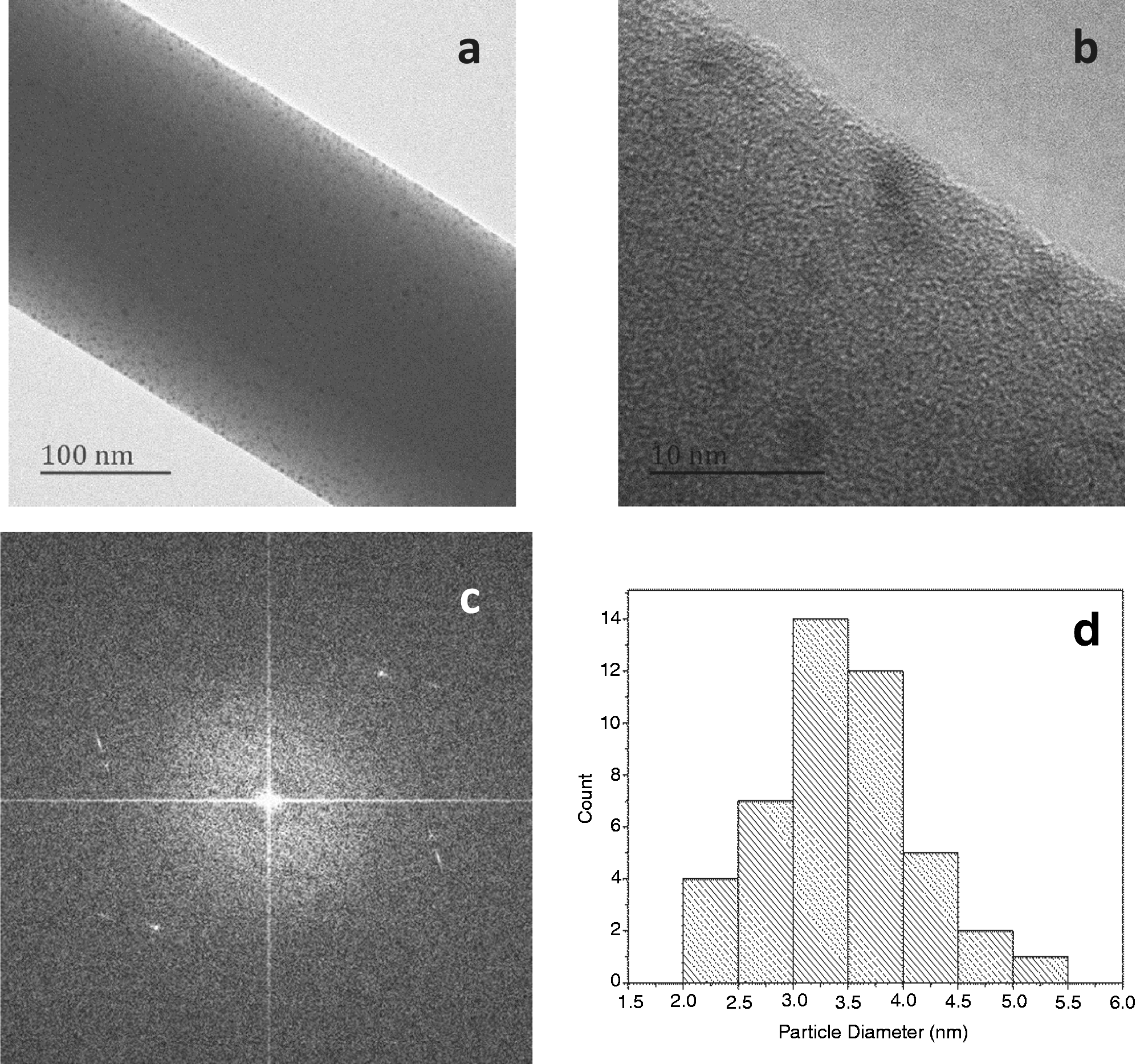
From the TEM image of Ag-chitosan/PEO nanofiber in Figure 2(a) and (b), Ag-NPs were distributed throughout the fiber, with the average diameters of these particles being 3.5 ± 0.6 nm (Figure 2(d)). The electron diffraction analysis of the particles was shown in Figure 2(c).
UV–vis spectra (a) and maximum absorption wavelength (b) of Ag-chitosan/PEO solutions irradiated by UV light for different time. TEM images of electrospun nanofiber from Ag-chitosan/PEO solution that has been UV irradiated for 30 min (a, b), and the electron diffraction image of the silver nanoparticles (c) and the histogram of silver nanoparticle diameter (d).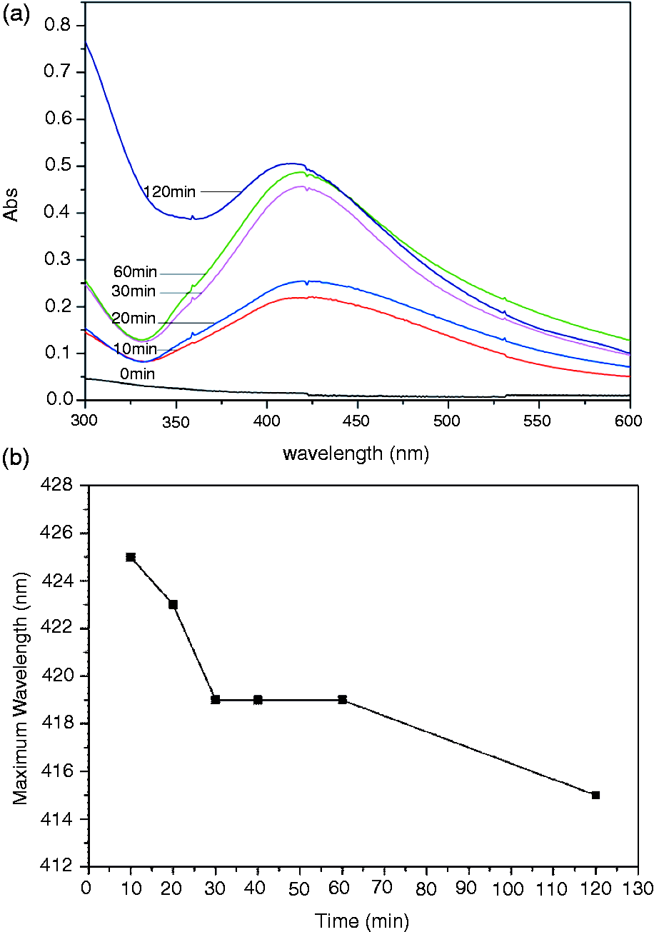
Characterization of Ag-chitosan/PEO nanofibers
As shown in Figure 3(1), the obtained fibers had a cylindrical appearance with no fiber bundles, which implied that the process parameters were suitable for fiber formation. In addition, the morphology of the electrospun nanofibers was slightly affected by the silver. As the content of AgNO3 increased, the average diameter first decreased and then increased. The decrease of the fiber diameter was likely attributed to the increased conductivity of the electrospinning solution resulted from the presence of silver ions. However, the viscosity became higher, when more AgNO3 was added into the solution, which was probably responsible for the increased diameter. The EDX spectrum of 8% AgNO3-chitosan/PEO nanofibers is shown in Figure 3(2). The results indicated that carbon, oxygen, and silver were the principal elements of the nanofiber composites. EDX analysis provided direct evidence that Ag-NPs were embedded in the chitosan/PEO nanofibers.
(1) SEM images of Ag-chitosan/PEO nanofibers prepared from solutions containing various content of AgNO3: 0% (a), 2% (b), 4% (c), 8% (d), and (2) EDX spectrum of nanofibers electrospun from (d).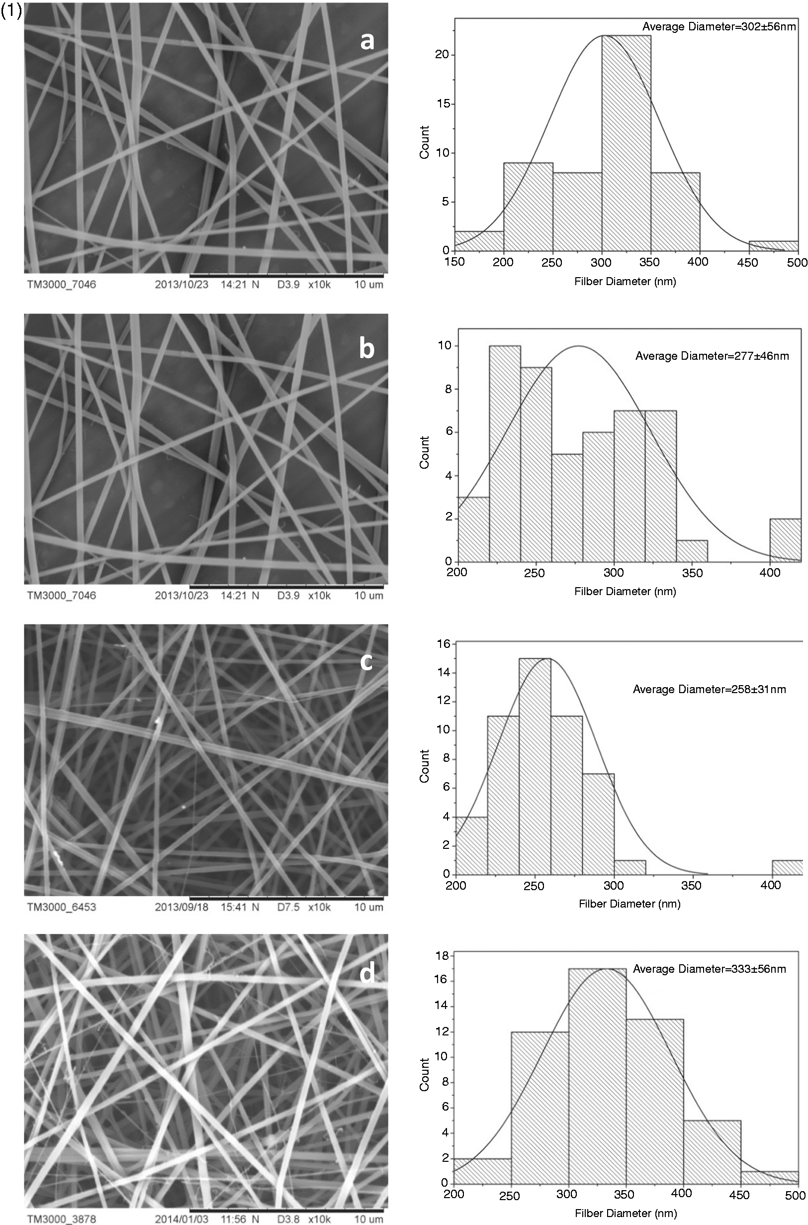
XRD was applied to confirm the presence of Ag-NPs and their crystal structure in the fibers obtained from Ag-chitosan/PEO solution after UV irradiation. The XRD spectra of chitosan powder, chitosan/PEO nanofiber, and Ag-chitosan/PEO nanofiber are displayed in Figure 5, respectively. From this figure, it can be seen that the peak with 2θ values at 20.3° was corresponding to the chitosan crystal structure (Figure 4(a)), which disappeared in chitosan/PEO blended nanofibers. It indicated the formation of hydrogen bonds between PEO and chitosan, and the excellent polymers compatibility in the blends (Figure 4(b)). The reflections with 2θ values at 36.24° and 45.8° were corresponding to the (111) and (200) crystal faces of silver, respectively.
XRD patterns of chitosan powder (a), chitosan/PEO nanofiber (b), Ag-chitosan/PEO nanofiber from 8% AgNO3-containing solution after UV irradiating for 30 min (c). FTIR spectra of chitosan/PEO nanofiber (a) and Ag-chitosan/PEO nanofiber prepared from the chitosan/PEO solution containing 8% AgNO3 (b).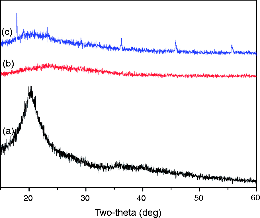
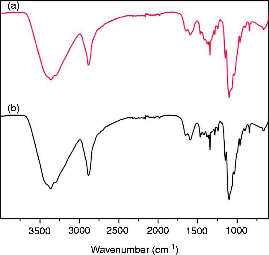
FTIR was employed to investigate the effect of silver on chitosan/PEO nanofibers. The FTIR spectra of chitosan/PEO nanofibers with and without silver are presented in Figure 5. All the peaks were almost identical except in the range of 1464–1281 cm–1, suggesting that the silver addition had little effect on the chemical structure of the resultant nanofibers.
Mechanical properties analysis
Mechanical properties of randomly oriented chitosan/PEO nanofiber membranes containing varied amount of AgNO3. Data are representatives of five independent trials and all the data were expressed as mean ± SD.
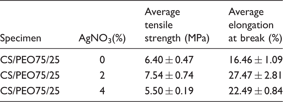
With more AgNO3 added, the elongation of the nanofiber composites increased, while the tensile strength first increased then decreased. The possible reason was that, with the appropriate increment in AgNO3, the excellent crystallinity of Ag-NPs enhanced the mechanical properties of composites. The nanofiber composite containing 2% AgNO3 showed the highest tensile strength (7.54 MPa) and the largest elongation (27.47%). The electrospun nanofibers exhibited good mechanical property, which could meet the demand of practical uses.
Antibacterial activities analysis
The antibacterial activities of chitosan/PEO nanofibers mats containing various amounts of AgNO3 were examined against Gram-positive S. aureus and Gram-negative E. coli by zone of inhibition tests.
As shown in Figure 6, only a narrow inhibition zone was observed around the nanofibers mat produced from the solution without AgNO3. However, there were clear and fair-sized inhibition zone around silver loaded nanofiber composites, which suggested that silver loaded nanofibers could inhibit the growth of bacteria. Firstly, the Ag-NPs entered the pathogenic bacteria combining with the cell membranes and then they combined with the thiol of the oxygen metabolism enzyme quickly to make the respiratory enzyme inactivation. The respiratory metabolism of the pathogenic bacteria thus is blocked and choked to death. As the particle diameter decreased, the particle specific surface increased, conducive to the adsorption of bacteria and action with the thiol to kill the bacteria. On the other hand, the decreasing of the silver particle was helpful for dissolution of silver ions and inhibition of the growth of bacteria.
(a) Inhibition zone of Ag-CS/PEO 75/25 nanofibers against S. aureus and E. coli, where SA stands for S. aureus and EC stands for E. coli (0) 0% AgNO3; (1) 4% AgNO3; (2), (3) 8% AgNO3. (b) Calculation of inhibition zone diameters in (a).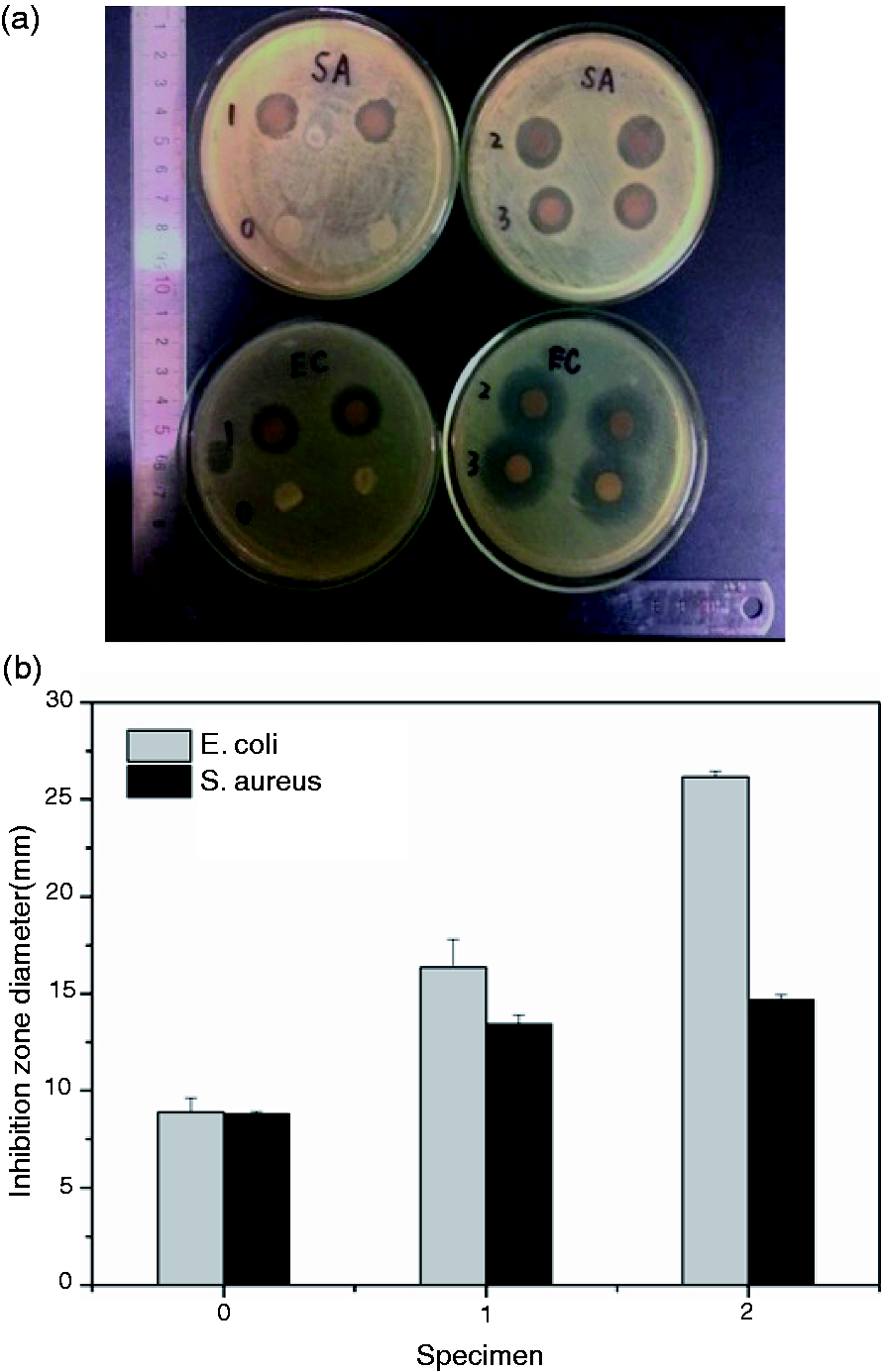
The results showed that the inhibition zone against E. coli was generally bigger than that against S. aureus, demonstrating that silver is more effective to inhibit E. coli. It is mainly because of the structure differences on the cell wall of the gram-positive and gram-negative bacteria. The cell wall outermost layer of gram-negative bacteria is a layer of lipopolysaccharide, and the inside is a thin layer of peptidoglycan (7–8 nm). The lipopolysaccharide is covalently connected by the lipids and polysaccharide. The overall cell wall of the gram-negative bacteria is lack of thickness and hardness. However, the cell wall of gram-positive bacteria is mainly composed of peptidoglycan. The linear peptidoglycan is connected into reticular cross-linked three-dimensional structure by a mass of small peptides, resulting in the thicker and denser cell wall than the gram-negative bacteria. The Ag-NPs are difficult to pass through the positive bacteria cell wall. Therefore, the inhibiting or killing actions of the Ag-NPs to gram-negative bacteria obviously are stronger than the gram-positive bacteria.
Moreover, the more silver was contained in the fibers, the bigger inhibition zone was formed around the specimen. It indicated that the nanofibers possessed the stronger antibacterial activity.
Release of silver from the Ag-chitosan/PEO nanofiber mats
The silver release activity from the chitosan/PEO nanofiber membranes was investigated over 72 h. The release profiles of silver are shown in Figure 7. The nanofiber mats loaded with different amounts of silver showed similar release profile trend, with a concentration above 0.1 ppb. Both of the mats exhibited an initial relatively fast release in the first several hours, and almost 10 h later, the release speed slowed down and went to steady along with the incubation time (72 h). The initial fast release was probably attributed to the diffusion of silver near the surface of the fibers, followed by dissolution of PEO. The initial fast release is helpful to kill or inhibit the growth and proliferation of pathogenic bacteria on the wound immediately. Silver remaining in the fiber would release slowly for a lasting antibacterial activity 10 h later.
In vitro release profiles of Ag-chitosan/PEO nanofibers from 4 and 8% AgNO3-containing solution after UV irradiating for 30 min.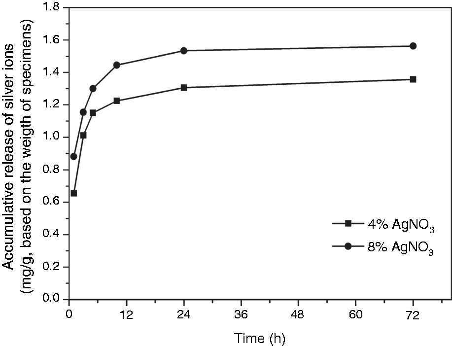
Cytotoxicity analysis
An ideal wound dressing should be nontoxic to the tissue, which can be evaluated through in vitro cytotoxicity assays. In the evaluation, tissue culture plates (TCPS) were used as positive reference. The results of ALB assay illustrating the viability of PIEC cultured with the specimen are shown in Figure 8. No significant differences were observed in cell activity of PIEC culture for 24 h in the presence of chitosan/PEO nanofiber mat and Ag-chitosan/PEO nanofiber mat in comparison with positive reference, although the average absorbance values were lower than that of the control condition. Both absorptions of chitosan/PEO and Ag-chitosan/PEO nanofiber mats were above 90% compared with that of the positive control. The results indicated that the obtained Ag-chitosan/PEO nanofiber mats were less toxic to PIEC.
Cytotoxicity assays of chitosan (CS)/PEO and Ag-chitosan (CS)/PEO nanofiber mats with positive controls. The data represented mean and standard deviations of 12 samples.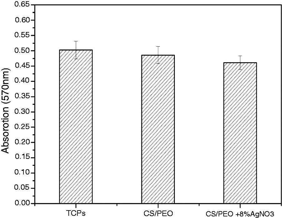
Conclusions
Nanofiber mats of high chitosan/PEO ratio 75/25 embedded Ag-NPs were successfully fabricated by electrospinning. PEO was miscible with chitosan and had good electrospinnability when blended with chitosan, which could not be electrospun alone. The addition of AgNO3 to the polymer blend solution of PEO and chitosan improved the electrospinnability of the blend. The formation of Ag-NPs with diameter about 3 nm dispersed throughout the e-spun fibers was confirmed using different analytical tools. The Ag-NPs showed obvious antibacterial activity and acted as a nucleating agent during cold crystallinity. The silver in the chitosan/PEO blend solutions resulted in an increase in the mechanical properties of the chitosan/PEO nanofiber mats. The antibacterial experiment indicated that the nanofibers mats of Ag-chitosan/PEO had good bactericidal activity against both the gram-negative bacteria E. coli and gram-positive bacteria S. aureus. The results of silver release from the nanofiber composites showed that the nanofibers exhibited an initial relatively fast release in the first several hours, which is helpful to inhibit the growth and proliferation of pathogenic bacteria on the wound immediately. The release speed slowed down and went to steady along with the incubation time (72 h) about 10 h later, which could be beneficial to a prolonged antibacterial activity. At last, the in vitro cytotoxicity assays indicated that both the chitosan/PEO nanofiber mats with or without silver were less toxic to PIEC. So the whole results suggested that the obtained Ag-chitosan/PEO nanofibers mats were good potential candidates as wound dressing.
Footnotes
Declaration of conflicting interests
The authors declared no potential conflicts of interest with respect to the research, authorship, and/or publication of this article.
Funding
This work was financially supported by the “China Postdoctoral Science Foundation” (NO. 2013M530165) and the “Fundamental Research Funds for the Central Universities.”