
Abstract
Polyelectrolyte complexes represent a special class of polymeric compounds consisting of stoichiometric equivalents of oppositely charged polyions interacting together spontaneously to yield a complex in different forms. The present study aimed at preparing coacervates of alginate and chitosan polymers ready for casting as wound dressing films. This was based on controlling the pH of solutions and the reactions speed through controlling the rate of mixing of the polymers solutions together without using any water-miscible solvents. Alginate was modified with radiation and oxidation, and the interactions of the resulting chains and chitosan chains were tested with FTIR spectroscopy and scanning of the resulting films with SEM. This work showed the ability to prepare a complex of highly connected polymeric chains for further biomedical applications. This complex in the form of hydrogel could enhance the proliferation of cells in vitro and the healing efficiency with accelerating the wound closure rate as evidenced through the histological observations.
Introduction
Wound healing is a complex and dynamic process that results in the restoration of anatomical continuity or at least securing the damaged tissues, and it functions through a predictable chain of molecular events involving complex interactions among extracellular matrix molecules, soluble mediators, resident, and infiltrating inflammatory cells. 1 Today, there is a wide variety of dressing products including film, simple island, nonadherent, moist (hydrogel and hydrocolloid), absorbent (hydro fiber and foam), and composite dressings. The polysaccharides, chitosan (Ch), and alginate (Alg) are ideal materials for constructing dressings suitable for accelerating wound healing during its various phases.
Ch is a pseudonatural glycosaminoglycan of superior biodegradability, low-toxicity with bioabsorbable, and easy chelating power. It possesses rare bioactivity and biocompatibility 2 and has become important biomaterial for wound management where it was observed to have hemostatic properties independent of the normal clotting cascade 3 and can promote normal histoarchitectural organization, enhance tensile strength of wounds, accelerate the infiltration of inflammatory cells, 4 and activate the migration and proliferation of fibroblasts in the wound area through the induction of interleukin release as well as keratinocytes. 5 Due to their antimicrobial activities, dressings of Chs with bacteriostatic and fungistatic activities were prepared.
Alg is an unbranched copolymer with repeated two kinds of 1 → 4 covalently linked monomers: β-
The polyelectrolyte complexes (PECs) have been studied since the early 1960s, and the nature and the extent of such reactions and their products were proven to be sensitive to the molecular properties of the reacting polymers including molecular weights, structures, and the complexation conditions (e.g. temperature, pH, moisture content, mixing ratio, ionic strength of the system, cross-linking agents, and period).10,11 One of the most common uses for PECs made of Alg/Ch is the synthesis of wound dressings, which may be in the form of Alg/Ch composite with cotton, Ca-Alg filaments coated with Ch, or sponge dressings. Based on previous reports,12–14 addition of water-miscible organic solvent (e.g. acetone, ethanol, or PEG200) into the Ch solution was proven effective to slow the rate of coacervation. They have lower polarity, and so the Ch chains will assume less extended conformation, and this will restrict the fast reaction between the two polyelectrolytes and so perfect complexation can occur.
The aim of the current study was to use the least number of chemicals, which may cause skin irritation, inflammation, or ulceration on application of the prepared hydrogel film to the wound with a main dependence on the physical activity for mixing the two polymers to produce a high quality of PECs through the coacervation process. Thus, controlling the reactions speed through controlling the rate of mixing of Ch and Alg solutions together along with certain solutions pH values is undoubtedly the pivotal point for success.
Materials and methods
Modification of sodium Alg
For a sharp decrease in molecular weight of Alg, high molecular weight (HMW-Na-Alg), medium viscosity, and M/G ratio = 1.56 (Sigma-Aldrich, UK) was mixed with H2O2, 10 wt % to obtain in the form of a paste that was then packaged tightly in polyethylene bags and subjected to γ-irradiation at 50 kGy at RT on air from Cobalt-60 (Gamma-Cell 220), Atomic energy of Canada Limited, installed at the Radioisotope Department of the Egyptian AEA, Cairo-Egypt with dose rate 5.6 kGy/h.
HMW-Na-Alg was oxidized according to the protocol of Bouhadir et al.
15
using Na-periodate (LOBA-Chemie, India) (0.25 M). NH2OH.HCl reagent (0.25 M) was prepared freshly according to Zhao and Heindel,
16
and the degree of aldehyde substitution was monitored through potentiometric titration that was determined by the following equation
Characterization of the resulting Algs
After dissolving in 0.01 M NaCl, the viscometric method for determining the viscosity of the resulting Alg solutions was applied, and the viscosity average molecular weight of the polymer (Mv) was calculated on the basis of Mark–Houwink–Sakurada equation, 17 while the number–average molecular weights (Mn) and weight–average molecular weights (Mw) of raw and modified Algs were measured by gel permeation chromatography 1100 Agilant instrument.
Preparation of Alg/Ch PEC membranes
Homogeneous Ch solution in acetic acid (Sigma Aldrich, UK, 0.5 wt%, pH 4.8) was prepared. Alg solutions of equal ratios of the non-modified, irradiated, and oxidized Algs (0.5 wt%, pH 6.84) were prepared in 10 mM NaCl. All preparations were fresh to avoid any intrinsic changes of the polymers solutions with time and were autoclaved before using. Twenty-five milliliters of Ch solution (0.5 wt%, pH 5) was added dropwise to 25 ml Alg with continuous gentle shaking till the ending of the coacervation reaction with the formation of PECs. Few drops of CaCl2 solution (2%) were added dropwise with continuous shaking to prepare the semi-IPNs. After the ending of reaction, selected suspensions were kept aside for some time till the removal of air bubbles, casted into glass Petri dishes, and allowed to dry at 40℃ for 2 days. The dried hydrogels were then packaged in polyethylene bags under nitrogen atmosphere and irradiated with γ-irradiation at 25 kGy.
Characterization steps for the resulting hydrogels
FTIR-spectroscopic characterization
KBr pellet method was used as previously described, and experimental spectra for the prepared films were obtained by mixing dried film with KBr. Pellets of the physical mixture of the used powder Algs and Ch (1:1) were also obtained with KBr.
Scanning electron microscopy
Representative air-dried samples from the normal and irradiated PEC sheets were analyzed by SEM. The films were sectioned with blade and mounted on an aluminum sample support by means of conductive and double-sided adhesive tape before sputter-coating with ultra-thin gold layer (300 Å) under vacuum in a Polaron E-coating apparatus. Morphologies of the film surfaces were then analyzed using JEOL-JSM-5400 Scanning Electron Microscope Japan at an accelerating voltage of 30 kV.
Fibroblasts cell culture
The ability of the PEC membranes to support cell growth and proliferation was determined by sterilizing small pieces and casting individually in 24-well culture plates. About two milliliters of culture medium containing human fibroblast cells (MRC-5) at a concentration (40,000 cells/ml) was seeded in each well with incubation at 37℃ in 5% CO2. The cells were observed with an inverted microscope after one and three days of incubation with the changing of the medium every 24 h. The cellular proliferation was assayed by 3-(4,5-dimethylthaizol-2-yl)-2,5-diphenyl-2 H-tetrazolium bromide (MTT) assay after one, two, and three days of incubation. 18 The amount of MTT–formazan product released was measured at 590 nm on a micro-plate reader with values reported as mean optical density ± SEM.
In vivo wound healing
All cooperated steps using adult male albino rat model (Rattus norvigicus) were in accordance with the National Institute of Health, Egypt. At first, the rats (weight: 120–150 g) were lightly anesthetized with diethylether, and then the dorsal surface skin was digitally shaved completely and cleaned with alcohol (70%). The rats were returned to their cages, each one in individual cage for 24 h. Application fields were then outlined with a marking pen, and single right full thickness wound of area (1 cm2) was created by circular excising in the dorsal skin and the underlying panniculus carnosus using toothed forceps and scissors. The day of skin excision was defined as D0, and the animals were then randomly assigned into the following two groups: rats of the first group were treated with the chosen hydrogel and fixed with plaster. Rats of the second one were only disinfected with ethanol and left untreated (Control group). Daily, the wounds of all groups were grossly monitored for infection, and rats with its signs were excluded. Photographing of the wounds was performed during the post wounding days from a standard height to monitor any changes during healing using a digital camera (Samsung, South Korea). The wound areas were measured daily using marker and tracing paper method. The wound tissues, including the scab and complete epithelial margin of the control as well as the dressed wounds with the hydrogel dressing remnants, were rapidly extirpated during different post-wounding days with the surrounding skin tissue at the edges, and a supporting layer of body-wall musculature as a mass was preserved in 10% (v/v) formal-saline for subsequent staining with haematoxylin (H), eosin (E), and Masson’s trichrome (MT).
Results and discussion
Characterization of the modified Algs
Mv, Mn, Mw, and aldehydes analyses (formyls/mol of Alg)) for the modified Algs.
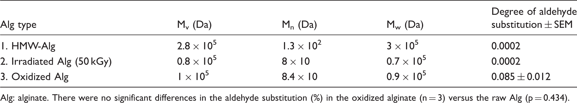
Alg: alginate. There were no significant differences in the aldehyde substitution (%) in the oxidized alginate (n = 3) versus the raw Alg (p = 0.434).
Characterization of the prepared hydrogels
FTIR spectroscopic characterization
FTIR spectroscopy has been used to test and explain the interactions between functional groups of the oppositely charged polyions. When two immiscible polymers are brought together, the resulting IR spectrum will be expected to be the sum of spectra for the individual compounds as they will have the same environment of their pure states.
19
When they are by contrary miscible, intermolecular interactions may occur, and this will be reflected in changes on the IR spectra of the mixture such as wavenumber shifts, band broadening, and the appearance of new absorption bands as evidences of the polymers miscibility. The FTIR-spectra for the physical mixture of Algs, Algs with Ch, and the irradiated PECs membranes are shown in Figure 1. Many free (Na-Alg) characteristic peaks appeared with the chart of the PEC (e.g. the asymmetric stretching mode of CH2 group at 2931 cm–1).
20
The peaks for C–O stretching vibrations of the PEC appeared at 1084 and 1039 cm–1 without any differences from their locations with the physical mixture of the polymers. Shifting occurred for the band 1432 cm−1 with the physical mixture of the polymers to 1415 cm−1 with the PEC chart indicating the deproteination of most Algs along with 1262 cm–1 band with the binding of Alg with NH3+ groups of Ch via hydrogen bonding. The characteristic amide III band of Ch at 1326 cm–1 disappeared with complexation. In addition, the irradiation activated the formation of free radicals, which induced the binding of -OH groups together as well as with NH3+ groups of Ch causing shifting for the stretching peak of -OH groups to appear at 3444 cm–1. No such absorption peaks were observed for the simple mixture of polyions.
FTIR spectra for alginates mixture, Algs/Ch mixture, and the PEC membrane. Alg: alginate; Ch: chitosan.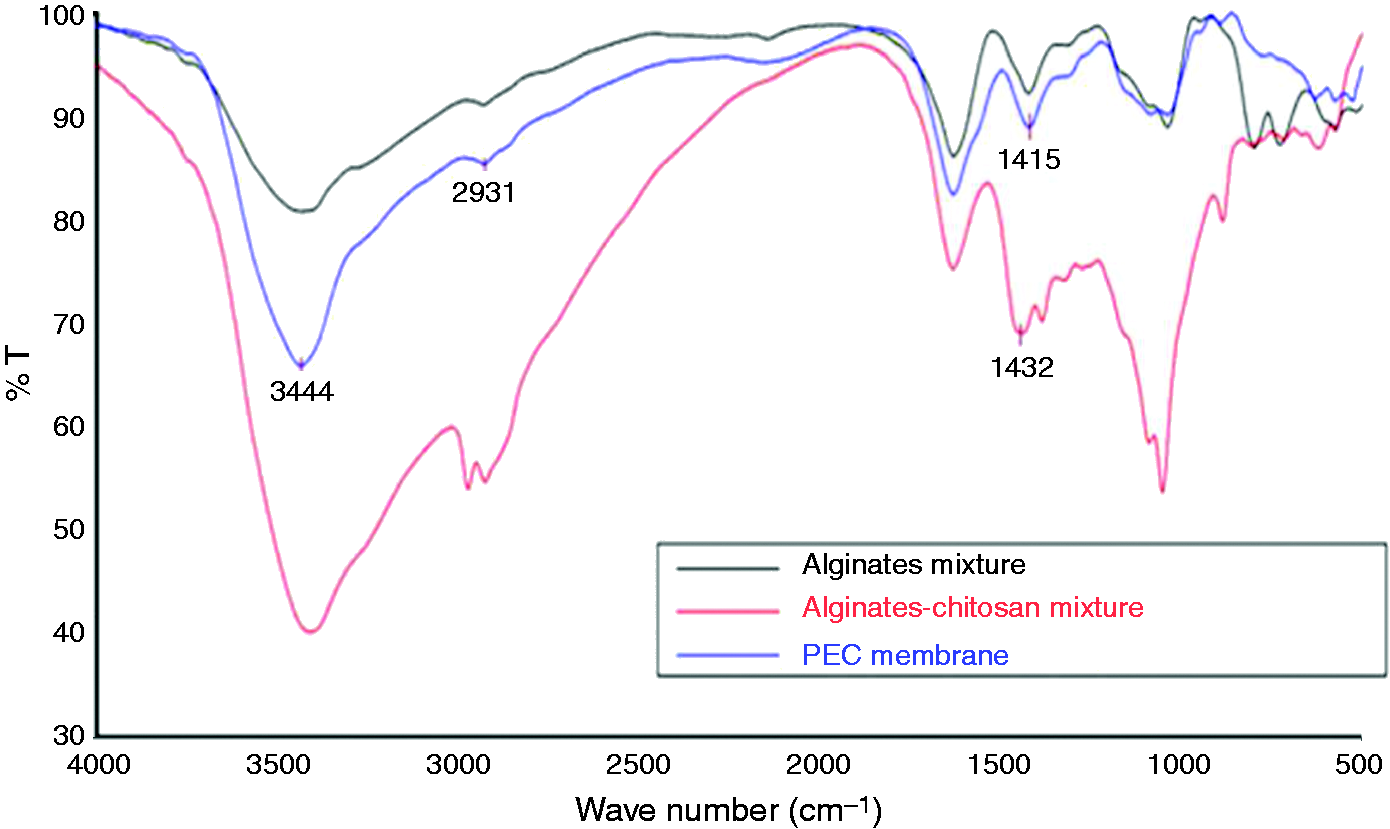
Scanning electron microscopy
The SEM images for the irradiated PECs (Figure 2) spot changes in the morphological appearance of the prepared membranes of F-IV and its corresponding Ca-free gel (denoted: F-IV′) with studying the effect of cross-linking. Figure 2(a) shows the micrograph of F-IV [Algs + (CaCl2) + Ch] membrane where changes in the surface were captured due to complex formation under irradiation using γ-rays. The dose of 25 kGy was applied beneficially for the presence of Ch in the structure as a natural polymer that might degrade under high doses. A rough, porous net structure was formed indicating the interaction of Ch with Alg under γ-rays with the formation of breathable structure indicating compatibility and proved that this membrane can withstand mechanical action for the dressing purposes. The scanned surface with the taken shot (mag. 1500; Figure 2(b)) may be due to rupture of the strong bonds between the polymers chains on preparation of the films for the SEM observation. Additionally, Figure 2(c) for the same formula membrane shows big to medium clusters, with porous layer form. Figure 2(d) illustrates post irradiated physical gel (F-IV′) membrane and shows small clusters in semi uniform distribution that implies interaction and slight homogeneity; however, increasing of the surface roughness was detected without the formation of a uniform permeable network with weak structure. In general, morphological differences could be spotted easily between the two membranes (Alg/Ch/CaCl2 and Alg/Ch/CaCl2-free) (Figure 2(a) to (d), respectively). Alg/Ch/CaCl2 membrane showed a more uniform network structure, while the other membrane indicates less homogeneity and porosity.
SEM micrographs showing the prepared membranes for the formulae: (a) F-IV, Mag. 1500, (b) F-IV, Mag. 1500, (c) F-IV, Mag. 750, and (d) F-IV, Mag. 1500.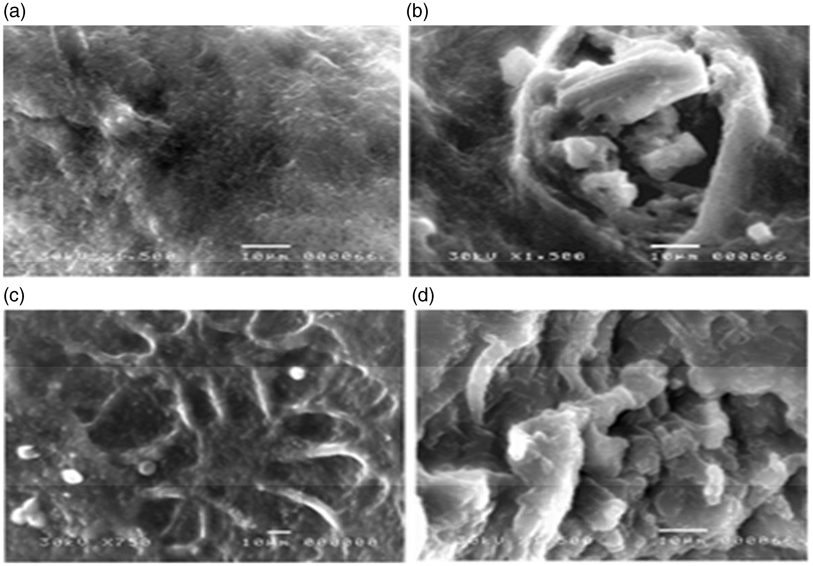
Polyelectrolyte titration method was used to prepare the PEC from Alg/Ch. Concentration of the polyelectrolyte in the beaker, Alg, into which the Ch is titrated, changes during titration. First, the solvent of the titrant dilutes the solution, and second, the Alg in the beaker is consumed by the ongoing complexation process. To counteract this dilution, the first polyelectrolyte in beaker sometimes has a 2–10-times higher concentration than the other one. To circumvent that during the study, the reaction was stopped directly after the observation of coacervates formation, and so the used volumes of Ch and Alg were not equal. Specific concentrations of the reactants were used to prevent the precipitation of either one out of the formed complex. Under certain dilution conditions, the prepared complex disperses homogeneously as small particles in solution, which finally grow to fine fibrous structures on allowing to stand for few days at certain temperature range. 21
Rapid coacervation for the reacting polymers at definite pH values induces the formation of dense interphasic PEC membranes that separate the polymer solutions and prevent further reaction, and this is the common employed technology to prepare microspheres of Ch– Alg PECs.22,23 This technology was not used in the present study as it was unsuitable to produce coacervates for casting into films. The extensive development of these intermolecular interactions during films preparation would generate viscosities too high to distribute or matrices too solidified to flow. To prepare coacervates for casting into homogeneous films, the rate of reaction between the two polymers must be sufficiently slow to prevent the formation of such membranes, so that the reaction can complete.
The high shaking rates with the titrant polymer solution addition promote foam formation with opaque sheets having impaired barrier properties, more permeability to gases, in addition to forming some suspensions of soluble complexes with no ability to coacervate. The weak speeds lead to fast coacervation with no ability to form gels. Accordingly, the moderate shaking and mixing speeds (about 50 RPM) with controlled addition rate (about 300 µl/min) were perfect to prepare ideal PEC suspensions of Alg/Ch despite taking some time. The resulting hydrogel films were transparent that allowed effective observation of changes in the wounds during skin repair.
Multifaceted structure may have been formed when a Ch drop of certain concentration fell into Alg solution at the desired pH values. Complexation may have occurred between the two oppositely charged polyelectrolytes through the electrostatic interactions. By this way, complexation between the common free (-COO−) groups of Alg chains and the Ch protonated amino groups (NH3+) occurred and Ch may have acted as the insolubilizing cation for the production of Alg–Ch physical complex.24,25 With the addition of CaCl2 into the mixture during complexation, it may have diffused into the Alg core more rapidly than Ch because of its lower molecular weight to form a gel core. Calcium cross-linking for Alg helped in its stabilization with forming a Semi Interpenetrating Network (Semi-IPN) of pH sensitivity and reversible properties that were proved later through the characterization tests.
pH is the most pivotal factor affecting the strength and directing the formation of Alg/Ch physical hydrogels with retaining the stoichiometric composition of strong charged polyelectrolyte (i.e. strong polyacid-polybase complex). 21 In addition, it controls balancing of charge thus limiting repulsion without net charge of zero to allow surplus electrical charging of the resulting coacervates, which is necessary for the solubilization of particles. 25 Generally, at pH < 3.5, the carboxylate groups of Alg become less ionized with protonation to form alginic acid physical gel clusters through inter-chain interactions. The increase in pH makes free (-COO−) on the polyanion chains. At pH > 8, the high concentration of Na+ ions restrains the extension of the tangled molecular chain on the hydrogel; therefore, its swelling may inhibit 26 Ch precipitates above its pKa value of 6.3. Charge density of the Ch molecules is reduced with increasing the pH of solution, and the polymer chains become less extended. 28 As pH increases from 4.5 to 6.2, the ionization degree of Ch decreases from 1.0 to 0.5, the amino groups become less charged with reduction in the charge density by almost 50% resulting in expected fewer ionic linkages between any negatively charged polymer and Ch. In contrast, the chains become less extended with these changes at pH 6.2 with smaller radius of gyration and enhanced diffusion coefficient into the (Alg/Ch network) causing stronger combination with (Alg-chains) during a specified reaction time than the linkages of the extended chains at pH 4.5. Accordingly, PECs formed of Ch at pH 6.2 have lower DS than the complex formed at 4.5. Raising of the pH of Ch solution from 4.5 to 5.5 with a corresponding decrease in its total charge was observed to have no significant effects on DS of the complex gel. 29 As a conclusion from our experimental trials, mixing Alg solution (pH 6.84) and Ch (pH 5) at equimolar compositions may lead to reaching a suspending solution (pH 5.28) at which the Ch protonated amino groups spontaneously associate with the negatively charged carboxylate groups of Alg to form the PEC in solution, which then mutually precipitates from it.
The stand-alone films from macromolecules are generally produced by extrusion, deposition, or casting/solvent evaporation. 30 For PEC-film formation, all the involved physical changes must occur in situ during the preparation in order to ensure the formation of the desired complex structure in place within the final film. Generating films by casting melted plastics or preparing bio-hydrogels involves spreading of the film-forming solution onto non-adhering, flat, and smooth substrates, followed by the removal of the solvent usually through a hot air flux. This is the mostly used method in research as it is simple, practical, does not require expensive equipment, and has no adverse effects on properties of the prepared films from bio-macromolecules on keeping under control. During the step of membranes drying, the hot air evaporates water from the outermost region of the nascent membrane to induce phase to coalesce and ultimately fuse to form dense skin, and then the underlying phase separation region tends to coalesce with time and fuse with the dense skin to increase its thickness. 14 The unreacted Alg and Ch molecules have supporting effect to help in forming the PEC coacervates that give the coherent flexible thin membranes. 14 The upper surface of the membrane is responsible for controlling the loss of water through evaporation, inhibition of body fluid loss, and protection from any external contamination.
Fibroblasts cell culture
For detecting the viability of cells on seeding on the prepared hydrogels, the fibroblasts were observed during different culture times (Figure 3). The fusiform appearance of cells was observed after one day of incubation, and they were able to continue proliferation with healthy growth during the third observation day. This may indicate the biocompatibility of the prepared PEC.
Bright field microscopy for cultured fibroblast after one day (a) and three days (b) of incubation with the sterilized PEC hydrogel. The scale bar corresponds to 200 µm.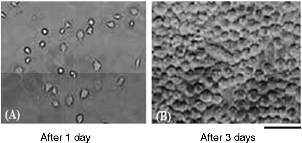
The MTT colorimetric assay was employed as a precise method to study the susceptibility of the fibroblasts to the hydrogel components and determine the viable cell number by measuring the metabolic activity of cellular enzymes. The MTT levels were significantly higher with the cells seeded with hydrogels than the control cells (p < 0.05) (Figure 4). There were higher steady increases in cell number with culture time on the hydrogel versus the cells without hydrogel (control cells).
Assessment of cell viability via MTT assay at days one, two, and three. Values are mean ± SEM (n = 5).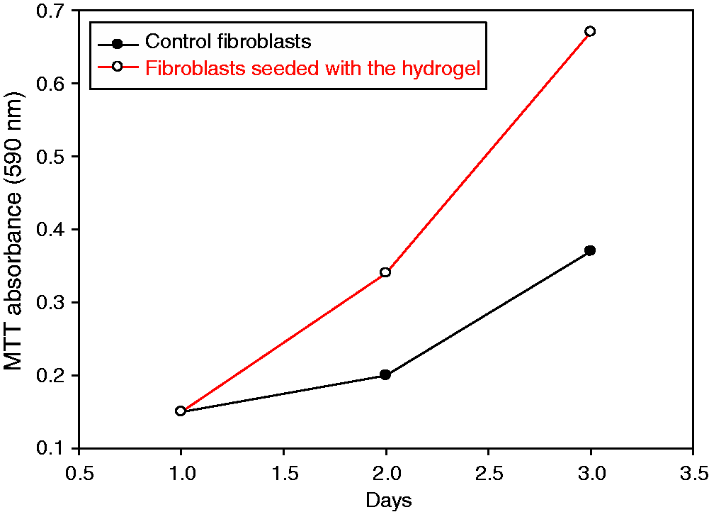
In vivo wound healing
The excision wound model was used. During the first two post-wounding days, no significant differences were observed in wound sizes between the treated wounds and non-treated ones (p > 0.05), but significant differences in rates of wound closure between the two groups appeared with the third day and continued in the following days (p ≤ 0.05). The % reduction for wounds treated with the dressing reached about 50% after three days, 90% after six days, and about 95% after 10 days (Figure 5). The % reduction was only 30% in the case of control wounds after three days and then showed weak reduction in size to reach its maximum (95%) after about two weeks. The faster contraction in size of wounds treated with the hydrogel than the untreated group may be due to the physical forces, initiated by adsorption of various protein molecules from the wound surface into the dressing components, especially Ch fraction.
Rate of closure of wounds, treated with the prepared dressing and the untreated wounds. (*p > 0.05 for control wound versus treated one, Φp ≤ 0.05 for control wound versus treated one).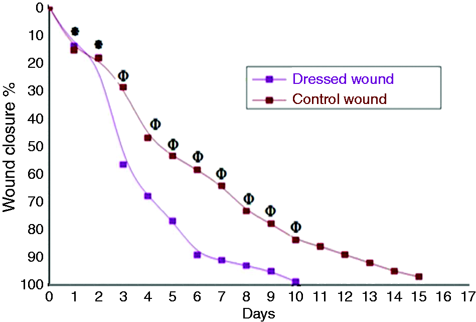
Figure 6 displays that wound healing was grossly normal with complete closure for rats of the Dre-group on the 11th post-healing day. Indeed, the growth of hair started on day three, and its density was significantly heavier in the center of hydrogel-treated wounds than in the surrounding non-wounded skin area at the end of healing days with continuous treatment. No repair occurred completely with the untreated wounds even after 12 post-wounding days. Some rats of this group showed signs of infection with pus secretion, and the skin was hemorrhagic with a few of them.
Representative digital photographs assessment of healing progression during the first 2 post-operative weeks (Con: untreated wounds; Dre: wounds treated with the hydrogel).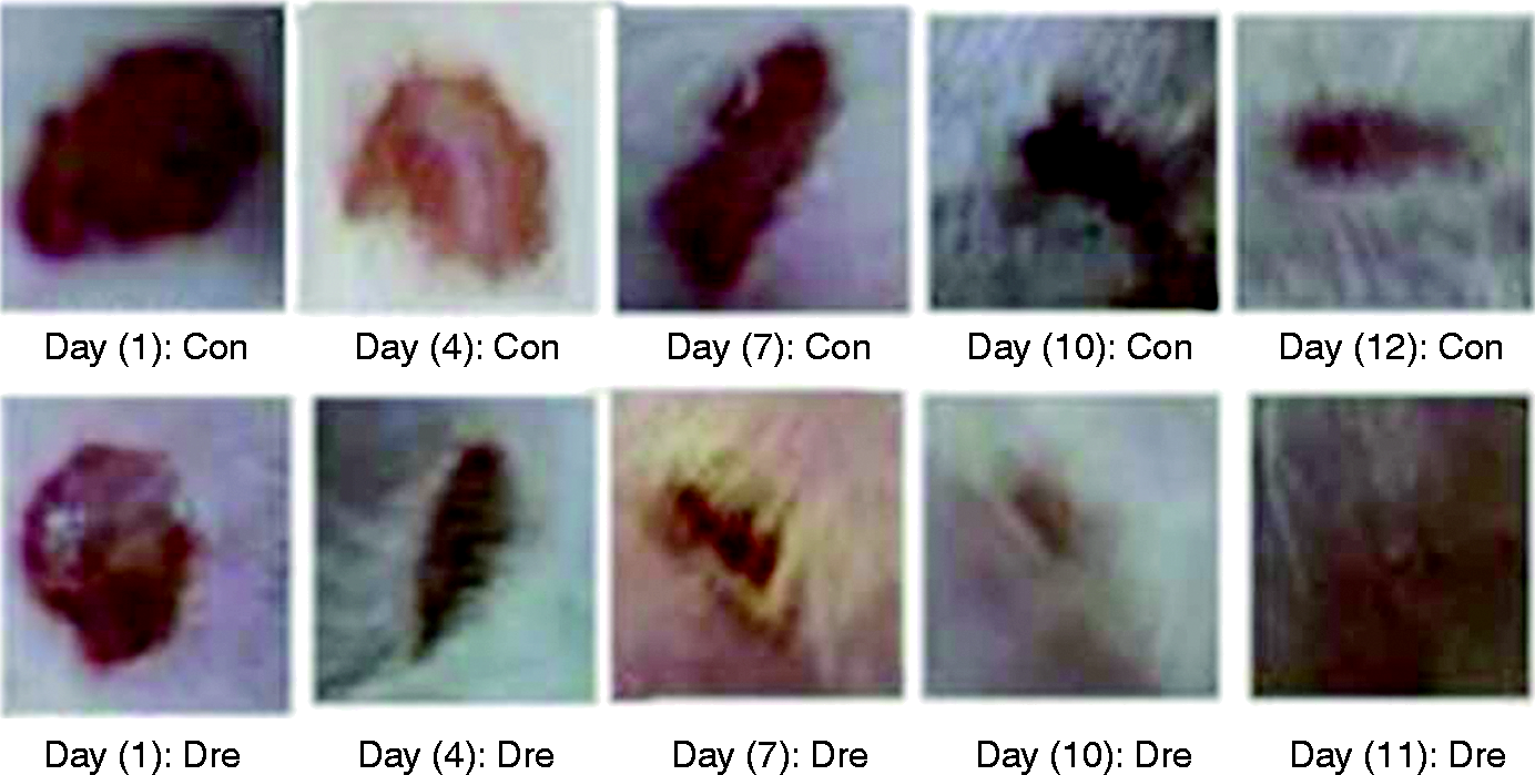
A quantitative assessment strategy for wound-healing cascades was established based on histological criteria. During the 11th observation day, granulation tissues were detected with the new skin in sections of 11C and 11D (Figure 7(a) and (d)). Moderate collagen deposition was observed in the stained 11D sections (Figure 7(e)), but the deposition of massive collagen fibers was observed in the case of stained control healed tissue (not shown) with healing patterns in the epidermal and dermal layers, but with penetration of hemorrhage to the collagen fibers of these stained sections.
Representative images of (H, E, and MT) stained wounds sections. (* refers to the inflammatory cells; arrows: keratinocytes; AT: adipose tissues.)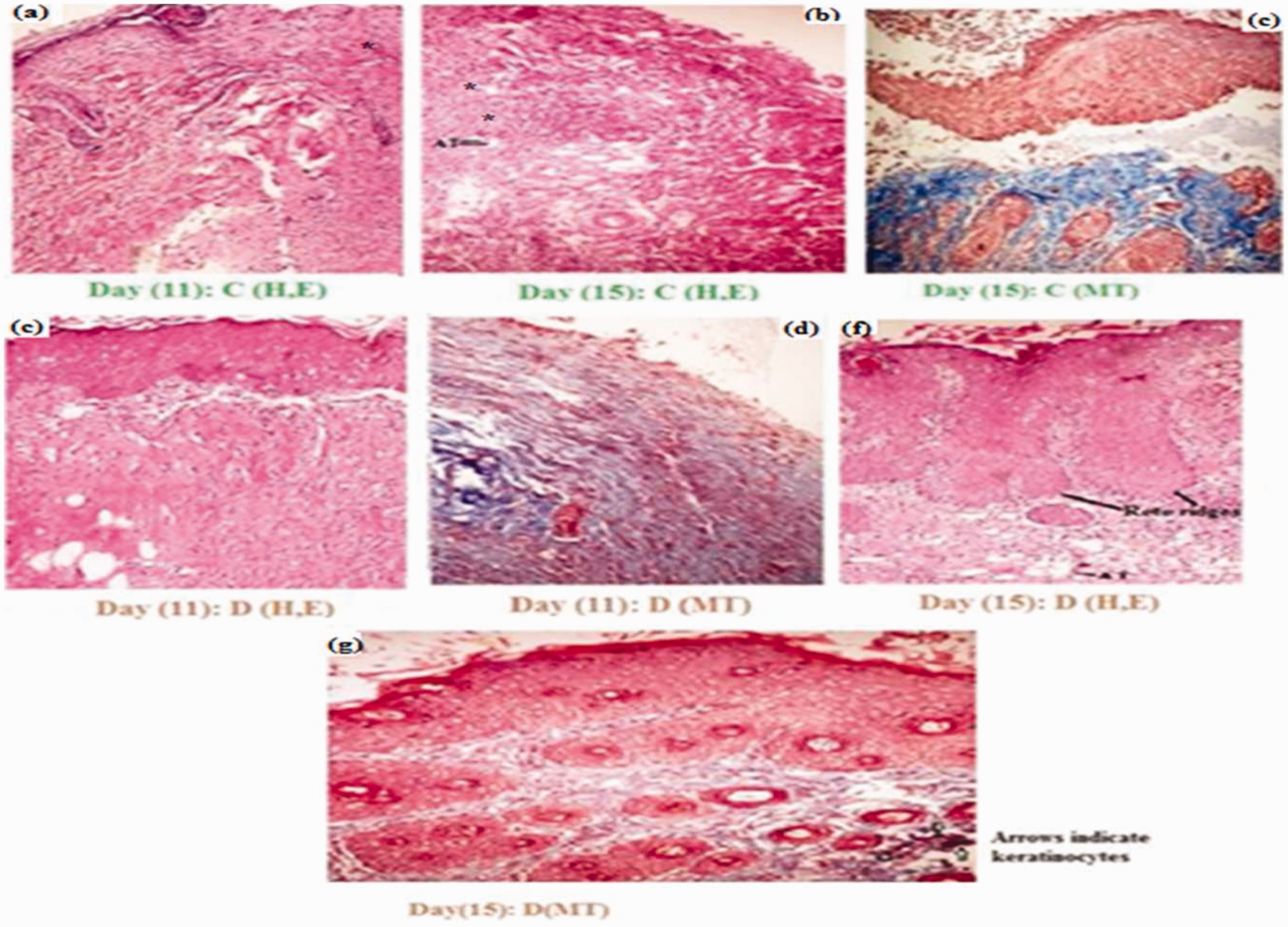
For the 15th day of observation, sections of 15C showed regenerated epidermal and dermal layers with inflammatory cells infiltration, granulation, and necrotic tissue formation (Figure 7(b)). Adipose tissues with sections from both wounds tissues types were observed. The defect area became smaller with the treated wounds with the observation of regenerating proliferated epidermal cells resting on granulation tissue (Figure 7(f)) and absence of inflammatory cells. Marked collagen fibers deposition was observed with the 15C sections of new skin within the hypodermis layer (Figure 7(c)), but few collagen fibers deposition was observed with the 15D sections (Figure 7(g)). These observations may meet an aim of the study for preparing a dressing that allows synthesis of thin collagen fibers with less scar tissue, which can result in a reduction in healing time. Although the collagen deposition with using the hydrogel dressing was massive in the early healing days, its degree decreased with time in accordance with the complete skin regeneration.
Conclusions
A hydrogel matrix of Alg/Ch in the form of PEC was prepared with controlling the mixing speeds between the two polymers solutions and their pH values. The resulting membrane is supposed to have new unique composite properties over the properties of each used one alone but with retaining some of their properties that are suitable for various biomedical and pharmaceutical interests such as enhancing the regeneration of new tissue with wound healing. Details of a biological study for the effects of the hydrogel on rat excisional model will be published elsewhere with a complete daily quantitative assessment strategy for wound-healing cascades based on histological examination for the wound tissue during healing days as well as testing the expression of angiogenic genes to give an overview of the mechanism of hydrogel action in vivo, its behavior with the angiogenesis cascade in wound bed and testing if it can act as a scaffold for the newly growing tissue.
Footnotes
Acknowledgements
The authors have received a research grant from the Egyptian Academy of Scientific Research and Technology (ASRT) as a part of the SNG program, which helped in funding the present research.
Declaration of Conflicting Interests
The author(s) declared no potential conflicts of interest with respect to the research, authorship, and/or publication of this article.
Funding
The author(s) disclosed receipt of the following financial support for the research, authorship, and/or publication of this article: The work was funded and supported by the Egyptian Academy of Scientific Research and Technology as a part of the SNG grant program.