
Abstract
Engineering whole organs from porcine decellularized extracellular matrix and human cells may lead to a plentiful source of implantable organs. Decontaminating the porcine decellularized extracellular matrix scaffolds is an essential step prior to introducing human cells. However, decontamination of whole porcine kidneys is a major challenge because the decontamination agent or irradiation needs to diffuse deep into the structure to eliminate all microbial contamination while minimizing damage to the structure and composition of the decellularized extracellular matrix. In this study, we compared four decontamination treatments that could be applicable to whole porcine kidneys: 70% ethanol, 0.2% peracetic acid in 1 M NaCl, 0.2% peracetic acid in 4% ethanol, and gamma (γ)-irradiation. Porcine kidneys were decellularized by perfusion of 0.5% (w/v) aqueous solution of sodium dodecyl sulfate and the four decontamination treatments were optimized using segments (n = 60) of renal tissue to ensure a consistent comparison. Although all four methods were successful in decontamination, γ-irradiation was very damaging to collagen fibers and glycosaminoglycans, leading to less proliferation of human renal cortical tubular epithelium cells within the porcine decellularized extracellular matrix. The effectiveness of the other three optimized solution treatments were then all confirmed using whole decellularized porcine kidneys (n = 3). An aqueous solution of 0.2% peracetic acid in 1 M NaCl was determined to be the best method for decontamination of porcine decellularized extracellular matrix.
Introduction
Kidney failure is among the leading causes of mortality and morbidity worldwide. According to the National Kidney Foundation, as of April 2015, there are 101,662 patients waiting for kidney transplants. 1 The use of decellularized extracellular matrix (dECM) scaffolds with patient-specific cells is a promising solution for kidney replacement. The dECM is obtained by removing all cellular material from an animal organ to leave the residual inert protein scaffold. Because of the conserved homology in the amino acid sequences of the proteins of various mammalian species, animal ECM proteins elicit a very minor immune response when transplanted into humans. 2 Decellularization of whole rat,3,4 porcine,5,6 and cadaveric human 7 kidneys has been shown to be feasible for producing intact dECM. For example, Song et al., 8 used HUVECs and rat neonatal kidney cells to repopulate whole rat kidneys that were functional after transplantation.
Although effective in removing eukaryotic cells, decellularization processes are not as effective in disrupting prokaryotes (bacteria) due to the cross-linked peptidoglycan surface of their cellular membrane. Therefore, the dECM must be decontaminated before recellularization with human cells. 5 Many decontamination techniques exist in the medical device industry. However, many of these existing techniques produce detrimental effects when applied to protein scaffolds. For example, traditional decontamination techniques such as heating may lead to extracellular matrix (ECM) protein denaturation and thus a loss of function. 9
For the decontamination of tissue, the most common methods are γ-irradiation, ethylene oxide, electrolyzed water, and peracetic acid.10–13 However, many of these methods are also known to cause damage to ECM proteins.14–16 Ethylene oxide can cross-link amino acids, and γ-irradiation can cleave the polypeptide chains.11,17–19 Certain components of the dECM are vital; such as glycosaminoglycans (GAGs), which are necessary for the retention of cellular signaling factors; and fibronectin, which is important for cell adhesion.20,21 To identify a decontamination method for the creation of useful dECM, we compared four methods which are applicable to whole intact renal dECM, and their impact on vital renal ECM components. It is our recommendation that results obtained in this article are only applicable to renal tissue. Methods for decontamination of other solid organs still need to be optimized based on organ-specific composition, structure, and function.
An extensive literature review was conducted and numerous methods for decontamination (e.g. supercritical CO2, electrolyzed water, ethylene oxide, and hydrogen peroxide) were identified. Four previously proposed decontamination methods that were deemed the most practical for our purposes: aqueous solutions of 70% (v/v) ethanol, 22 0.2% (v/v) peracetic acid in 1 M NaCl, 23 0.2% (v/v) peracetic acid in 4% (v/v) ethanol 4 ; and γ-irradiation 6 were chosen for evaluation and applied to small pieces (pieces of kidney were chosen to maintain consistency for comparison tests) of dECM. The resulting collagen and GAGs content and their distribution were compared to determine the treatments that caused the least amount of damage. The potential of decontaminated dECM to support cell adhesion and proliferation was assessed using human renal cortical tubular epithelium (RCTE) cells.
Materials and methods
Kidney retrieval
Porcine kidneys were harvested from 6-month-old slaughter-weight swine at a local abattoir immediately after exsanguination. The kidneys were perfused via renal artery catheterization with 1× phosphate-buffered saline solution (1× PBS) containing 10 U/ml heparin to prevent blood clot formation, and then preserved at −20℃ until decellularization.
Decellularization of whole porcine kidneys
Frozen kidneys (n = 15) were thawed slowly at room temperature and cannulated via the renal artery with white nylon tubing (Value Plastics MTLS210-1, male luer slip to 200 series barbed coupler, 1/16″ tube ID). Kidneys (n = 15) were then perfused at room temperature with 0.5% (w/v) sodium dodecyl sulfate (SDS) under constant pressure of 50 mmHg for 5–7 h at 10 ml/min, depending on the kidney weight. The flow rate was then increased by 2 ml/min every 15 min while the pressure was kept constant. After complete decellularization and achieving a completely white kidney matrix (Figure 1), the kidneys were washed with deionized (DI) water for 2 days at 10 ml/min and 50 mmHg to remove all remaining detergent from the structure and to avoid cytotoxic effects at the time of recellularization.
(a) Schematic diagram of decellularization method. (b) Whole porcine kidneys were decellularized via 0.5% (w/v) SDS perfusion through the inherent vasculature. Completely white kidneys devoid of any cellular components were achieved after 7 h. H&E staining (10× magnification) of native and dECM showed complete removal of all nuclear and cytoplasmic components (insets are 40× magnification). (c) Residual DNA in dECM was less than 50 ng/mg and (d) residual DNA length was less than 200 bp, which are criteria for preventing an immune response (n = 6, data are Ave ± SD).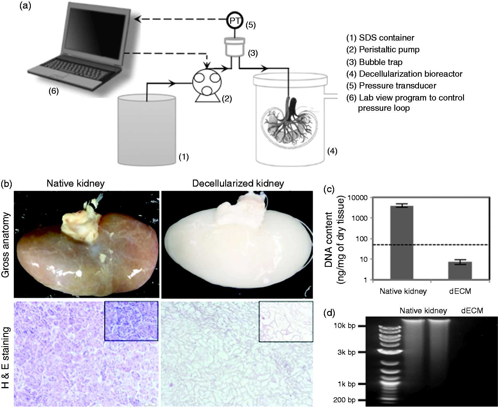
To evaluate cell removal, samples of renal cortex (n = 6) were excised from native and decellularized kidneys, vacuum dried, and weighed. Residual DNA was extracted from samples with the Qiagen DNeasy Blood and Tissue Kit (Qiagen Inc., Valencia, CA) and quantified by Quant-iT PicoGreen dsDNA assay kit (Invitrogen Corp., Carlsbad, CA) according to the manufacturer’s instructions. Briefly, approximately 3 mg of dry sample was solubilized with proteinase K overnight and DNA extracts were collected and washed using several centrifugation steps. Then, PicoGreen reagent was added to each sample and the fluorescence of all samples was determined using a Synergy 2 Multi-Mode Microplate Reader. To characterize the residual DNA in the structure of the ECM, gel electrophoresis was performed for DNA extracts loaded on 1% agarose gel.
Decontamination methods
Slices of renal cortex (7 mm in diameter and 2 mm in depth) were subjected to four decontamination treatments (n = 60 for each method), including γ-irradiation and three decontamination solutions. The solutions are as follows: 70% (v/v) ethanol in DI water, pH: 7.4; 0.2% (v/v) peracetic acid (peracetic acid solution 32% (w/v) in dilute acetic acid, Sigma Aldrich, St Louis, MO) in 1 M NaCl aqueous solution, pH: 3.1, which was freshly made just before each experiment; and 0.2% (v/v) peracetic acid in 4% (v/v) ethanol in DI water, pH: 3.0. Slices of tissues were soaked in 50 ml of the decontaminating solutions under mild shaking for various times (0.5, 1, 2, 3, 4, and 5 h) at room temperature. For γ-irradiation, samples were submerged in DI water in plastic tubes and irradiated at room temperature with intensities ranging from 1 to 10 kGy with a J.L. Shepherd Mark I model 22 self-shielded irradiator. To decontaminate whole porcine dECMs, three whole kidney dECMs that were cannulated through the renal artery were submerged in 3 litres of each type of solution. Then, the solutions were perfused through the kidneys with flow rates of 10 ml/min for 1, 2, and 3 h. The treated dECMs were then perfused 5 times with fresh autoclaved DI water to remove any remaining agent.
Samples were aseptically excised from the renal cortex and incubated in cell culture media to check for contamination. The decontaminated samples as well as non-decontaminated controls were washed with autoclaved DI water and were incubated in Dulbecco’s Modified Eagle Medium (DMEM, Gibco®) supplied with 10% (v/v) fetal bovine serum (GeneMate FBS, Bioexpress, UT) at 37℃ for at least 14 days. The presence of contamination was checked visually under the microscope. The shortest duration of decontamination time or irradiation intensity that produced acceptable samples was used for the remaining tests and comparisons.
Collagen and GAGs quantification
To compare the optimum conditions of the four decontamination treatments, soluble collagen was measured using the Sircol Soluble Collagen Assay Kit (Biocolor Ltd., Newtownabbey, UK) and sulfated glycosaminoglycans (sGAGs) were measured using the Blyscan sGAGs Assay Kit (Biocolor Ltd., Newtownabbey, UK) for all treated and non-treated (control) samples. For soluble collagen, 3 mg of lyophilized tissues (n = 5) were digested overnight in acid/pepsin solution at 4℃. Then a dye–collagen complex was formed and after dissociation of the dye–collagen complex, the absorbance was recorded at 555 nm. For sGAGs determination, each sample (n = 5) was incubated at 65℃ in papain extraction solution for 3 h. Then, the Blyscan dye reagent was added to all samples to precipitate the sGAGs–dye complex and after dissociation, the absorbance was measured at 659 nm using a microplate reader.
Histology and immunohistochemistry
Non-treated control samples and treated decellularized renal cortex biopsies (n = 24) from the four optimized decontamination treatments were prepared and fixed in 4% (v/v) paraformaldehyde overnight and then partially dehydrated in 30%, 50%, and 70% (v/v) ethanol prior to complete dehydration in a graded alcohol series. Afterward, samples were immersed in xylene and embedded in paraffin. Samples in paraffin blocks were sectioned with a microtome into 5 -µm thin slices and placed on slides. Slides were then incubated overnight at 60℃, dehydrated, deparaffinized, and then stained either with Orcein (O7380, Sigma-Aldrich), Picro-sirius red stain (Direct Red 80, Sigma-Aldrich), or IHC WORLD NovaUltra Alcian Blue/PAS Staining Kit (IHC WORLD Inc., Woodstock, MD) according to the manufacturer’s instructions.
Immunohistochemistry was performed on the incubated slides. After being dehydrated and deparaffinized, slides were blocked for 2 h in a 2% (v/v) goat serum blocking solution (Vector Laboratories Inc., Burlingame, CA) on a shaker. A primary antibody against Collagen IV (Abcam, Cambridge, MA, ab6586) was diluted at a concentration of 1:500 in blocking solution and applied to the tissue sections. The following day, slides were incubated with the appropriate secondary antibody diluted to 1:200 in rabbit α-goat serum (Vector Laboratories Inc., Burlingame, CA). Imaging of all stained samples was performed using a standard light microscope equipped with a digital camera (Olympus America Inc., Center Valley, PA) at magnifications of 10× and 20×.
Standard hematoxylin and eosin (H&E) was also used to uniformly stain samples for porosity measurements and scoring the microstructural damage. Porosity fractions of stained samples (n = 5) were determined using ImageJ (NIH) software. 24 Briefly, five random images of each sample were chosen and after adjusting the threshold, the area fraction of void area was calculated using the Analyze plugin. The averages of the final results were reported as porosity fraction of samples. A histologist, blinded to each treatment, performed the evaluation of structural damage using H&E-stained slides. Each slide was examined for general histology, tubules, and glomeruli in five high-powered (20×) fields (three slides per sample). The damage was scored as follows: (5) no structural damage, (4) outline visible/minimal damage, (3) outline visible/moderate damage, (2) outline visible/marked damage, and (1) no outline visible/marked damage.
Scanning electron microscopy
Five-cubic millimeter samples (n = 10) of optimally treated dECMs were incubated in 2% (w/v) glutaraldehyde in Millonig’s phosphate buffer (MPB) at pH 7.3 for 24 h at 4℃. Samples were washed 6 times for 15 min in MPB (pH 7.3). Samples were then left in a cryoprotectant consisting of 25% (w/v) sucrose and 10% (w/v) glycerol in a 0.5 M PBS solution for 2 h. After replacing the solution with fresh cryoprotectant, samples were frozen and fractured in liquid nitrogen. The buffer wash procedure was repeated, followed by 1.5 h in a 1% (w/v) OsO4, MPB (pH 7.3) solution. Samples were subjected to graded dehydration using ethanol baths [10, 30, 50, 70, 95, 100% (v/v)]. Each sample was submerged for 15 min up to the 70% concentration ethanol solution and left overnight at 4℃. The dehydration process was completed in critical dryer baskets with 70%, 95%, and three steps of 100% (v/v) ethanol solution baths. Samples were then placed in a CO2 critical point dryer and subsequently mounted on aluminum stands. Samples were sputter-coated with 15 nm of a Gold–Palladium (Au-Pd) alloy and imaged with a scanning electron microscope (Phillips/FEI XL30ESEMSEG, Hillsboro, OR). Fibers in 40,000× magnified images were characterized using DiameterJ (NIH) software. 25 Briefly, five random images of each sample were chosen and segmented using Segment Mixed plugin. The most appropriate segmented images were selected and characterized by DiameterJ 1.011 plugin and the final results of fiber diameter and intersection densities were averaged for each sample.
Swelling test
Water absorption was measured to determine the structural integrity and swelling properties of the dECM after treatment with the optimized conditions for each method. Lyophilized samples (n = 6) treated via the four decontamination methods were weighed and submerged in 1× PBS. The amount of absorbed water was measured every hour for 6 h and the swelling ratio was calculated as
Fourier transform infrared (FTIR) spectroscopy
Decellularized renal cortex samples were prepared by each of the four optimized decontamination treatments and then lyophilized. A lyophilized tissue sample of a decellularized renal cortex that wasn’t decontaminated was also prepared as a control. Background spectra were collected using a Smart Orbit single-reflection, diamond ATR accessory (Thermo Scientific) in a Nicolet 6700 FTIR Spectrometer (Thermo Scientific). Then, 100 mg of lyophilized tissue was applied to the crystal surface. A consistent maximum pressure was applied to the tissue sample using an adjustable pressure tower. The resulting IR spectra were a composite of 20 scans imaged from 4000 cm−1 to 400 cm−1 wavenumbers. The absorptions represented a tissue penetration depth of 2.0 µm at 1000 cm−1. An ATR correction was performed using OMNIC software. Peak assignments were made using integrated library searches within the OMNIC software.
Cytotoxicity of treated samples
To check the cytotoxicity of treated samples, human RCTE cells (provided by Feinberg School of Medicine, Northwestern University, Chicago, IL) were grown in contact with dECM samples treated with the optimum conditions of each decontamination method. Treated samples were rehydrated in cell culture media containing DMEM supplemented with 10% (v/v) FBS and 1% (v/v) Pen-Strep (Gibco®) at 37℃ and 5% (v/v) CO2 for 2 days in six-well plates to allow any potentially toxic materials to be released into the cell culture media, thus increasing the reliability of the cytotoxicity assay. Then, 1 × 10⁵ RCTE cells were added to wells containing treated dECMs with media and a well without dECM as a control. The tissue samples (n = 3) were removed after 3 days and the Biotium® Viability/Cytotoxicity Assay (Hayward, CA) was used for attached cells to tissue culture plastic according to the manufacturer’s instructions to determine whether there were any toxic materials released by the treated samples into the culture media. Briefly, adhered cells were washed with autoclaved 1× PBS and 1 ml of solution containing 2 µM calcein AM and 4 µM EthD-1 was added to each well. After 45 min incubation at room temperature, imaging was performed using a fluorescence microscope (FLoid Cell Imaging Station, Life Technology, Grand Island, NY).
Cell adhesion and proliferation on treated dECMs
Small pieces of decellularized renal cortex (7 mm in diameter) were excised with a punch tool and treated by the four optimized decontamination methods (n = 12 for each method). The samples were then washed with autoclaved distilled water and soaked in cell culture media (DMEM supplemented with 10% (v/v) FBS and 1% (v/v) Pen-Strep (Gibco®)) at 37℃ and 5% (v/v) CO2 for 2 days in a 96-well plate. Equal volumes (150 µl) of solutions containing 1 × 10⁵ RCTE cells were used to repopulate all tissues. Samples were removed after 1 and 3 days incubation at 37℃ and 5% (v/v) CO2. The DNA contents were extracted from the recellularized tissue samples using the Qiagen DNeasy Blood and Tissue Kit (Qiagen Inc., Valencia, CA) and quantified using the Quant-iT PicoGreen dsDNA assay kit (Invitrogen Corp., Carlsbad, CA).
Statistical analysis
All reported data are Ave ± SD. Statistical analysis was performed using SAS JMP® 11 (Cary, NC) software. Two-tailed p-values were calculated for comparison of data. Student’s T-test was used to determine the significance of comparisons. A p-value < 0.05 was considered as significantly different, whereas a p-value > 0.05 signified no difference.
Results
Decontamination is essential to prepare decellularized scaffolds for recellularization and transplantation; however, the decontamination process may damage the structure and chemical composition of the dECM. Decontamination is challenging for whole solid organs such as porcine kidneys because care needs to be taken in order to minimize damage to the structure during the treatment. In this study, the optimal conditions for decontamination treatments for whole porcine renal dECM scaffolds were identified.
Optimization of decontamination conditions for each applied method.
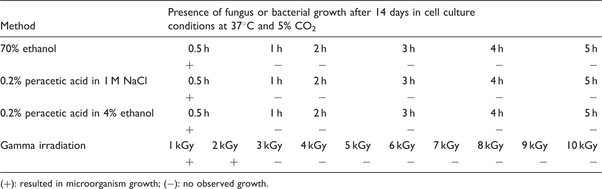
(+): resulted in microorganism growth; (−): no observed growth.
Summary evaluation of decontamination methods.
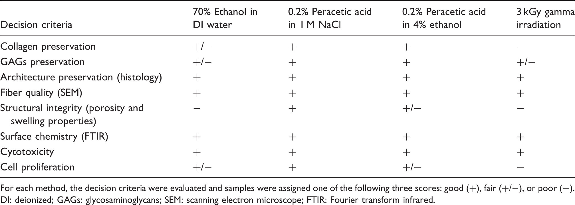
For each method, the decision criteria were evaluated and samples were assigned one of the following three scores: good (+), fair (+/−), or poor (−).
DI: deionized; GAGs: glycosaminoglycans; SEM: scanning electron microscope; FTIR: Fourier transform infrared.
Decellularization of whole porcine kidneys
Whole decellularized renal ECMs were obtained in less than 7 h by perfusion of the organ with 0.5% (w/v) SDS solution under constant pressure while steadily increasing the flow rate (Figure 1(a)). H&E staining of native and renal dECM verified complete cell removal after 7 h SDS exposure while preserving the microstructure (Figure 1(b)). More than 99% of genomic DNA was removed (Figure 1(c)) as a result of decellularization and the length of remaining DNA fragments were all below 200 bp (Figure 1(d)), which is one proposed criteria for producing no immunogenic reaction due to DNA. 26
Decontamination of dECM slices
Slices of renal cortex were decontaminated by four methods: 70% (v/v) ethanol, 0.2% (v/v) peracetic acid in 1 M NaCl, 0.2% (v/v) peracetic acid in 4% (v/v) ethanol, and γ-irradiation. Various exposure times and irradiation intensities were studied to achieve optimum conditions (Table 1). It was determined that 1 h of solution exposure was sufficient to completely decontaminate slices of renal tissue. For samples immersed in DI water at room temperature and exposed to γ-irradiation, 3 kGy was the minimum required intensity to eliminate bacterial or fungus growth. The decontaminated tissues along with non-decontaminated control tissues were then incubated in media containing 10% (v/v) FBS at 37℃ for at least 14 days and the media was viewed under a microscope to check for the presence of contaminating microorganisms. As the media contained all required nutrients for viable proliferation of fungi and bacteria and 37℃ being the optimum growth temperature, this incubation method was deemed appropriate for confirming decontamination.10,22 The control sample was contaminated after 12 h, verifying no inhibition of microorganism growth under these culture conditions.
Decontamination of whole decellularized kidneys
Whole kidney decontamination was also performed with the three treatment solutions (n = 3 for each method) and the absence of contamination was determined after incubation of randomly selected pieces (n = 3) of tissue in cell culture media for 14 days. Two hours of exposure to the three treatment solutions was determined to be optimal for decontaminating whole renal dECM. As 3 kGy γ-irradiation was the most damaging method for slices of kidneys, and we did not intend to pursue this method for whole organ decontamination, γ-irradiation was not tested on whole organs; however, it has been reported elsewhere that 10 kGy6 to 25 kGy 27 irradiation is required for decontamination of whole porcine ECM.
Essential ECM components preservation
To compare the impact of the four decontamination treatments on collagen, the soluble collagen content of decellularized renal cortex samples was determined and the results are shown in Figure 2. As the assay kit quantified the soluble collagen, an increase in collagen content after treatment indicated more exposed and susceptible collagen fibers. However, the collagen content was not an absolute value and an observed increase for some samples might have occurred due to the removal of some other materials (e.g. for peracetic acid, these materials could be lipids, nucleic acids, and/or other types of proteins).
28
Among all of the decontamination methods, γ-irradiation reduced the collagen content by more than 50%, and was considered the most damaging method. Ethanol and peracetic acid treatments were equivalent in preservation of general collagen.
Top panel: Collagen quantification of optimally treated and control non-treated samples (n = 5). There was no significant difference between solution-treated and non-treated samples, signifying that no damage to soluble collagen occurred during treatment by ethanol or peracetic acid. However, γ-irradiated samples had significantly (**) less collagen, showing greater damage to collagen fibrils. Bottom panel: GAGs quantification of optimally treated and control non-treated samples (n = 5). Optimally treated means 1-h solution treatments and 3 kGy γ-irradiation. All treatment except peracetic acid in 1 M NaCl caused significant reduction in GAGs content. NS means no significant difference (p-value > 0.05) and ** means significant difference (p-value < 0.05).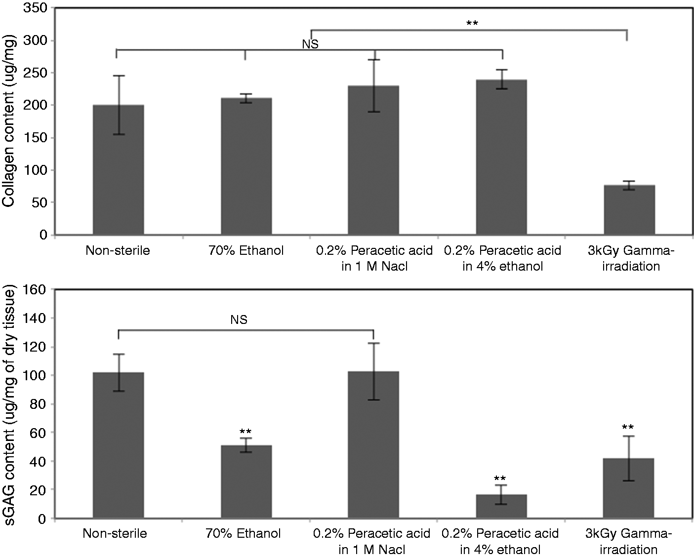
GAGs are the other important components of ECM that bind growth factors and retain water due to their hydrophilic structure. Preservation of these components is required for recellularization to create a whole solid organ. Sulfated GAGs contents of all samples (including non-treated and treated samples obtained from the four decontamination treatments) were determined by the Blyscan sGAGs assay and the results are presented in Figure 2. Peracetic acid in 4% (v/v) ethanol resulted in the most removal of sGAGs among all methods. Gamma-irradiated samples also showed significant reduction in sGAGs contents, which is in complete agreement with previous research that demonstrated depolymerization and degradation of GAGs after γ-irradiation. 29
The distribution of essential ECM components before and after treatment was examined using staining (Figure 3(a)). No significant detectable changes were observed in elastin and collagen IV staining. However, in images of Sirius staining, representative of general collagen, more collagen damage and increased void space were observed in γ-irradiated tissue. The damage to the collagen of γ-irradiated samples correlated with increased swelling properties and decreased soluble collagen contents. The microstructural scoring of tissues treated with different methods by a histologist blinded to each method showed that all methods except 70% ethanol could cause further damage to tubule and glomeruli structure (Figure 3(b)).
(a) Distribution of essential components of the ECM (n = 3). Sirius, Orcein, Immuno, and PAS staining were used to detect general collagen, elastin, collagen IV, and GAGs, respectively. (b) The scoring system is based on preservation of the microscopic architecture of optimally treated dECM examined by a histologist blinded to the decontamination treatment. Results are reported as Avg ± SD (n = 5). In the charts, 70% EtOH, PAA-NaCl, PAA-EtOH, and γ-irradiation represent 1-h exposure to 70% ethanol in DI, 0.2% peracetic acid in 1 M NaCl, 0.2% peracetic acid in 4% ethanol, and 3 kGy γ-irradiation, respectively. NS means no significant difference (p-value > 0.05) and ** means significant difference (p-value < 0.05).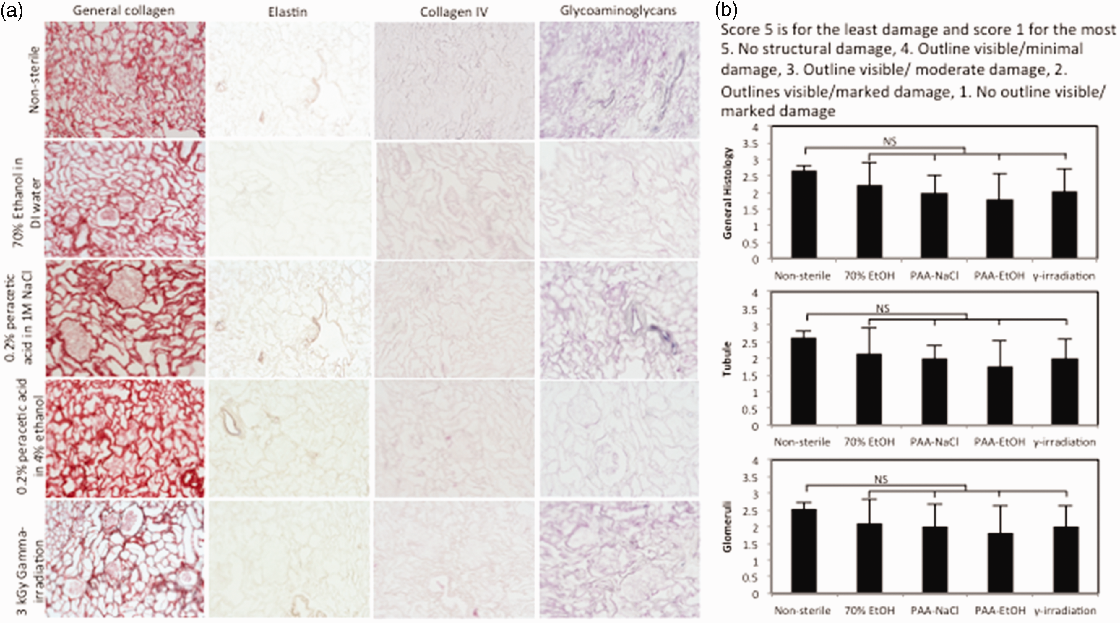
Scanning electron microscopy and fiber characterization
Scanning electron microscopy imaging of treated samples is shown in Figure 4. As demonstrated in this image, the filtration units of renal tissue (nephrons), which are required for functional kidneys and are considered the most important physical structures for cell migration and differentiation, were well preserved. Fiber diameter (Figure 4(e)) and intersection density (Figure 4(f)), the important factors for morphology and phenotype of cells in 3D cell culture, were similar for all decontamination treatments.
Scanning electron micrographs of samples treated for 1 h with (a-1, a-2) 70% ethanol; (b-1, b-2) 0.2% peracetic acid in 1 M NaCl; (c-1, c-2) 0.2% peracetic acid in 4% ethanol; and (d-1, d-2) 3 kGy γ-irradiation. In (1) and (2), scale bars represent 20 µm and 500 nm, respectively, and white arrows show glomeruli structure. Fiber diameter (e) and intersection density (f), measured using DiameterJ software (n = 3), showed no significant difference between decontamination methods.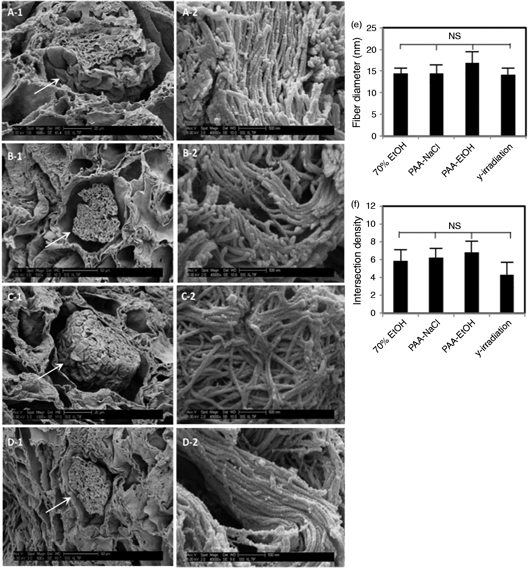
Swelling properties of ECM
To determine the possible damage to collagen fibrils of renal tissue during decontamination, the swelling ratio of treated samples was measured and the results are shown in Figure 5(a). Gamma-irradiated samples demonstrated the greatest water absorption, which might have been caused by cross-link elimination, increased void fraction, or structural damage to collagen fibrils. Peracetic acid in 1 M NaCl solution preserved the integrity of structures more than any other solution. Peracetic acid in ethanol caused more damage to structural cross-linking, purportedly due to the acidic pH. These results corroborate the collagen quantification values and further confirm the relationship between collagen degradation and swelling ratio. Increased porosity of γ-irradiated samples also confirmed structural disruption that might be caused by irradiation (Figure 5(b)).
(a) 1× PBS absorption of optimally treated samples (n = 6) representative of swelling and cross-linking damage during decontamination. (b) Thin sections of optimally treated and non-treated tissue (5 µm) were stained with H&E and used for porosity evaluation using ImageJ software. Optimally treated means 1-h solution treatments and 3 kGy γ-irradiation. NS represents no significant difference with p-value > 0.05 and **represents significant difference with p-value < 0.05.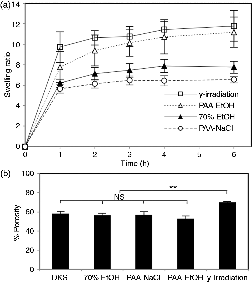
Surface chemistry of treated samples
Protein denaturation on the surface of non-treated and treated samples obtained from the four decontamination methods was examined by FTIR. The resulting spectra are shown in Figure 6. Amide I, II, and III bands are the most important determinants of the secondary structure of collagen on the surface of ECM, which has significant effects on cell–ECM interactions.
30
No significant shift could be observed in these bands suggesting no further denaturation due to decontamination after decellularization, which is comparable with previous studies.
31
FTIR spectra of non-treated (control) and optimally treated dECM samples. No significant differences were observed between the various decontamination methods and spectra showed no shifting in amide bands, signifying that surface proteins were not further denatured during decontamination. Optimally treated means 1-h solution treatments and 3 kGy γ-irradiation.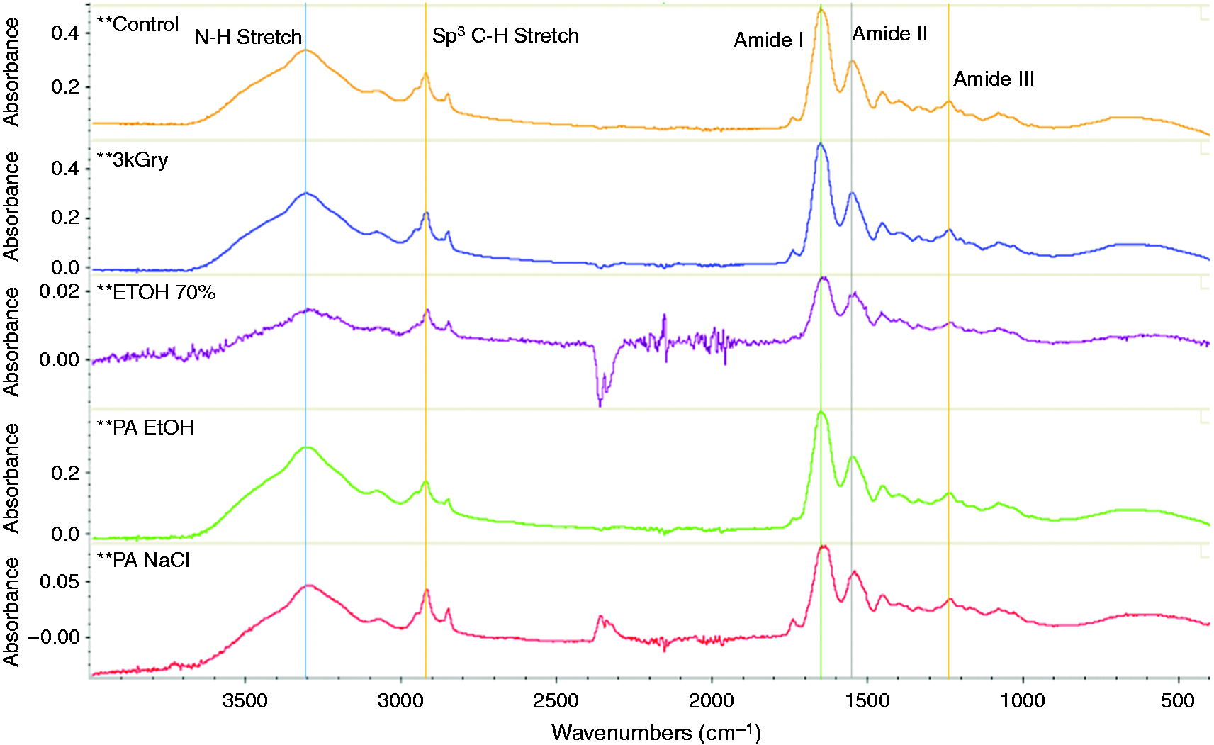
Cytotoxicity of treated samples and potential for support of cell growth
The various decontamination treatments can produce several by-products that could be toxic to cells and induce necrosis. To remove cytotoxic by-products, treated samples were washed with autoclaved distilled water before adding cell culture media. To compare cytotoxicity, RCTE cells were grown in six-well plates in the presence of pieces of tissues treated with one of the four decontamination methods. A Live/Dead assay was used to determine the number of viable and dead cells after 3 days and the results are shown in Figure 7. Cells grown in media containing treated samples were as viable as cells in control media (no dead cells were detected), which indicates that no toxicity caused by treated samples was observed after 3 days of cell growth.
Cytotoxicity of optimally treated samples (n = 3) was compared using human RCTE cells and Live/Dead assay. No dead cells were observed after 3 days of cell culture in the presence of treated dECM, and cell proliferation was unhindered. DNA content of recellularized samples of treated tissues (n = 6) was measured after 1 and 3 days. Cell adhesion and growth of samples treated with ethanol or solution containing peracetic acid in NaCl were significantly higher than the other two samples; especially, γ-irradiated samples showed considerably less capability to support cell adhesion and growth. Optimally treated means 1-h solution treatments and 3 kGy γ-irradiation.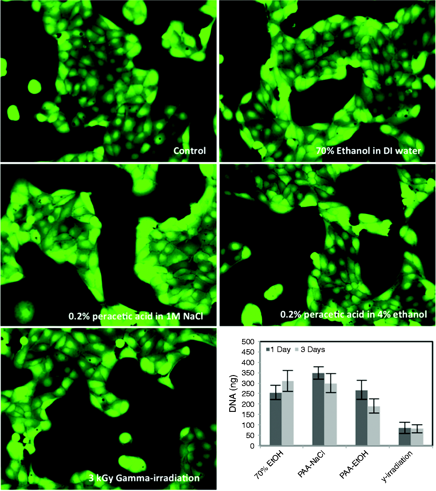
The most important attribute of dECM that must be preserved during decellularization and decontamination is the capability to support cell adhesion and growth during recellularization. To examine this potential, RCTE cells were used to repopulate treated dECM samples. DNA contents were measured after 1 and 3 days (Figure 7), as an indirect method for determination of cell number. Residual DNA after decellularization and incubation in cell culture medium was less than 5 ng/mg of dry tissue; therefore, the quantified DNA after repopulation was attributed to the RCTE cells and representative of cell adhesion and growth. The γ-irradiated samples demonstrated considerably less cell adhesion potential than the three solution decontamination treatments.
Discussion
Whole organ regeneration is an active area of research in tissue engineering and regenerative medicine. The hope of this technology is that organs can be built from porcine dECM and human cells to replace the need for donor organs and reduce the risk of organ rejection by a patient’s immune system. 32 Various steps are necessary before a scaffold is ready for recellularization. First, cadaveric or animal organs are harvested and decellularized. Then, a gentle decontamination method is applied to remove contaminants that can cause infection and/or inflammatory response at the time of recellularization and transplantation. Decontamination of complicated solid organs such as kidneys is challenging because common methods, such as ethylene oxide that requires diffusion through solids, are not applicable. 33
In this study, decellularization of whole porcine kidneys with perfusion of 0.5% (w/v) SDS solution through the inherent vasculature was performed using an improved method in less than 7 h. Previous researchers have accomplished the decellularization of whole porcine kidneys using less efficient methods. For example, Orlando et al. 27 perfused kidneys for 48 h with 0.5% (w/v) SDS under a constant flow rate and Sullivan et al. 6 studied whole porcine kidney decellularization using 0.5%, 0.25% (w/v) SDS, and 1% (v/v) Triton X-100 in 36 h. By freezing kidneys at −20℃, as suggested by several previous authors, and increasing flow rate under constant pressure, we were able to reduce the decellularization process time.34,35 Four different decontamination treatments were compared using slices of kidney tissue. All of the experiments were performed with uniform sizes of tissues, which were obtained from the same kidney for each set of comparison tests. To extrapolate these results to whole decellularized organs, and provide a safety margin, it was determined that 2-h perfusion with the decontamination solutions; or 10–25 kGy γ-irradiation, as suggested by previous researchers,6,27 were needed to decontaminate whole kidneys.
Previous researchers have used sterile PBS containing 10,000 U/ml penicillin G, 10 mg/ml streptomycin, and 25 µg/ml amphotericin B to sterilize whole rat kidneys 8 and whole rat hearts.36,37 However, their method was not effective under the conditions in which kidneys in this study were harvested at an abattoir in a non-aseptic manner. Stronger decontaminants were required to remove all contaminating agents from porcine organs. Other researchers have proposed similar decontamination methods, such as peracetic acid for rat kidneys, 4 γ-irradiation for porcine kidneys, 6 and the combination of peracetic acid and chloroform gas for rat livers. 21
According to our results, 70% (v/v) ethanol in DI water has a great potential to decontaminate porcine renal tissue. It is also very commonly used to decontaminate equipment and surfaces during cell culture experiments. However, ethanol is not considered strong enough to remove hydrophilic viruses and bacterial spores and therefore is not recommended for clinical use. 38 It has also been reported that ethanol can denature proteins, dehydrate ECM, and affect cell–ECM interactions. 33 The 70% ethanol solution was included in this study as a comparator.
Gamma-irradiation is another widely used method to sterilize biological tissues, however, substantial damage to the microstructure and composition have been reported.39,40 The degradation effect of γ-irradiation on different types of collagen has been previously reported for decellularized lung tissue, 41 human dermis, 19 and porcine pulmonary valves. 40 Gamma-irradiation can cause scission of alpha polypeptide bonds or cross-linking in the presence of free radicals. 33 In addition, γ-irradiation can induce fragmentation of GAGs by production of hydroxyl, carbonate, and nitrogen dioxide radicals. 29 It has also been shown that γ-irradiation intensity greater than 2 kGy could cause ECM instability for dermis tissue and decrease the denaturation temperature of ECM below the temperature of the body. 19 In contrast to these results, Uriarte et al. 41 reported that resistance and elasticity of decellularized lungs were improved and modified after γ-irradiation. It has been reported that for the decontamination of an organ as large as a porcine kidney, at least 10 kGy intensity of γ-irradiation would be required. 6 Even with 3 kGy intensity, we observed considerable structural damage (Figures 2, 3, and 5) and a significant reduction in RCTE cell adhesion and proliferation (Figure 7) on slices of dECM kidney tissue. We do not recommend the use of γ-irradiation for decontamination of whole organs.
Peracetic acid is a strong oxidizing agent that can disrupt the cellular membrane and cause cell death. 42 It was observed that peracetic acid in low concentrations was able to inactivate viruses, fungi, and bacteria with the creation of non-toxic by-products such as water, oxygen, and carbon dioxide. 43 Previous researchers have used peracetic acid to sterilize decellularized lungs, 14 human heart valves, 44 decellularized anterior cruciate ligaments, 45 human spongiosa cuboids, 46 and whole rat kidneys. 4 It has been suggested that it is better to use peracetic acid in neutral pH solutions or high ionic strength to preserve collagenous structures without any swelling damage. Low concentrations of peracetic acid in ethanol can lead to dissolution of collagen and swelling in collagen fibrils because of low pH. 23 Our findings here also confirmed that peracetic acid in ionic solution could preserve microstructure, surface chemistry, and cell adhesion sites more than peracetic acid in ethanol solution or any other applied decontamination treatments.
Different tissues have unique compositions and are used for a variety of purposes. The decontamination process must be optimized according to tissue type and the tissue’s specific purpose. A summary of the results of this study is reported in Table 2. An aqueous solution of 0.2% (v/v) peracetic acid in 1 M NaCl solution performed the best among the tested decontamination treatments by preserving essential components of the dECM that are necessary to promote increased cell adhesion and proliferation. We suggest that a combination of rapid decellularization with a gentle and reliable decontamination treatment of peracetic acid in aqueous ionic solution is the optimal method to prepare a whole porcine kidney scaffold for recellularization.
Conclusion
Decontamination of decellularized porcine renal tissue was performed using common treatment methods such as 70% (v/v) ethanol, peracetic acid, and γ-irradiation. The required exposure time and intensity were selected to produce completely decontaminated decellularized tissue slices and whole organs. Gamma-irradiation was found to be the most damaging decontamination treatment, with reduced cell adhesion, proliferation, mechanical properties, and altered microstructure of the ECM. The results of this study suggest that 0.2% (v/v) peracetic acid in 1 M NaCl is the best method to completely decontaminate renal tissue and preserve essential components of the ECM that are required for cell–ECM interaction.
Footnotes
Acknowledgments
We thank Edwin Jackson for his assistance with the γ-irradiation tests. We also thank Jason A. Wertheim, MD, PhD, at Northwestern University for providing RCTE cells. We acknowledge the laboratory assistance of Spencer Baker, Robert M. Fuller, Evan M. Buckmiller, Blake J. Cannon, and Daniel Thomas.
Declaration of Conflicting Interests
The author(s) declared no potential conflicts of interest with respect to the research, authorship, and/or publication of this article.
Funding
The author(s) received no financial support for the research, authorship, and/or publication of this article.