
Abstract
Purpose
A functionalized graphene oxide-based thermosensitive hydrogel loaded with docetaxel for intratumoral delivery was designed to enhance therapeutic efficacy and alleviate system toxicity.
Methods
First, graphene oxide was functionalized with chitosan to acquire high stability in physiological solutions. And then docetaxel–graphene oxide/chitosan gel was formed by mixed docetaxel–graphene oxide/chitosan suspension with hydrogel which was made from Poloxamer 407 and Poloxamer 188. Cellular uptake, antitumor effect in vitro and in vivo, cell apoptosis, and biodistribution of docetaxel–graphene oxide/chitosan gel were investigated, compared with the docetaxel solution.
Results
Graphene oxide/chitosan was stable in physiological solution, and docetaxel released much slower from docetaxel–graphene oxide/chitosan gel with a pH-responsive feature. Compared with free docetaxel, docetaxel–graphene oxide/chitosan could afford higher antitumor efficacy in Michigan Cancer Foundation-7 (MCF-7) cells in vitro. Furthermore, docetaxel–grapheme oxide/chitosan gel which was injected within tumor could afford higher concentration and longer resident time in tumor tissues of mice in vivo, without obvious toxic effects to normal organs. Meanwhile, the combination of near-infrared laser irradiation at 808 nm significantly enhanced tumor inhibition in vitro and in vivo.
Conclusions
Docetaxel–graphene oxide/chitosan gel in combination with 808 nm near-infrared laser irradiation had great potential for cancer chemo-photothermal therapy.
Introduction
Docetaxel (DTX), a member of taxane family of antineoplastic agents, has demonstrated antitumor activity in the treatment of patients with various cancer types. 1 Despite its clinical activity, DTX has several drawbacks including poor solubility, short half-life time, and nonspecific distribution throughout the body. 2 Moreover, the solvent of ethanol and Tween 80 (polysorbate 80) in the commercial DTX formulation (Taxotere®) has been associated with a number of toxicities like acute hypersensitivity reactions and the alterations in DTX pharmacokinetic profiles. 3 Due to these disadvantages, sustained drug delivery system such as hydrogels combined with targeting intratumoral (i.t.) injection has been invented to circumvent problems accompanied with DTX pharmacotherapy.4–6
Intratumoral delivery may target chemotherapeutic agents directly to tumor and reduce the exposure at normal tissues to avoid toxicity and improve efficacy. 7 In situ gel systems can go sol–gel transition after local administration under physiological conditions, such as pH change, ionic cross-linkage, and temperature modulation, which makes them increasingly important in drug delivery.8–10 Controlled release from in situ gel for injection has been attractive in recent years, especially the vehicle made by Poloxamer 407 (P407). The physical properties, biocompatibility, and recent advancement of injectable P407 thermosensitive in situ gel have been widely used in pharmacy field.11,12 Despite the success of current treatment for several types of cancer, multidrug resistance remains a major limitation to many chemotherapeutic agents.13,14
The tremendous development of nanotechnology is bringing us closer to the dream of clinical application of nanoparticles in photothermal therapy (PTT) of tumors. 15 PTT is a noninvasive modality for treating cancer, which utilizes photoabsorbing agents to generate a high localized temperature for ablating tumor without damaging surrounding healthy tissues under near-infrared (NIR) light irradiation. 16 Due to its ultrahigh surface area and easy surface functionalization, graphene oxide (GO) has been intensively explored for drug delivery. Utilizing their intrinsic high NIR absorbance, GO and its derivatives have been found to be excellent candidates for cancer photothermal and chemo- and/or photodynamic therapies.17–19 GO was soluble in water but aggregated in solutions rich in salts or proteins, to impart aqueous stability and prevent biofouling, we sonicated the GO/chitosan (CS) to make them into small pieces and conjugated CS to the carboxylic acid groups on GO via carbodiimide-catalyzed amide formation.20,21
In the present study, we set out to develop a functionalized GO-based thermosensitive hydrogel loaded with DTX for i.t. delivery to enhance therapeutic efficacy and alleviate system toxicity. For this purpose, functionalized GO and DTX–GO/CS gel were prepared and in vitro release of DTX–GO/CS gel was investigated. The design ideas of DTX-GO/CS gel are shown in Figure 1.22 In addition, the antitumor efficacy in MCF-7 cells in vitro and in S180 tumor-bearing mice in vivo and the biodistributions of DTX–GO/CS gel were evaluated. Furthermore, histological analysis of hematoxylin and eosin (H&E) staining and TUNEL immunohistological assay were further employed for antitumor efficacy assessment of DTX–GO/CS gel.
Experimental section
Materials
GO (powder, purity >95%) were purchased from Zhengzhou Shier Chemicals Co. Ltd (Zhengzhou, China). DTX (purity >98%) was purchased from Wuhan Yuanchen Gongchuang Technology Co Ltd. Fluorescein isothiocyanate (FITC), sulforhodamine B (SRB), dimethyl sulfoxide (DMSO), and NIR fluorescent dye IR-783 were obtained from Sigma Aldrich (St Louis, MO, USA). Penicillin, streptomycin, and fetal bovine serum were bought from Life Technologies (Carlsbad, CA, USA). Other reagents were acquired from China National Medicine Corporation Ltd. MCF-7 cell line and S180 cell line were all obtained from the Chinese Academy of Sciences Cell Bank. BALB/c mice (8–10 g, ♀) were provided by the Henan Laboratory Animal Center.
Synthesis of GO/CS
In total, 100 mg of GO and 500 mg of CS (the deacelation degree was about 75%) were added into 50 ml of 0.1 mol/l of 2-(N-morpholino)ethanesulfonic acid hydrate (pH 5.0), then the mixture was bath sonicated for 1 h to obtain evenly dispersed suspension. After that, 0.652 g of N-(3-dimethylamino ropyl-N0-ethylcarbodiimide) hydrochloride (EDC·HCl) and 0.782 g of N-hydroxysuccinimide (NHS) were added to the suspension, then sonicated for 6 h and followed by stirring at room temperature for 16 h. To remove unreacted CS, EDC·HCl, and NHS, the mixture was put on 0.2 µm microporous membrane filter and washed with ultrapure water until the solution was neutral. The filter cake was scattered with 20 ml of ultrapure water and then was freeze dried to obtain GO/CS. 23
FT-IR spectra of GO, CS, and GO/CS were recorded on a Nicolet iS10 spectrometer (Thermo). The morphological, particle size, and zeta potential of GO/CS were characterized by transmission electron microscopy (TEM) (JEM-1400 Electron Microscope, JEOL Ltd, Japan) and dynamic light scattering (DLS) (Zeta Sizer Nano ZS-90, Malvern, UK), respectively. For photothermal conversion performance of GO/CS, an 808 nm laser emitter was chosen as the NIR light source (Changchun laser station of the Chinese Academy of Sciences) with a power density of 2.5 W/cm2. GO/CS solution at 0, 0.1, 0.3, 0.5, 0.7, and 1.0 mg/ml was exposed to NIR laser irradiation, and the temperature was measured and recorded.
Preparation of DTX–GO/CS gel
Five milligram of GO/CS was dissolved in 5 ml of ultrapure water, and the mixture was bath sonicated at 400 W for 40 times (work 5 s and apart 5 s every time) to obtain GO/CS dispersion. DTX was dissolved in DMSO to get a 20 mg/ml of DTX solution. Dropped GO/CS dispersion into 0.25 ml of DTX solution, and then sonicated the mixture using an ultrasonic cell disruption system (400 W, 20 times) at room temperature to get an excellent aqueous dispersion. To remove excess DMSO, the dispersion was centrifugated at 4000 r/min for 10 min, and the supernatant was freeze dried to obtain DTX–GO/CS. 0.2 g of P407 was completely dissolved in 4 ml of ultrapure water which was precooled at 4℃, 1.1 g of Poloxamer 188 (P188) was added, and the mixture was put in refrigerator at 4℃ overnight for full swelling. Ultrapure water was added to the mixture until the final volume was 5 ml and then P407/P188 gel was formed. DTX–GO/CS solution was dropped into the P407/P188 gel (1:1), and the mixture was stirred until it was uniform at room temperature. The gelation temperature of the gel was 37.2 ± 0.5℃ which was determined with test tube inversion method. 24
The morphology of DTX–GO/CS gel was observed by scanning electron microscopy (SEM) (JSM-7500F Electron Microscope, JEOL Ltd, Japan). Briefly, the sample to be observable in SEM was dropped onto the silicon wafer and followed by natural drying. The drug loading ratio of DTX in DTX–GO/CS was determined using ultra-filtration method. Briefly, took 0.2 ml DOX–GO/CS gel nano-suspension into ultra-filtration tube (MWCO = 10 KDa), centrifuged at 1000 r/min for 2 h. The concentration of DTX was determined by HPLC. The mobile phases, methanol:water (75:25), were run at a flow rate of 0.8 ml/min. The column effluent was monitored with a UV detector at λ = 230 nm.
For release study, 25 DTX solution and DTX–GO/CS gel were placed into dialysis bags (molecular weight cutoff MWCO = 8000–14,000), which were dialyzed in 50 ml PBS (pH 5 and 7.2, respectively) at 37.0 ± 0.5℃ with a stirring rate of 100 r/min. 0.2 ml of dialyzate was drawn at various time points, being replaced by the same volume of aqueous buffer solution. The concentration of released DTX was quantified by HPLC, and the cumulative percentage of drug release was calculated.
Antitumor effect of DTX–GO/CS in vitro
Cell viability
MCF-7 cells were cultured in normal RPMI-1640 culture medium with 10% FBS and 1% penicillin/streptomycin in 5% CO2 at 37℃ in a humidified incubator. MCF-7 cells (5 × 10³ cells per well) were seeded in 96-well plates and incubated for 24 h. Then taken off the medium and treated with medium containing various concentrations of DTX and DTX–GO/CS for 24, 48, and 72 h. Growth inhibition was measured by the SRB assay. In addition, the cells were also treated with different concentrations of GO/CS for 72 h to investigate the cytotoxicity of the carrier. The cells were then subjected to 808 nm NIR laser irradiation (2.5 W, 2 min) and then incubated at 37℃ in 5% CO2 for 24 h. Growth inhibition was measured by standard SRB assay. 26
Cellular uptake
To evaluate the intracellular uptake capacity of DTX–GO/CS by MCF-7 cells, flow cytometry analysis was used. 27 DTX–GO/CS was labeled with a fluorescence probe, FITC. MCF-7 cells (6 × 10⁵ cells per well) were seeded in six-well plates for 24 h. Then discard the original culture medium and wash three times with PBS. Two milliliters of medium containing DTX–GO/CS was added into each well, followed by culturing for 1, 2, and 4 h, respectively, and discarding the culture medium, washing three times with PBS, followed by fixing in ice cold 75% ethanol under 4℃ for 2 min, 4′,6-diamidino-2-phenylindole (DAPI) was added into each well, followed by culturing for 10 min, and washing three times with PBS. The cells were observed under fluorescence microscopy (Zeiss LSM 510) and the images were recorded. In order to further research the cellular uptake quantitatively, the collected cells were measured using a flow cytometer (Partech GmbH, Germany), and the results were analyzed by the Flow Jo software (version 7.6.1).
Cell apoptosis
The Annexin V-FITC/PI apoptosis detection kit (KeyGEN BioTECH, Nanjing, China) was used to detect the apoptotic cells. MCF-7 cells (6 × 10⁵ cells per well) were seeded in six-well plates for 24 h and then exposed to DTX and DTX–GO/CS for 24 h. For Laser and DTX–GO/CS + Laser group, the treatment was the same as described above with the exception that the 808 nm laser irradiation (2.5 W) was performed toward the six-well plates for 2 min in 4 h after each administration. After 24 h of incubation, the cells were trypsinized and collected by centrifugation, then washed twice with PBS, and gently resuspended in 100 µl binding buffer. Then 5 µl of Annexin V-FITC and 10 µl of PI solution were added, after incubation in the dark at room temperature for 15 min according to manufacturer’s instruction, cell apoptosis was analyzed with a flow cytometry (Partech GmbH, Germany). Annexin V+ and PI− cells were scored as early apoptotic, and double-stained cells were considered as late apoptotic. 28
Antitumor effect of DTX–GO/CS gel in vivo
Xenograft tumor mouse model
All animal experiments were performed under a protocol approved by Henan Laboratory Animal Center. The S180 tumor models were generated by subcutaneous injection of 2 × 10⁶ cells in 0.1 ml saline into the right shoulder of female BALB/c mice (18–20 g). The mice were used for in vivo antitumor experiment while the tumor volume reached ∼100 mm3 (about seven days after tumor inoculation).
Antitumor activity
S180 tumor-bearing mice were divided into seven groups (six mice per group): (1) N.S., (2) DTX, (3) GO/CS gel, (4) DTX–GO/CS gel, (5) N.S. + Laser DTX–GO/CS gel, (6) GO/CS gel + Laser, (7) DTX–GO/CS gel + Laser. The mice were administrated different preparations with the same dosage of DTX (10 mg/kg) every three days for four times. 29 For the Laser group, the treatments were the same as described above except that 808 nm NIR laser irradiation (2.5 W) was performed for 1 min once a day during the therapy. The body weight and tumor sizes were measured before administration, and tumor volume (V) was calculated according to the formula: V = [length × (width)2]/2. At the end of experiment, the mice were sacrificed and heart, liver, and tumor tissues were taken out and weighed.
Then, the tissues of each group were soaked in 10% formalin solution, embedded with paraffin for H&E staining. 30 Morphological changes were observed under a microscope (Eclipse 80i, Nikon, Japan). For the detection of apoptotic cells in situ, the methodology described previously has been processed. 31 Briefly, tumor sections were stained with the TUNEL agent (KeyGEN BioTECH, Nanjing, China).
Biodistribution assay in vivo
For FX imaging in vivo, GO/CS gel was labeled with the NIR fluorescent dye, IR783, to monitor biodistribution in vivo and tumor accumulation ability. 32 Noninvasive optical imaging system was utilized. When the tumor volume reached approximately 100 mm3, mice were injected with free IR783 by vein as the control and GO/CS gel-labeled IR783 was injected in situ with same dosage of IR783 (2 mg/kg) as the control and GO/CS gel-labeled IR783 (2 mg/kg) via within tumor injection. NIRF imaging experiment was performed at different time postinjection using a Kodak in vivo imaging system FX PRO (Kodak, USA) equipped with an excitation bandpass filter at 700 nm and an emission at 830 nm. Images were analyzed using the Kodak Molecular Imaging Software 5.X. 33
To quantitatively assess the biodistribution of the drug delivery system, tumor-bearing mice were treated with DTX solution which was injected i.v. or DTX–GO/CS gel which was injected in situ with the same dosage of DTX (10 mg/kg). At 0.5, 1, 3, 6, and 12 h after injection, six animals per time point were killed and tumor tissues were homogenized in saline with W/V 1:3, then 200 µl of sample was placed into 5 ml centrifuge tubes. Eight-hundred microliters of ether was added to the above tubes and centrifuged after mixing for 5 min by vortex. After centrifugation at 3000 r/min for 10 min, organic layer was transferred to a new 1.5 ml tube and evaporated to dryness under nitrogen. Finally, the residue was dissolved in 100 µl methanol and a 20 µl aliquot of each sample was injected for HPLC analysis.
Statistical analysis
All the data were presented as mean ± SD from three to 10 independent measurements in separate independent experiments and analyzed using descriptive statistic and single-factor analysis of variance.
Results and discussion
Synthesis and characteristics of GO/CS
Figure 2(a) shows photos of GO and GO/GS in different solvents, where it is possible to observe a better dispersibility and stability of the latter. As shown in Figure 2(b), in the spectrum of GO, the peaks at 1729.11 and 1622.07 cm−1 are characteristics of the C=O and O–H bands, respectively. In the spectrum of CS, there are two characteristic absorbance bands centered at 1652.18 and 1595.67 cm−1, which correspond to the C=O and N–H, respectively. Compared with pure CS and GO, both peaks at 1595.67 cm−1 related to −NH2 and at 1729.11 cm−1 belonging to C=O stretch of the carboxylic group disappear in the spectra of GO/CS.
34
Moreover, the band corresponding to the C=O characteristic stretching band of the amide group shifted to a lower wave number. The FT-IR analysis of GO/CS clearly indicated that GO interacted with CS through intermolecular hydrogen bonds, so there should be good miscibility between GO and CS.35,36
The design of DTX–GO/CS gel for chemo-photothermal therapy.22 Characterization of GO/CS. (a) Photos of GO (a1) and GO/CS (a2) in different solvents, (b) FT-IR spectrum, (c and d) TEM photos and energy spectrum of GO/CS, (e) size distribution of GO (e1) and GO/CS (e2), (f) zeta potential of GO (f1) and GO/CS (f2), (g) temperature curve of GO/CS under 808 nm NIR laser irradiation.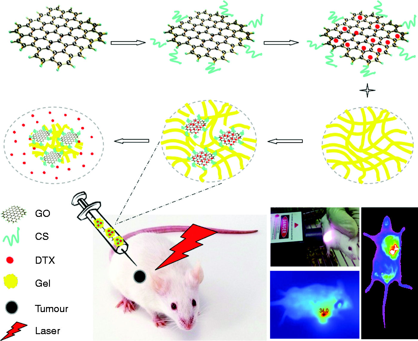
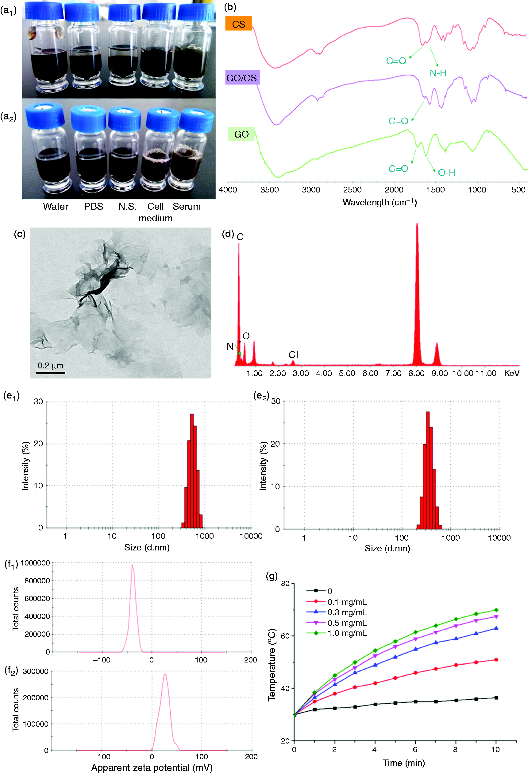
TEM with energy dispersive X-ray (EDX) was used to observe the morphology and component elements of GO/CS. TEM photo demonstrated clearly the graphene layers (Figure 3(c)), EDX showed that there were C, N, and O elements in GO/CS nano-material (Figure 2(d)). The dynamic light scattering analysis showed that the size of GO and GO/CS was 922.5 ± 8.4 nm (Figure 2(e1)) and 536.2 ± 6.2 nm (Figure 2(e2)), respectively; the zeta potential for them were −38.6 ± 6.2 mV (Figure 2(f1)) and 25.0 ± 9.6 mV (Figure 2(f2)), respectively. The change of the zeta potential between GO and GO/CS might be due to the CS, which was positively charged. GO/CS could generate heat under 808 nm NIR laser’s irradiation in a concentration-dependent manner (Figure 2(g)).
Characterization of DTX–GO/CS gel. (a) Drug loading rate of DTX–GO/CS, (b) SEM photos of P407/P188 gel (b1) and DTX–GO/CS gel (b2), (c) drug release curve (c1: pH 7.4; c2: pH 5.0, and c3: DTX–GO/CS gel).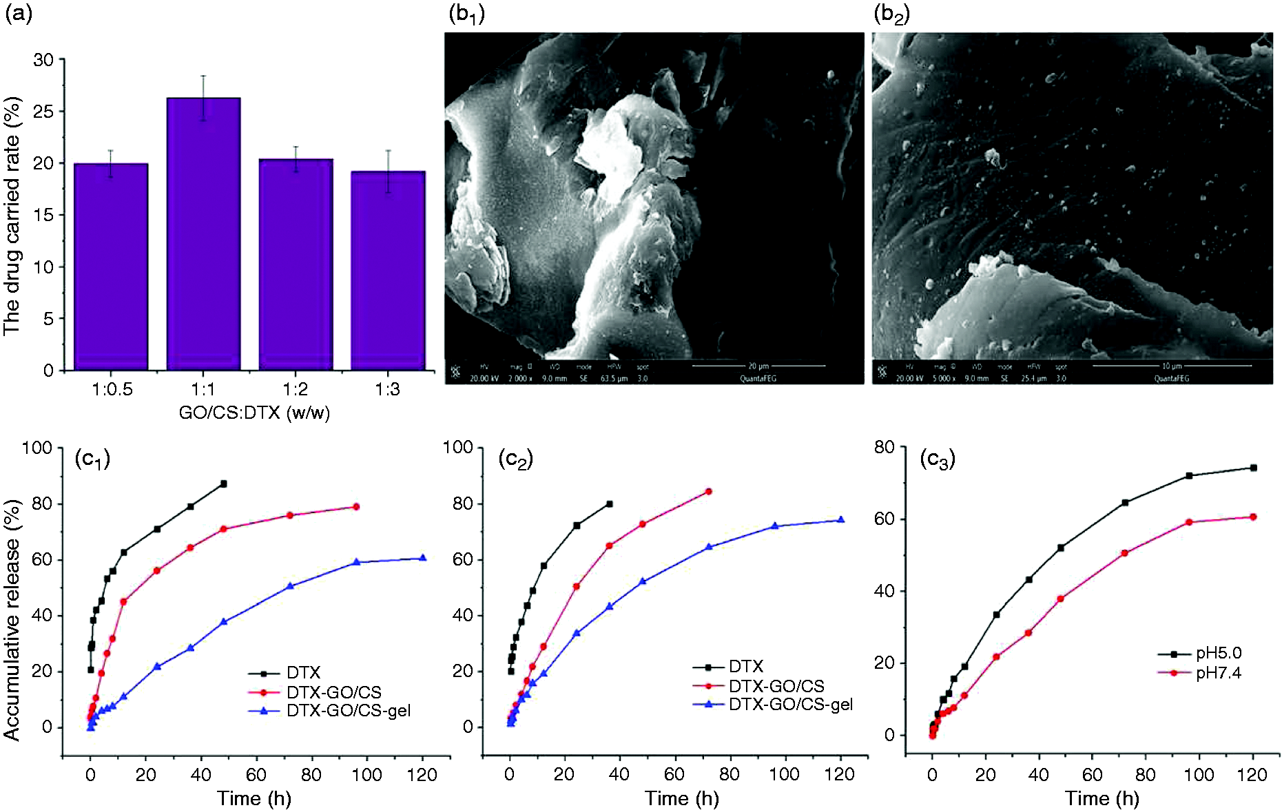
Preparation and characteristics of DTX–GO/CS gel
Figure 3(a) showed a high drug loading rate (26.28 ± 2.16%) when the weight rate between GO/CS and DTX was 1:1. Figure 3(b) shows the SEM photos of P407/P188 gel and DTX–GO/CS gel, respectively. The surface of P407/P188 gel was loose and smooth, and DTX–GO/CS gel was also loose but porous. Figure 3(c1) showed that DTX released much slower from DTX–GO/CS gel than that of DTX solution and it demonstrated a pH-responsive feature (Figure 3(c2)). So due to the tumoral low pH targeting characteristics, it was possible to realize controlled release of the drug in vivo.
Antitumor effect of DTX–GO/CS in vitro
Cell viability
The cytotoxicity study of GO/CS on MCF-7 cells was carried out at different concentrations of GO/CS. The cell viability remained above 95.2% when the concentration was 0.001–10 µg/ml (Figure 4(a)). So we consider that GO/CS has no obvious toxicity to MCF-7 cells after 72 h of incubation while the concentration was below 10 µg/ml. As seen from Figure 4(b), NIR laser irradiation only has little effect on the cell proliferation inhibition, while DTX inhibits the cell growth with concentration dependence. The inhibition rate of MCF-7 cells treated with DTX–GO/CS + Laser significantly increased (Figure 4(b)), for example, when the DTX concentration was 0.01 µg/ml, the cell inhibition rates of DTX–GO/CS and DTX–GO/CS + Laser groups were 21.56 ± 1.8 and 32.15 ± 3.3%, respectively. And there was significant difference between DTX–GO/CS and DTX–GO/CS + Laser (P < 0.05). As shown in Figure 4(b) and (c), with the concentration of DTX increasing, the inhibition rate of DTX and DTX–GO/CS groups increased constantly at 24, 48, and 72 h. On the whole, DTX–GO/CS group had on MCF-7 cells than DTX group in the same dosages. However, the difference between the two groups decreased with the concentration of DTX increased.
Cytotoxicity and inhibition rate on MCF-7 cells (n = 6). (a) Cytotoxicity test, (b) inhibition rate, (c) photothermal therapy.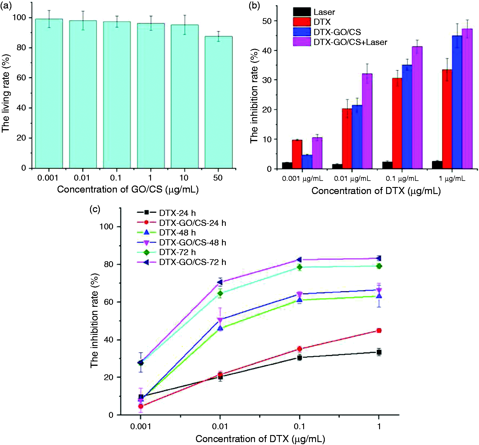
Cellular uptake
To investigate intracellular uptake by MCF-7 cells, DTX–GO/CS was labeled with FITC and DAPI. As seen in Figure 5, DTX–GO/CS could enter into MCF-7 cells at 1, 2, and 4 h. In addition, cellular uptake increased as the incubation time increased from 1 to 4 h. We next made quantitative measurements of the cellular uptake of various DOX formulations using flow cytometry, and the uptake ratios at 1, 2, and 4 h were 47.4 ± 2.52, 76.8 ± 3.35, and 92.1 ± 3.84%, respectively. This result indicated that DTX -GO/CS could be highly uptaken by MCF-7 cells, and the cellular uptake ratio increased with time increasing.
The uptake of DTX–GO/CS by MCF-7 cells.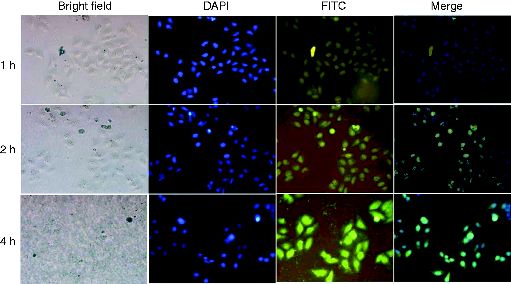
Cell apoptosis
Treatment of DTX–GO/CS with laser showed strong antitumor efficacy on MCF-7 cancer cells in vitro due to the combination of special chemotherapy mechanism of DTX and the PTT properties of GO/CS. The mode of cell death was further investigated and verified with cell test by flow cytometry. As shown in Figure 6, the blank cells exhibited a good state (Figure 6(a)), while early stage apoptotic cells and late stage apoptotic cells were both observed after treatment with different preparations. DTX and DTX–GO/CS could increase the percentage of apoptosis cells, and the percentages of cells were 18.02 ± 0.35% (Figure 6(b)) and 14.96 ± 0.22% (Figure 6(c)), respectively. Laser irradiation only did induce some MCF-7 cells apoptosis, but the effect was weaker than that of DTX or DTX–GO/CS group. And there were more apoptotic cells in DTX–GO/CS + Laser group (18.34 ± 0.12%, Figure 6(d)), suggesting that tumor cells death was caused through apoptotic pathway after DTX–GO/CS with NIR Laser treatment.
Quantitative apoptotic measurement of MCF-7 cells. (a) Blank, (b) DTX, (c) Laser, (d) DTX–GO/CS, (e) DTX–GO/CS + Laser, (f) percentage of apoptosis cells in different groups.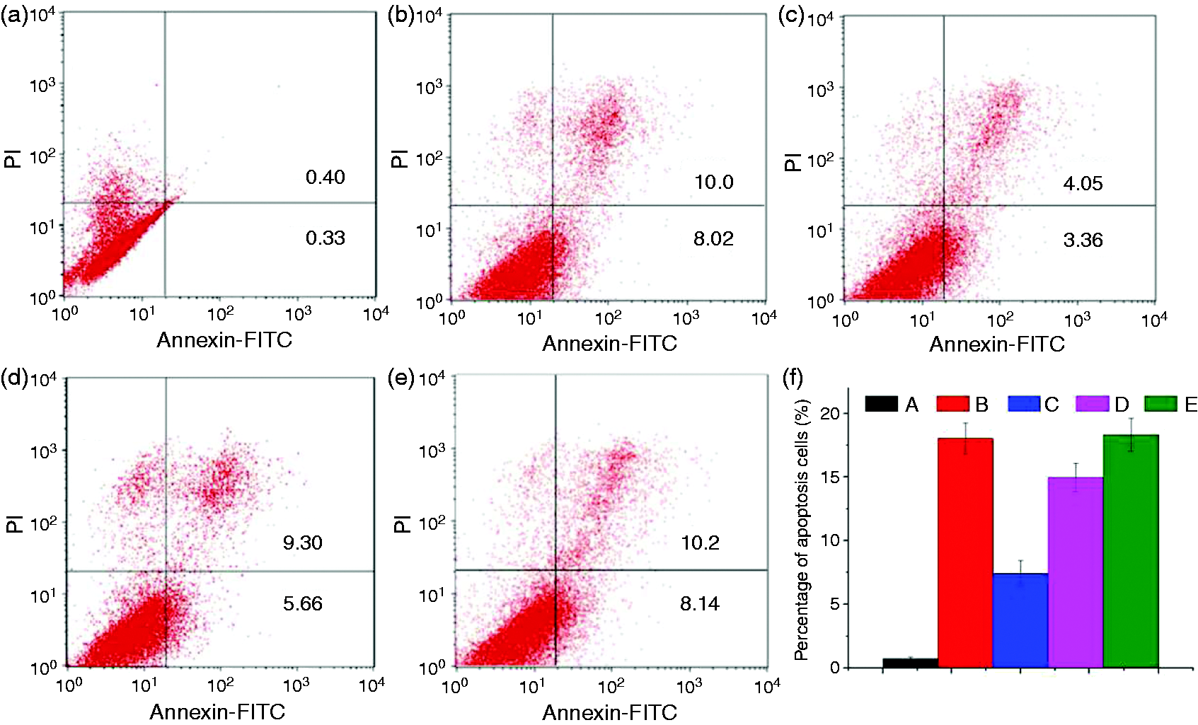
Antitumor effect of DTX–GO/CS gel in vivo
As seen in Figure 7(a1) to (
a2
), S180 tumor-bearing mice could be heated under 808 nm NIR Laser, and compared with the N.S. group, the tumor temperature increased obviously after the mice was injected DTX–GO/CS gel into the tumor with 808 nm NIR Laser. After 12 days of treatment (Figure 7(d)), the relative tumor volumes (V/V0) of N.S., DTX, GO/CS gel, DTX–GO/CS gel, N.S. + Laser, GO/CS gel + Laser, and DTX–GO/CS gel + Laser groups were 2.56 ± 0.25, 1.40 ± 0.28, 4.77 ± 0.48, 3.69 ± 0.28, 1.51 ± 0.20, 2.23 ± 0.52, and 0.54 ± 0.46, respectively. The tumor tissue weight of the seven groups at the 12th day was 0.43 ± 0.15, 0.25 ± 0.10, 0.64 ± 0.08, 0.48 ± 0.03, 0.40 ± 0.13, 0.26 ± 0.06, and 0.11 ± 0.02 g (Figure 7(b)), respectively. These results indicated that DTX–GO/CS gel combined with NIR Laser could inhibit the tumor growth. Allowing for high toxicity usually leads to weight loss,
37
body weight of the mice for all groups was measured during the treatments, and no weight loss was observed (Figure 7(c)), implying that the toxicity of treatments was not obvious.
The pharmacodynamic experiment results of DTX–GO/CS gel. (a) Tumor was heated under 808 nm NIR Laser (a1: N.S. a2: DTX–GO/CS gel), (b) tumor weight at last, (c) body weight curve of mice, (d) relative tumor volume curve.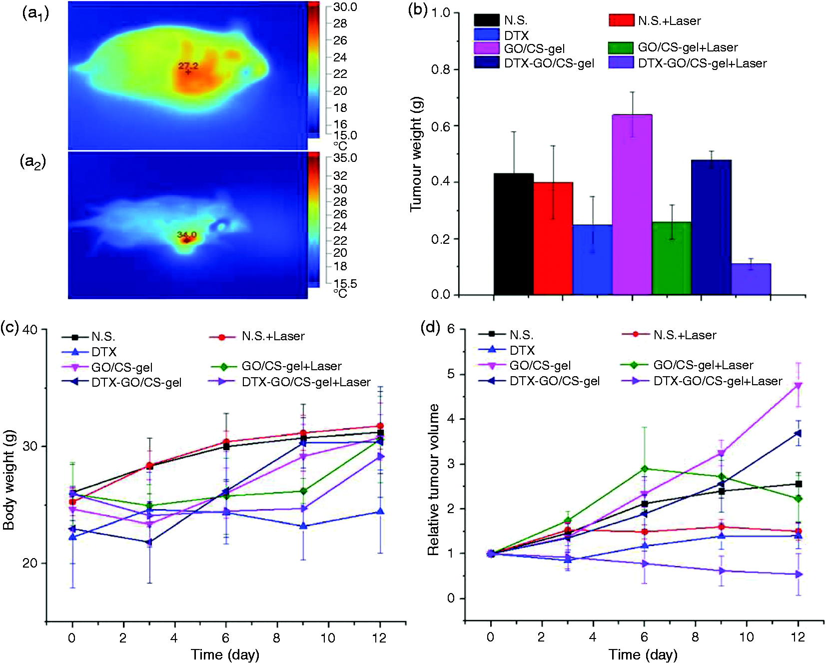
Tumor tissues were stained with H&E for pathology analysis or TUNEL agent to detect apoptotic cells. As seen in Figure 8, tumor cells in N.S. Group and GO/CS gel group showed vigorous growth, a tight arrangement, and visible pathological karyokinesis phase, while cell necrosis, lysis, and fragmentation occurred to a certain extent in N.S. + Laser, GO/CS gel + Laser, DTX, and DTX–GO/CS gel groups. In DTX–GO/CS gel + Laser group, cell fusion occurs and a large number of cell debris, which may declare that the damage degree of tumor tissue is more serious than the other groups. The results of TUNEL assay in this study (Figure 8) indicated that a few and even more apoptotic cells were found in N.S. + Laser, GO/CS gel + Laser, DTX, and DTX–GO/CS gel groups, respectively. However, with NIR Laser, the amount of apoptotic cells increased obviously. It indicated that DTX–GO/CS gel delivery system could induce cell apoptosis, and TUNEL-positive cells increased significantly under PTT treatment.
H&E stained and TUNEL apoptosis images of different tissues (400×). (a) N.S., (b) GO/CS gel, (c) DTX–GO/CS gel, (d) DTX, (e) N.S. + Laser, (f) GO/CS gel + Laser, (g) DTX–GO/CS gel + Laser.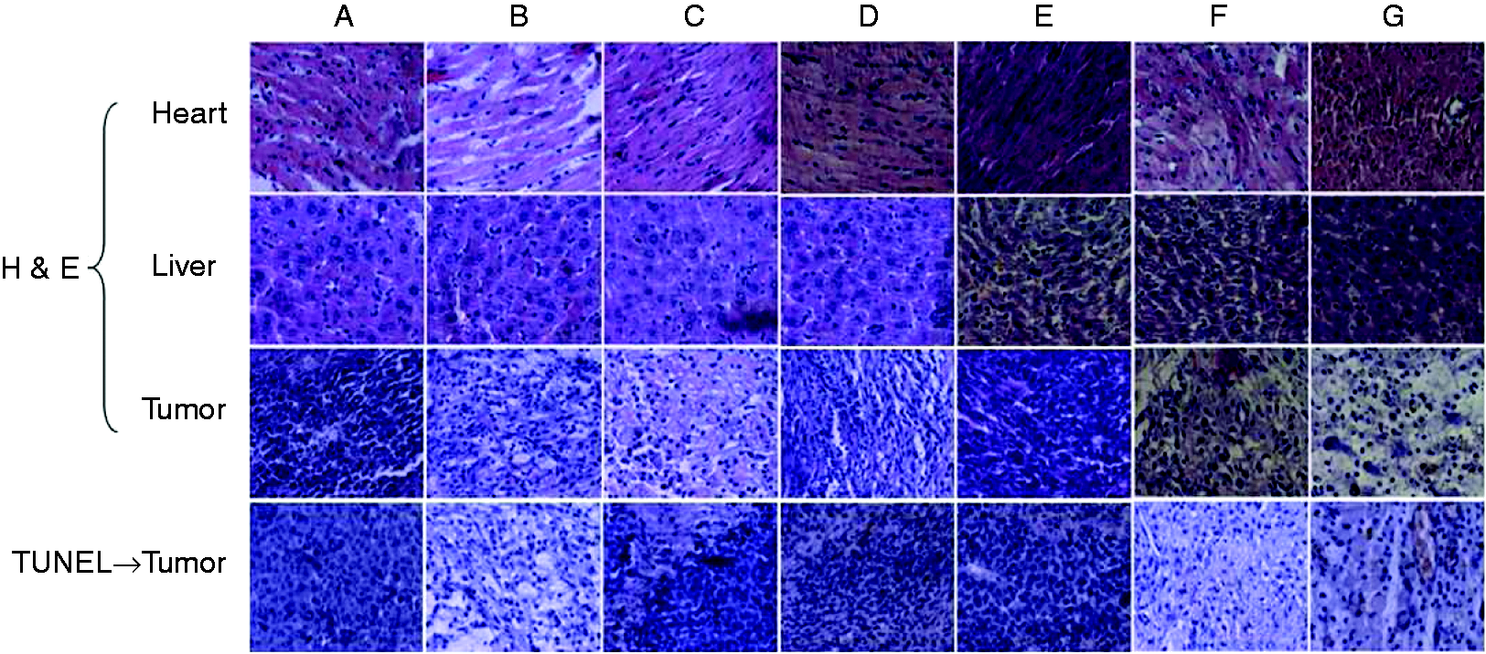
Biodistribution assay in vivo
In this study, the biodistribution of DTX–GO/CS gel was evaluated using FX imaging in vivo. Figure 9(a) showed the real-time images of IR783-labeled DTX–GO/CS gel (i.t.) and the free IR783 (i.v.) as the control in the tumor-bearing mice. After IR783-labeled DTX–GO/CS gel was given in situ, considerable fluorescence signals were detected mainly in tumor tissues of the mice. On the contrary, for the free IR783, the fluorescence signals were detected in the whole body of the mice and decreased more quickly. In particular, compared with free IR783, the fluorescence intensity of the IR783-labeled DTX–GO/CS gel in the tumor region was much higher at any time postinjection ranging from 0.5 to 24 h. This result was consistent with the quantitatively biodistribution assay (Figure 9(b)). DTX concentration in the tumor tissues of DTX–GO/CS gel group was obviously higher than the DTX group at any time postinjection, for example the concentrations at 6 h for DTX and DTX–GO/CS gel group were 17.67 ± 1.6 and 2.79 ± 0.38 µg/g, respectively. These results indicated that DTX–GO/CS gel injected in situ could significantly enhance the accumulation of DTX in tumor site and reduce distribution to normal tissues in a way, thus reducing the toxicity of DTX.38–40
Time-dependent biodistribution assay in vivo of S180 tumor-bearing mice. (a) FX imaging in vivo (a1: IR 783, i.v. a2: DTX–GO/CS gel, i.t.), (b) the drug distribution of b1: DTX (i.v.) and b2: DTX–GO/CS gel (i.t.) in tumor tissues (n = 6).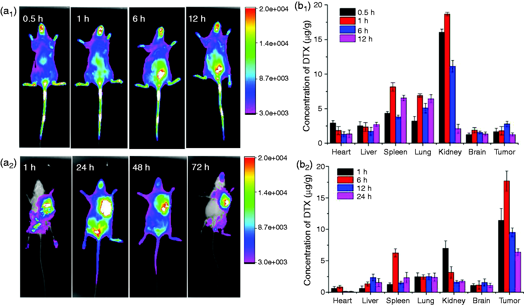
Conclusions
In conclusion, we have demonstrated successfully the chemical synthesis of GO/CS. GO/CS was stable in physiological environments, and the formation of this functionalized GO was characterized by FT-IR, TEM, and EDX. In this paper, DTX–GO/CS could successfully transfect into MCF-7 cells and cause significant tumor cell inhibition in vitro combination with 808 nm NIR laser irradiation. DTX–GO/CS gel could achieve selective killing of cancer cells in localized regions in vivo with minimal side effects, demonstrating that DTX–GO/CS gel may be promising for tumor PTT therapy in future.
Footnotes
Authors’ contribution
Zhu X and Zhang Y contributed equally to this work.
Declaration of Conflicting Interests
The author(s) declared no potential conflicts of interest with respect to the research, authorship, and/or publication of this article.
Funding
The author(s) disclosed receipt of the following financial support for the research, authorship, and/or publication of this article: This work was supported by the National Nature Science Foundation of China (No. 81273451).