
Abstract
Scaffolds in tissue engineering should be rationally designed to become an adhesion substrate friendly to cells. Schwann cells play an important role in nerve regeneration and repair. Previous studies have suggested that surface chemical groups have effect on many types of cells. However, there have hitherto been few reports on Schwann cells. In this study, we investigated cell adhesion, survival, proliferation, and neurotrophic actions of Schwann cells cultured on glass coverslips modified with different chemical groups, including methyl, carboxyl, amino, hydroxyl, mercapto, and sulfonic groups. Schwann cells on amino and carboxyl surfaces had higher attachment rate, presenting good morphology, high proliferation, and strong neurotrophic functions, while on methyl surfaces, few cells can survive, cells shrunk into round shape, exhibiting poor proliferation and weak neurotrophic functions. Growth of cells on other groups was between methyl and amino, carboxyl, and had little difference among them. Our data indicated that chemical groups can regulate behavior of Schwann cells, indicating a way to design new scaffolds for peripheral nerve regeneration.
Introduction
Peripheral nerve injury affected people’s health and life seriously.1,2 To repair peripheral nerve, various kinds of natural and synthetic biomaterials have been used to make tissue-engineered nerve grafts.3–7 For some shortcomings of autografts,8,9 artificial scaffolds are necessary to bridge peripheral nerve gaps and promote peripheral nerve regeneration. Artificial scaffolds can provide suitable space and neurotrophic support for the regenerated axons; meanwhile, they can guide axon growth.10,11 Though much effort has been devoted to prepare artificial tissue-engineered grafts for peripheral nerve repair and some success has been already made, the repair effects were still not satisfied with the clinical requests, especially for long nerve defects. 12 In recent years, the surface properties of biomaterials such as surface topography,13–15 surface chemistry,16,17 biochemical cues,18,19 and electrical activity20,21 were found to be important factors for tissue regeneration, including cell attachment, migration and proliferation, protein adsorption, etc., which decided the success or failure of the corresponding implanted scaffolds. Thus, it is very important and necessary to study the effect of surface properties of biomaterials on nerve regeneration.
Numerous studies have shown that cell adhesion to surface of biomaterials is influenced by the underlying substrate properties. Different surface chemistries will result in different cellular responses to the substrates. Effects of terminal chemical groups on growth of many kinds of cells have been studied, such as neural stem cells, 16 MC3T3-E1 cells, 22 human umbilical vein endothelial cells, 23 human fibroblasts, 24 mesenchymal stem cells. 25 The effects of chemical groups on cells will help us to understand the regulation mechanism of biomaterials on cells.
Schwann cells are a kind of glial cell in peripheral nerve system, which can guide the development and regeneration of injured axon. Schwann cells are able to enwrap axons to form compact myelin sheaths. Moreover, it was found that Schwann cells could produce various neurotrophic factors and growth factors, which are beneficial for promoting axonal growth.26–28 So, it is necessary to know more about biocompatibility between Schwann cells and the artificial materials in order to seek suitable substrates to enhance the cell survival and proliferation.
Therefore, the aim of this study was to investigate the relationships between chemical functional groups and the behaviors of Schwann cells. For this purpose, a set of substrates modified with different functional end groups, including methyl (–CH3), carboxyl (–COOH), amino (–NH2), hydroxyl (–OH), mercapto (–SH), and sulfonic (–SO3H) were prepared by chemical reaction. Adhesion, survival, proliferation, and neurotrophic actions of Schwann cells were then examined to characterize the effects of chemical functional groups on Schwann cells.
Materials and methods
Preparation of model surfaces on glass coverslips
Before being introduced functional groups on the surface, glass coverslips were pretreated with sodium dodecyl sulfate (SDS) solution and hydrochloric acid solution followed by a rinse with deionized water. Surface modification was carried out as previously described. In brief, to get glass coverslip modified with –OH, 29 clean coverslips were dipped into fresh Piranha solution made of concentrated H2SO4 and 30% H2O2 at 80℃ for 45 min, then rinsed with deionized water and stored in deionized water. To introduce –NH2 and –SH groups, 30 the –OH coverslips were dipped into 1% (3-aminopropyl)-triethoxysilane (Sigma, USA) or (3-mercaptopropyl)-trimethoxysilane (Aladdin, Shanghai, China) alcohol solution under vibration for 30 min followed by being rinsed in ethanol and deionized water and then dried at 100℃ for 1 h. To introduce –CH3 group, 25 the –OH coverslips were dipped into dimethyldicholorosilane (Aladdin, Shanghai, China) for 15 s, and rinsed with toluene and ethanol, respectively, dried at 100℃ for 1 h. To introduce –COOH group, 31 the –NH2 coverslips were dipped into succinic anhydride/dimethylformamide solution for 30 min, rinsed with dimethylformamide and deionized water and then dried in nitrogen. To introduce –SO3H group, 16 the –SH coverslips were dipped into concentrated nitric acid solution for 24 h, rinsed with deionized water and then dried in nitrogen.
Characterization of substrate contact angle measurement
The surface chemical states of coverslips were determined by static (sessile drop) water contact angle and X-ray photoelectron spectroscopy (XPS). Static water contact angles were measured by a contact angle system DCAT21 (Dataphysics, Germany). 32 Samples were fixed to a sample platform and then 3 µL of ultrapure water was added to the surface. The water contact angle was detected with a horizontal microscope. Each sample was examined three times at different locations and the mean value was calculated. XPS spectra were recorded using a Thermo Scientific K-Alpha spectrometer (Thermo Scientific, USA) equipped with a monochromatic Al-Kα X-ray source.
Rat Schwann cells isolation
Primary rat Schwann cells (rSCs) were isolated from sciatic nerves and dorsal root ganglia (DRG) of neonatal Sprague-Dawley rats (1–2 days) obtained from the Laboratory Animal Center of Nantong University, Jiangsu Province, China. First, the obtained tissues were sheared into fragments and digested in 0.25% trypsin at 37℃ for 30 min, DMEM supplemented with 10% fetal bovine serum (FBS) and 100 U/ml penicillin and 100 µg/ml streptomycin (complete medium) was then added to end the digestion process. The cells obtained were planted onto poly-L-lysine pre-coated dishes with complete medium, followed by incubation at 37℃ in a humidified 5% CO2 atmosphere. After 24 h, the medium was replaced by fresh complete medium together with 10 µM cytosine arabinoside (Sigma, USA) and the incubation was continued for another 48 h. Then rSCs were cultured in complete medium containing 2 µM forsklin (Sigma, St Louis, MO) and 2 ng/ml heregulin (Sigma, USA). For further purification, cells were treated with anti-Thy1 antibody (1:1000, AbD Serotec, Raleigh, NC, USA) on ice for 2 h, followed by treatment with complement (Jackson Immuno, West Grove, PA, USA) for another 40 min. The purified rSCs were cultured with complete medium containing growth factor until the cells were sufficient to seed on the glass coverslips.
rSCs culture
The prepared glass coverslips were sterilized with 70% alcohol and rinsed extensively with sterilized phosphate-buffered saline (PBS). Then all samples were put in 24-well culture plates and cell suspension was added in. The seeding cell density was 1 × 10⁵ cells/well. At different times of culture, the morphological changes of Schwann cells on these different glass coverslips were observed under an inverted light microscope.
Immunostaining of Schwann cell
To characterize the cell purity, primary cultured Schwann cells were fixed in 4% paraformaldehyde and allowed to incubate with mouse anti-S100 antibody (1:400, Sigma) at 4℃ for 24 h, followed by further reaction with FITC (fluorescein isothiocyanate)-labeled anti-mouse 488 (1:200 dilution, Proteintech Group, Chicago, IL) at 4℃ overnight. Then cells were incubated with a 5 µg/mL Hoechst 33342 (Sigma) for 15 min at room temperature. Cell samples were observed under an inverted light microscope.
Quantification of cell adhesion
After 4 h of seeding, Schwann cells started to adhere to each substrate; poorly attached and non-adherent cells were removed by washing the wells with PBS gently. The adherent cells were fixed with 4% (w/v) paraformaldehyde and stained with 0.1% (w/v) crystal violet for 30 min at room temperature and then 10% (v/v) acetic acid was added to release the dye. The absorbency was measured at 570 nm by a microplate reader (Bio-Tek Inc., USA). The relative number of adherent cells was determined in terms of the absorbance value.
Morphology observation
The morphology of Schwann cells cultured on different coverslips was examined by Philips XL-30 scanning electron microscope (SEM, Eindhoven, The Netherlands). First, all samples were dehydrated at increasing alcohol concentrations (50, 70, 90, and 100%; Valcohol/Vdd H2O) in succession and then dried naturally. Before SEM tests, the samples were coated with gold using a JEOL JFC-110E Ion Sputter.
MTT assays
The amount of cells on different substrates was determined using colorimetric MTT assay (Bio-Tek Inc., USA). MTT (3-(4,5-dimethylthiazol-2-yl)-2,5-diphenyltetrazolium bromide) gives a yellowish aqueous solution, which is reduced by mitochondrial dehydrogenases in living cells. After Schwann cells were cultured on different samples for 24 h and 72 h, MTT solution was added to replace the cell medium at a final concentration of 0.5 mg/ml to allow cell incubation at 37℃ for 4 h. Then SDS solution was added to dissolve the formazan precipitate for 20 h, and the absorbance was measured at 570 nm by an ElX-800 micro-ELISA reader (Bio-Tek Inc., USA).
EdU incorporation assay
EdU (5-ethynyl-2′-deoxyuridine) incorporation assay was performed to further investigate the impact of chemical groups on the proliferation of Schwann cells according to the manual of an EdU labeling/detection kit (Ribobio, Guangzhou, China). In brief, 50 µM EdU labeling medium was added to cell culture to allow incubation for 12 h at 37℃ under 5% CO2. Then cells were fixed with 4% (w/v) paraformaldehyde for 30 min and incubated with glycine for 5 min. After being washed with PBS, cells were worked with anti-EdU working solution for 30 min at room temperature. Afterwards, cells were washed with 0.5% TritonX-100, methanol, and PBS in turn, then the cells were incubated with 5 mg/ml Hoechst 33342 dye at room temperature for 30 min, followed by observation under a scanning laser confocal microscopy (Leica, Heidelberg, Germany). The percentage of EdU-positive cells was calculated from 10 random fields in three wells.
Western blot analysis
Schwann cells were rinsed twice with ice cold PBS and put in lysis buffer and then incubated for 30 min on ice. The cell lysates were centrifuged (12,000 r/min) at 4℃ for 10 min. The resultant samples were electrophoretically separated on a 10% SDS-polyacrilamide gel (PAGE) and transferred to a polyvinylidene fluoride (PVDF) membrane. The membrane was incubated with a mouse anti-N-cadherin antibody (1:2500) or a mouse anti-β-catenin antibody (1:500) (both from BD Transduction Laboratories, San Diego, CA) at 4℃ overnight. β-actin (1:2500) was used as an internal control. Then the membranes were further incubated with a secondary antibody, anti-mouse IgG (H&L) (goat), IRDye800 conjugated (1:4000, Rockland Gilbertsville, CA) at room temperature for 2 h, followed by image scanning with Odyssey infrared imaging system (LICOR, Lincoln, NE). The band density was quantified by the software of Quantity One (Bio-Rad).
Quantitative real-time RT-PCR
Total ribonucleic acid (RNA) was extracted from the cell lysate using a Trizol kit (Invitrogen, Carlsbad, CA) and transcribed into complementary DNA (cDNA) using an Omniscript RT kit (Qiagen, Valencia, CA) according to the supplier’s instructions. To detect the mRNA level of nerve growth factor (NGF) and brain-derived neurotropic factor (BDNF), quantitative real-time RT-PCR was performed in a StepOne real-time polymerase chain reaction (PCR) system (Applied Biosystems, Foster City, CA). A 20-µL reaction mixture contained 1 µL of cDNA from samples, 10 µL of 2 × Fast EvaGreen® qPCR Master Mix, 2 µL of 10 × ROX of the assays-on-demand kit, 1 µL primer, and 6 µL of RNase/DNase free water. PCR procedures were: incubation at 94℃ for 5 min, 25 cycles at 94℃ for 25 s and 64℃ for 25 s. The data were analyzed by a specific software supplied by the vendor (Applied Biosystems). The primers used for amplification were as followed: GAPDH, forward: 5′-GCA AGT TCA ACG GCA CAG-3′, reverse: 5′-CGC CAG TAG ACT CCA CGA C-3′ (141 bp); NGF, forward: 5′-GCT GGA CCC AAG CTC AC-3′, reverse: 5′-CCC TCT GGG ACA TTG CTA T C-3′ (179 bp); BDNF, forward: 5′-CAG GGG CAT AGA CAA AAG-3′, reverse: 5′-CTT CCC CTT TTA ATG GTC-3′ (153 bp).
Enzyme-linked immunosorbent assay
After Schwann cells were seeded on the samples and incubated in medium at 37℃ for one and three days, respectively, then the supernatant was collected and stored at −20℃ before use. NGF and BDNF released by Schwann cells were measured by a sandwich enzyme-linked immunosorbent assay (ELISA) kit (Chemicon, Temecula, CA) according to the package insert.
Results
Characterization of different model surfaces and Schwann cells
Static water contact angles measured by the sessile drop method on modified glass surfaces are shown in Figure 1. Among all test surfaces, the –OH surface was the most hydrophilic with a contact angle of 15.5 ± 1.4, while the –CH3 surface had the highest contact angle of 103.0 ± 3.5, which is the most hydrophobic surface in these test substrates. Order of contact angle of other four surfaces is: –COOH < –SO3H < –NH2 < –SH.
Contact angle measurements of different glass surfaces. Results are the means ± SD of three independent experiments. (Each in duplicate).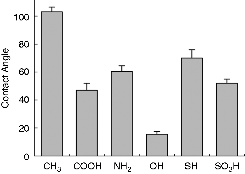
The XPS spectra of the coverslips surface modified with different chemical functions are shown in Figure 2. Compared with blank coverslip (no modification), a single weak peak at 400.1 eV is consistent with the N–H bond type, showing existing of –NH2 (Figure 2(A, d)). S 2 p peak at 169.4 and 170.3 eV reveals the presence of S on –SH and –SO3H surface (Figure 2(A, f) and (A, g)). On –COOH surface, the C = O component was positioned at 287.4 eV; O–C = O at 289.0 eV (arrow in Figure 2(b)). No obvious difference was seen between CH3, OH, and the blank coverslip maybe because C–H and O–H exist in every sample.
XPS survey spectrum of coverslips modified with different chemical groups. (A): (a) blank (no modification), (b) –CH3, (c) –COOH, (d) –NH2, (e) –OH, (f) –SH, (g) –SO3H; (B): high-resolution C 1 s spectrum of (a) blank, (b) –COOH.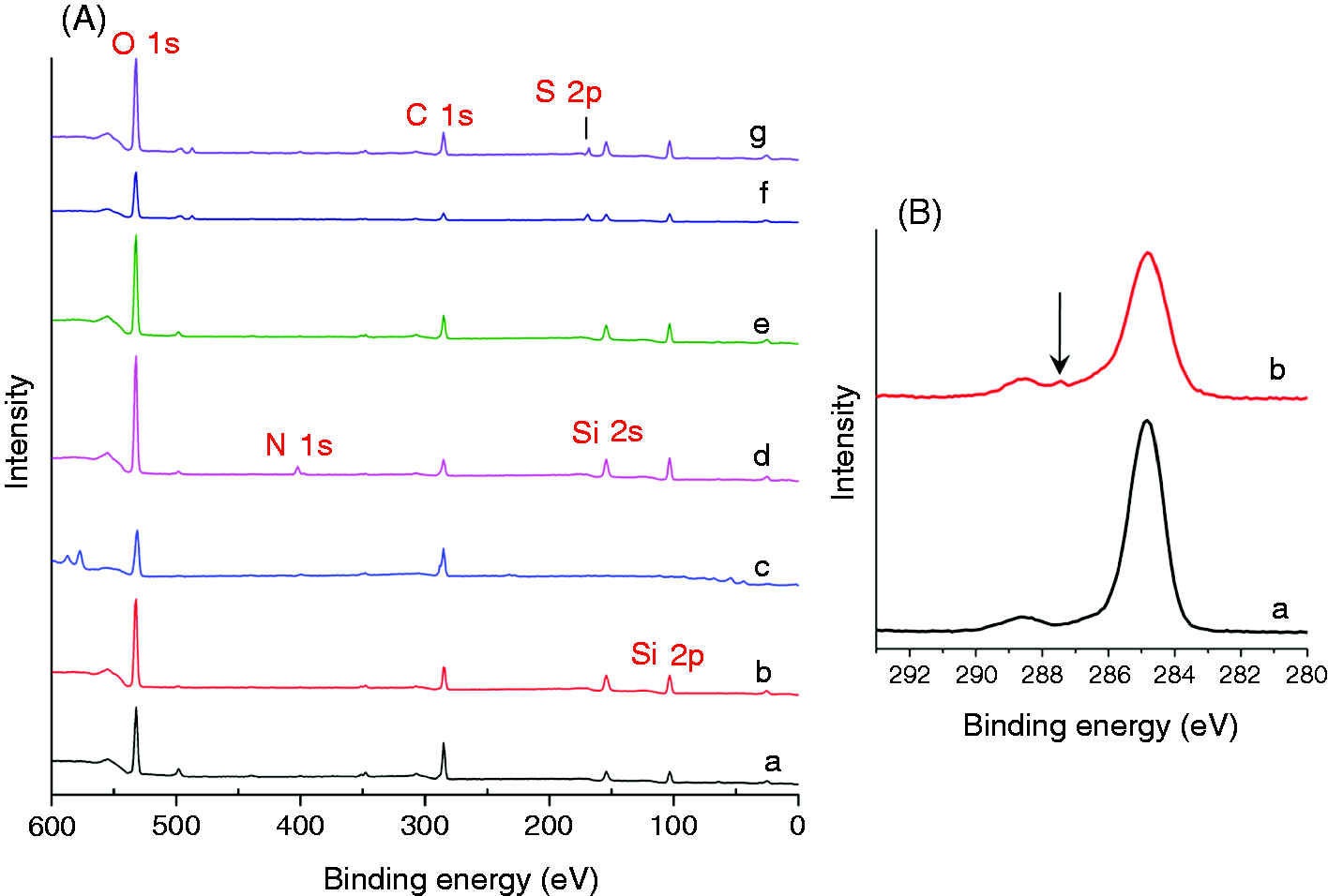
After isolation and purification, pure Schwann cells were procured. Figure 3 shows immunocytochemistry of Schwann cells with anti-S100.
Immunocytochemistry of Schwann cells: left, S100 (a), nuclei labeled with Hoechest 33342 (b), merge (c).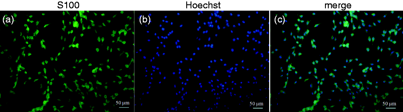
Adhesion of Schwann cells on substrates with different groups
Supporting cell adhesion is principal for biomaterials due to their intimate interactions with cells and tissues. After 4 h of plating, the differential adhesion of Schwann cells on different substrates was investigated, and quantitative data indicated that the relative number of adherent Schwann cells on the –NH2 surface was a little larger than –COOH surface, but significantly larger than four other surfaces. Schwann cells showed very low attachment on –CH3 surface, so the relative number was obvious lower than other groups (Figure 4(a)).
Adhesion and spreading of Schwann cells on substrates modified with different chemical groups. (a) The relative number of Schwann cells adhered on substrates after 4 h of seeding, as determined by the absorbance of released crystal violet dye at 570 nm. Data were presented as means ± SEM of three independent experiments (each in duplicate). *P < 0.05 compared to substrates modified with –NH2, #P < 0.05 compared to substrates modified with –CH3. (b) Brightfield images of Schwan cells grown on different samples for 48 h. Scale bar, 50 µm.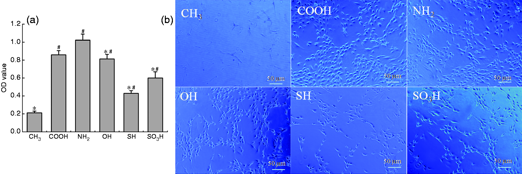
After 48 h culture, on –NH2 surface, Schwann cells exhibited the best adhesion and spreading, most cells displayed a spindle-like morphology with long filopodia at both ends, filopodia of different cells connected to each other and formed a network, showing strong cell–cell interaction. On –COOH, –OH, and –SO3H surfaces, many cells also exhibited spindle-like morphology with clear edges, but the cell extension was shorter than that grown on –NH2 surface. On –CH3 surface, number of cells was significantly less compared to other surfaces, in addition, most cells displayed round shape with no projection, showing poor attachment and spreading. On –SH surface, also some cells showed rounded cell body with short processes, but number of cells was larger than that on –CH3 surface and some cells exhibited ovoid shape (Figures 4(b) and 5(a)). For each group, as time went, branched structure appeared more and more obvious. Numerous filopodias grew out from the body. Especially on –NH2, –COOH, and –OH surface interactions between cells were more significant (Figure 5(a)). The extent of cell elongation on different surface was evaluated by aspect ratio of cells (Figure 5(b)). Cells on –CH3 had the least aspect ratio which is consistent with the morphology observation above. Cells on –NH2 surface had the largest ratio, indicating that substrates with –NH2 group can attribute to cell spreading, elongation, and grow. From 24 h to 72 h, the aspect ratio decreased slightly. There were many cells on –COOH surface, but they grew disorderly and the processes extent in all directions.
Morphology of Schwann cells cultured on coverslips modified by different chemical groups for 24 h and 72 h. (a) SEM of cells, scale bar = 100 µm. (b) length/width of cells, *P < 0.05 compared to substrates modified with –CH3.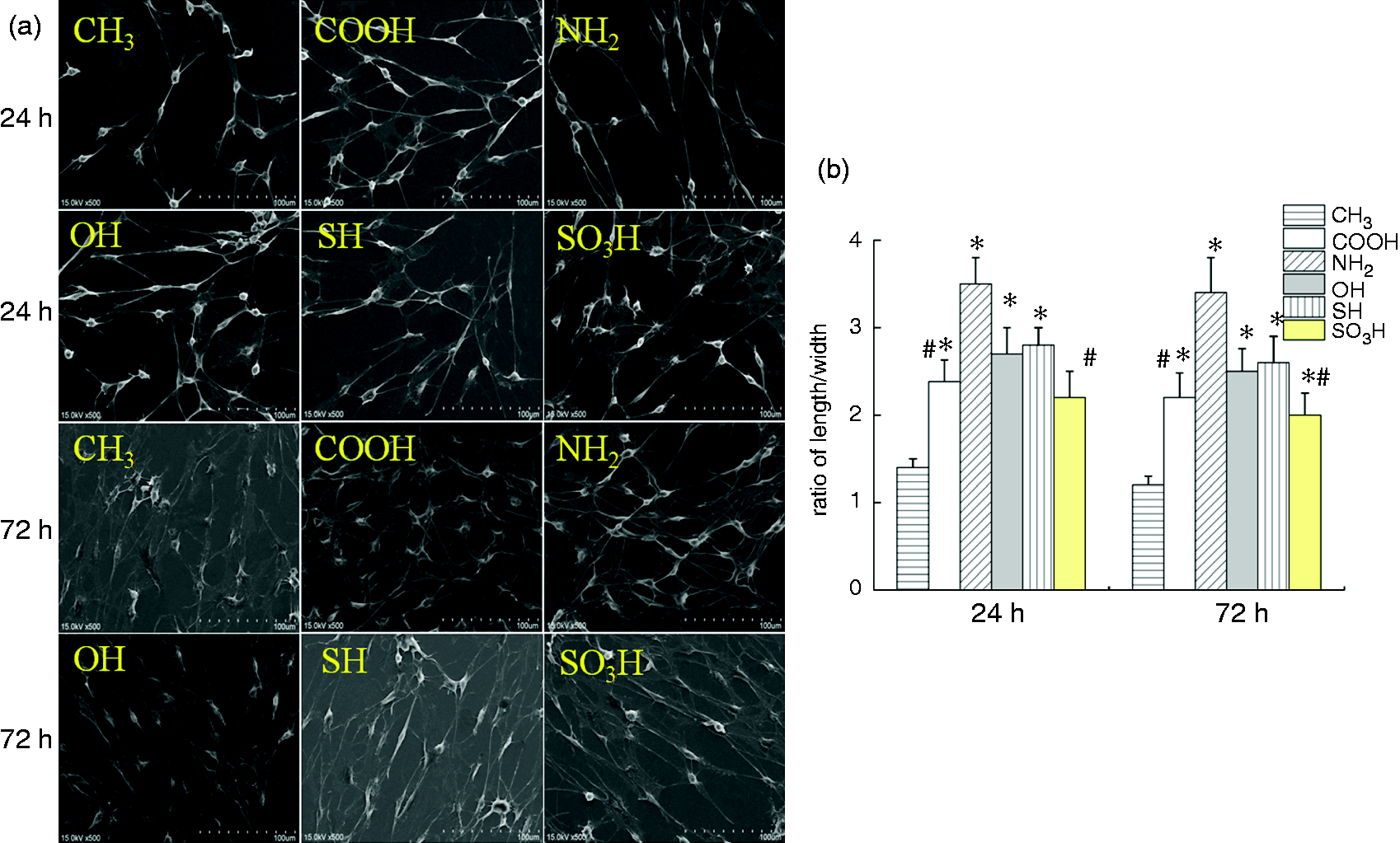
Survival of Schwann cells on substrates with different groups
The cell viability of Schwann cells on the samples was quantitatively investigated by an MTT assay after culture for 24 and 72 h, respectively. It could be found from Figure 6 that at 24 h, the cell viability of Schwann cells grown on the –NH2 surface was significantly higher than that on –CH3, –SH, and –SO3H surfaces, but a little higher than that on –COOH and –OH surfaces. There was no significant difference between –COOH, –OH, –SH, and –SO3H surfaces. Number of survival cells on –CH3 surface was obviously less than other surfaces. At 72 h, order of cell viability on all surfaces was the same as that at 24 h, but advantages of –NH2 surface was more obvious, the cell viability of Schwann cells grown on the –NH2 surface was significantly higher than that on all other surfaces, and the cell viability on –CH3 surface was still very low. Number of cells on all surfaces other than –CH3 increased from 24 h to 72 h, indicating better cell proliferation on these samples.
The changes in the cell viability of Schwann cells after they were cultured on different substrate for 24 h and 72 h, as measured by MTT assay.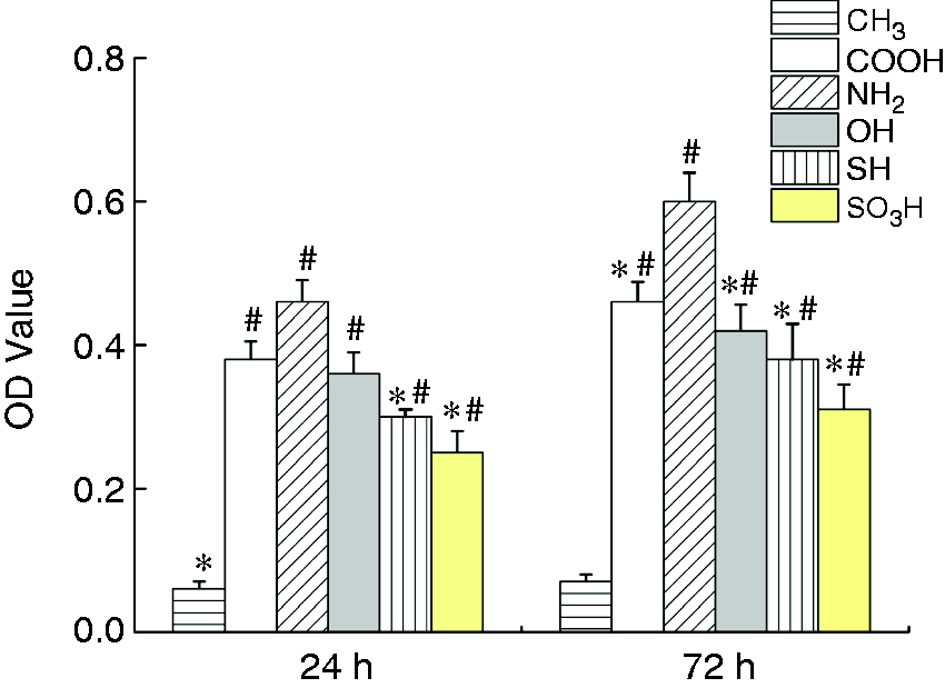
Proliferation of Schwann cells on substrates modified with different chemical groups
EdU incorporation was further performed to determine the effect of chemical groups on Schwann cell proliferation. From Figure 7(a) and (b), we can see that the number of EdU positive cells on –NH2 surface was the highest, indicating that the –NH2 surface led to prominent enhancement on cell proliferation among all substrates; there was no significant difference among –COOH, –OH, and –SO3H surfaces; –CH3 surface had the lowest proliferation as always.
(a) Representative EdU staining image. Scale bar, 50 µm. (b) The effect of chemical group on cell proliferation of Schwann cells in culture for 48 h, as determined by EdU staining. Data were presented as means ± SEM of three independent experiments (each in duplicate). *P < 0.05 compared to substrates modified with –NH2, #P < 0.05 compared to substrates modified with –CH3.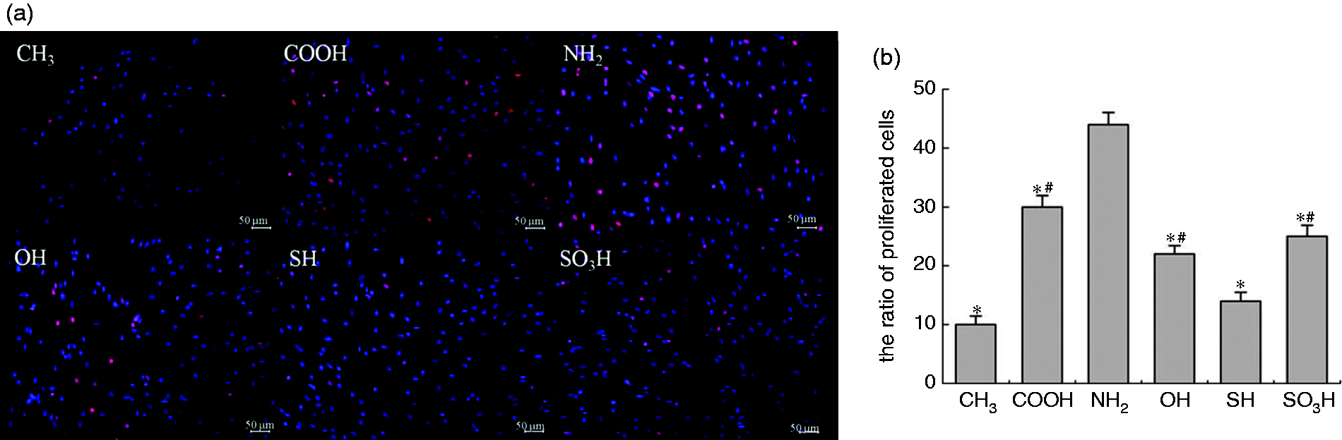
Expression of adhesion-related proteins in Schwann cells on substrates modified with different chemical groups
For tiny amount of protein was collected from Schwann cells cultured on –CH3 surface, expression of proteins in cells on –CH3 sample was not shown here. Western blot analysis indicated that expression of β-catenin was significantly higher in Schwann cells on the –COOH surface than that on –OH, –SH, and –SO3H surfaces. On –NH2 surface, expression of β-catenin was also higher than –OH, –SH, and –SO3H surfaces but a little lower than –COOH surface. The expression of β-catenin in cells adhered to different substrates followed the sequence: –COOH > –NH2 > –OH > –SH > –SO3H. Expression of N-cadherin was significantly higher in Schwann cells on the –NH2 surface than that on other surfaces. The order was: –NH2 > –COOH > –OH > –SH > –SO3H. Expression of two proteins was slightly higher at 72 h than that at 24 h, no obvious difference was observed (Figure 8).
Protein expression levels of N-cadherin and β-catenin in Schwann cells on different substrates for 24 h (a) and 72 h (b) culture. Also shown is the representative Western blot image. β-actin served as a loading control. Data were presented as means ± SEM of three independent experiments (each in duplicate). *P < 0.05 versus the substrates modified with –NH2 and –COOH. #P < 0.05 compared to substrates modified with –NH2.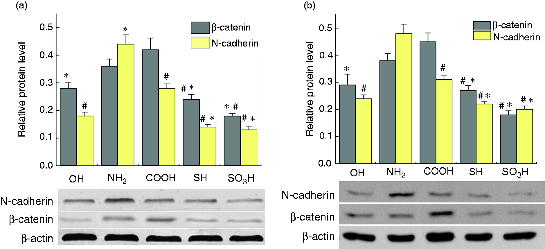
Neurotrophic actions of Schwann cells on different substrates
Since the chemical properties of substrate could affect the phenotype changes of Schwann cells, the possible influences of the chemical groups on Schwann cell function also deserved to be studied. Quantitative real-time PCR showed that the mRNA level of NGF and BDNF was significantly higher in Schwann cells grown on the –NH2 surface than in Schwann cells grown on other substrates, while the mRNA level of two factors was much lower in Schwann cells grown on –CH3 surface compared to that on other surfaces (Figure 9(a)). ELISA assay was used to determine the protein level of NGF and BDNF released by Schwann cells. We also found that after culture for 24 h and 72 h, Schwann cells on the −NH2 surface could release the higher level of NGF and BDNF than Schwann cells on other substrates, while the amount of NGF and BDNF secretion on –CH3 surface was much lower than that on other surfaces. However, there was no significant difference for mRNA level or release of two growth factors of cells on substrates modified by groups other than –NH2 and –CH3 at the same time point (Figure 9(b) and (c)).
Gene expression and release of growth factors in Schwann cells on different substrates. (a) Histogram showing that the mRNA levels of NGF and BDNF in Schwann cells cultured for 48 h. (b, c) Histograms showing the growth factors, NGF and BDNF, released by Schwann cells cultured for one and three days, respectively, as measured by ELISA assay. Data were presented as means ± SEM of three independent experiments (each in duplicate). *P < 0.05 compared to substrates modified with –NH2, # P < 0.01 versus the substrate modified with –CH3.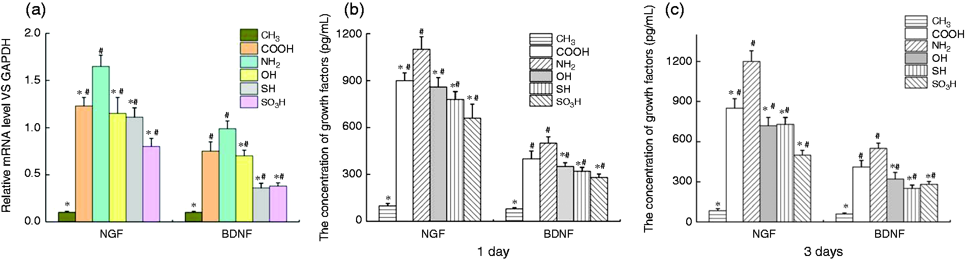
Discussion
Growth and behavior of cells are affected by a variety of factors. Many efforts have been made to promote cell adhesion and subsequent activities. For example, Li et al. 33 studied effects of chitosan scaffolds with surface micropatterning and inner porosity on Schwann cells. In Wohlrab’s study, the engineered recombinant spider silk protein was modified with the integrin recognition sequence RGD (arginine-glycine-aspartic acid) to enhance cell adhesion and proliferation. 34 Lee et al. 35 synthesized substrates with both amine functional group and electroactivity, which could promote adhesion of both Schwann cells and fibroblasts. Matsumine et al. 36 developed a biodegradable nerve conduit with PLA non-woven fabric and found it has comparable ability to induce peripheral nerve regeneration following autologous nerve transplantation.
Among these complex cues, surface chemistry property has been a concern.23,37,38 However, few studies have been performed to investigate effect of surface chemistry on Schwann cells. Therefore, in this study, we aimed to investigate Schwann cell behavior seeded on glass coverslips modified with different chemical groups.
In our study, the results from cell adhesion, cell viability, and micrographic observation designated samples with –NH2 and –COOH groups were suitable to be used to prepare substrates responsible for such cell behaviors. On –SO3H surface, cells also had a better adhesion. Though these three groups had similar wettability based on our contact angle measurement, there were differences in cell adhesion and proliferation among them, indicating that cell behavior are affected by various factors rather than depending only on hydrophilicity. Previous studies also reported that samples with same hydrophilicity but different charged functional groups might have obvious effect on cell adhesion, differentiation, and proliferation.
39
Substrates with –NH2 surface were most beneficial to cell adhesion probably owing to the
Schwann cells play a pivotal role in peripheral nerve development and regeneration. Non-myelinating Schwann cells are involved in maintenance of axons and are crucial for neuronal survival. In myelinated axons, Schwann cells wrap around axons of motor and sensory neurons to form myelin sheath. During the process of myelination, Schwann cells proliferate rapidly and provide a channel for the axon to grow along.40,41 Therefore, the speed of proliferation is closely related to myelin formation. In our study, Schwann cells cultured on –NH2 surface led to prominent enhancement on cell proliferation among all substrates, implying that scaffolds made of biomaterials with –NH2 may promote nerve regeneration.
Besides important role in myelination, Schwann cells are able to produce various growth factors and provide neurotrophic support for neurons. 42 NGF and BDNF are all neurotrophic factors. NGF can promote the survival and neurite outgrowth of sympathetic and sensory neurons. 43 BDNF can promote neurite outgrowth as well as induce motor axonal outgrowth.44–46 Neurotrophins mediate their effects by binding to specific receptors. In our study, the highest amount of NGF and BDNF was produced by Schwann cells cultured on –NH2 surface, cells on –COOH, and –OH surface also produced more NGF and BDNF. This result is in consistence with effect of chemical groups on cell behavior of Schwann cells.
Cytoskeletal proteins of Schwann cells contain N-cadherin. Cadherins are a class of transmembrane proteins, playing important roles in cell adhesion, forming adherens junctions to bind cells within tissues together.47,48 β-catenin is a subunit of the cadherin protein complex and acts as an intracellular signal transducer in the Wnt signaling pathway. It regulates the coordination of cell–cell adhesion and gene transcription. 49 In the present study, the –NH2 surface showed the higher expression of these two adhesion-related proteins than other substrates, which can explain the best adhesion of Schwann cells grown on –NH2 surface. Our data of western blot analysis suggest that sample with –NH2 surface may be an ideal substrate that mediates cell–substrate and cell–cell interactions.
Conclusion
In summary, our study offers evidence that the type of chemical group on substrates has effects on adhesion, viability, proliferation, and protein expression of Schwann cells. Positively charged –NH2 surfaces not only sustained the greatest amount of viable cell adhesion, proliferation, but also promote neurotrophic factor secretion, and expression of some adhesion-associated proteins. –COOH and –OH surfaces also benefited growth of cells. These results developed a new parameter for designing ideal scaffolds in nerve tissue engineering.
Footnotes
Declaration of Conflicting Interests
The author(s) declared no potential conflicts of interest with respect to the research, authorship, and/or publication of this article.
Funding
The author(s) disclosed receipt of the following financial support for the research, authorship, and/or publication of this article: This study was supported by the National Natural Science Foundation of China (No. 81371687 and 81171457), the National Science Research Program of Jiangsu Education Department (No. 14KJB 180020).