
Abstract
Arsenic trioxide (As2O3, ATO), a FDA approved drug for hematologic malignancies, was proved of efficient growth inhibition of cancer cell in vitro or solid tumor in vivo. However, its effect on solid tumor in vivo was hampered by its poor pharmacokinetics and dose-limited toxicity. In this study, a polyacrylic acid capped pH-triggered mesoporous silica nanoparticles was conducted to improve the pharmacokinetics and enhance the antitumor effect of arsenic trioxide. The mesoporous silica nanoparticles loaded with arsenic trioxide was grafted with polyacrylic acid (PAA-ATO-MSN) as a pH-responsive biomaterial on the surface to achieve the release of drug in acidic microenvironment of tumor, instead of burst release action in circulation. The nanoparticles were characterized with uniform grain size (particle sizes of 158.6 ± 1.3 nm and pore sizes of 3.71 nm, respectively), historically comparable drug loading efficiency (11.42 ± 1.75%), pH-responsive and strengthened sustained release features. The cell toxicity of amino groups modified mesoporous silica nanoparticles (NH2-MSN) was significantly reduced by capping of polyacrylic acid. In pharmacokinetic studies, the half time (t1/2β) was prolonged by 1.3 times, and the area under curve) was increased by 2.6 times in PAA-ATO-MSN group compared with free arsenic trioxide group. Subsequently, the antitumor efficacy in vitro (SMMC-7721 cell line) and in vivo (H22 xenografts) was remarkably enhanced indicated that PAA-ATO-MSN improved the antitumor effect of the drug. These results suggest that the polyacrylic acid capped mesoporous silica nanoparticles (PAA-MSN) will be a promising nanocarrier for improving pharmacokinetic features and enhancing the anti-tumor efficacy of arsenic trioxide.
Introduction
As the active ingredient of traditional Chinese medicine of arsenic, arsenic trioxide (ATO) was applied to treat cancers in the 1970s and approved by FDA for the treatment of acute promyelocytic leukemia (APL) in 2000.1,2 Recent studies showed that ATO can induce the apoptosis and inhibit the growth, migration of solid tumor cells.3–6 Despite its attractive pharmacological activities, the therapeutic potential of ATO has been significantly restricted for its rapid renal clearance of arsenic metabolites, 7 low concentration in tumor sites and dose-limited adverse reactions, such as skin reactions and liver dysfunction. 8 To improve the therapeutic efficacy of As2O3 in cancer treatments, various drug delivery system (DDS) such as As2O3 loaded polylactic acid(PLA)/magnetic nanoparticles, arsenic loaded multiple drug mesoporous silica nanoparticles and [Ni(HAsO3)]-loaded polymer-caged nanobin were conducted.9–11 However, few of them focused on the features of the DDS in vivo. Therefore, our group explored the in vitro/vivo characteristics of ATO after it was loaded by functionalized DDS.
Given the intrinsically inorganic nature of ATO, MSN, a novel inorganic nanomaterial with advantages of large surface area and pore volume, versatile possibility for further functionalization and excellent biocompatibility,12–15 are considered as promising nanocarriers for loading ATO. More importantly, MSN are suitable for loading toxic drugs and releasing them sustainedly.16–18 Despite these superiorities, the nanocarriers still facing the limitation of releasing the cargo in a burst fashion during its using period.19,20 Recently, stimuli-responsive mesoporous silica nanoparticles capping with light-, enzyme-, temperature- or pH-responsive materials could specifically release the drug by the stimulation of the internal/external condition.21–24 Among different kinds of stimuli, pH-responsive systems are most widely utilized in drug delivery for cancer therapy.25,26 As is known, the pH value in tumor sites (pH 5.5–6.5) is lower than that in normal tissue (pH 7.4).27,28 Therefore, it is meaningful to develop an ATO-loaded pH-responsive DDS to enhance the therapeutic effect on solid tumor.
As one of the most commonly used pH-responsive materials, polyacrylic acid (PAA) with low toxicity, good biocompatibility and carboxyl group has shown great potential in various areas for biomedicine for its unique physical and chemical properties.29,30 It has been grafted on biocompatible materials which were used as drug carriers frequently.31–33 In acidic conditions, PAA is likely to protonate to be relatively hydrophobic; on the contrary, it will deprotonate in neutral or alkaline conditions to be hydrophilic. Therefore, its solubility becomes poor and even collapses entangled together to precipitate with the decrease of pH. 34 In this paper, amino groups modified MSN (NH2-MSN) was prepared to load acidic ATO by electrostatic interactions. Then PAA-capped ATO-MSN was designed and synthesized via a condensation between NH2 group of NH2-MSN and carboxyl group of PAA. The PAA layers could not only conceal the large amounts of naked amino on NH2-MSN which may cause undesirable side effects to normal cells and organs 35 but also suppress the burst release action and control the release of ATO in acidic microenvironment in solid tumor.
Herein, we introduced a facile grafted-onto strategy to prepare pH-triggered MSN to achieve sustained release of ATO (Figure 1). And its cytotoxicity study showed low toxicity of blank nanocarriers (PAA-MSN) on SMMC-7721. The in vitro test of antitumor efficacy and cell cycle experiment exhibited strongest antitumor efficiency of PAA-ATO-MSN. In this paper, a method by inductively coupled plasma mass spectrometry (ICP-MS) with a low quantitative limit was applied to detect the drug concentrations in pharmacokinetic study. The PK results showed that the prepared DDS really prolonged the t1/2β and mean residence time (MRT) of ATO. What is more, with the advantages of pH-triggered and sustained release features, PAA-ATO-MSN significantly increased the antitumor efficiency in mice. In our studies, morphological evaluation employed transmission electron microscopy and mesoporous structure was characterized by N2 adsorption-desorption isotherms and small angle X-ray diffractometer. Furthermore, in vitro release behavior, pharmacokinetics and pharmacodynamics of the smart nanosized DDS were investigated. The obtained results indicated that PAA capped pH-responsive sustained release DDS was prepared successfully and it could improve the pharmacokinetic features of ATO. Otherwise, our functionalized MSN-based pH-triggered nanocarriers may have important significance as a carrier for poor pharmacokinetics and/or dose-limited toxic drug to enhance their therapeutic effect of solid tumor.
Schematic representation of the constructing PAA-ATO-MSN.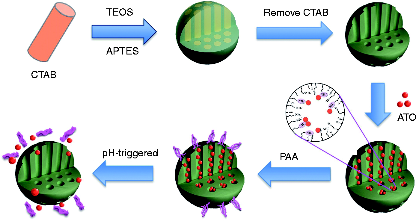
Materials and methods
Materials
Arsenic trioxide (ATO, 90%) was purchased from Alfa Aesar (Shanghai, China). Arsenic standard solution (1000 mg·L−1) was purchased from Beijing century audiocodes biological technology Co., Ltd. (Beijing, China). Tetraethyl orthosilicate (TEOS), hexadecyltrimethylammonium bromide (CTAB), ammonia propyl triethoxy silane (APTES) and polyacrylic acid (PAA, Mw = 1800) were obtained from Sigma Co., Ltd. (St.Louis, MO, USA). SMMC-7721 cell line was provided by the Department of Pharmacology, Zhejiang Chinese Medical University. Dulbecco’s modified Eagle medium (DMEM), fetal bovine serum (FBS), 3-(4,5-Dimethylthiazol-2-yl)-2,5-diphenyltetrazolium bromide (MTT), penicillin G sodium and streptomycin sulfate were all obtained from Gibco BRL (Gaithersberg, MD, USA). Sprague-Dawley (SD) rats (220–260 g, half male and half female) and Kunming mice (25–30 g, male) were supplied by the Laboratory Animal Center of Zhejiang Chinese Medical University. All other chemicals were of analytical grade and used as received without further purification.
Preparation of PAA-ATO-MSN
NH2-MSN was prepared with modified Stober method according to the exiting methods. 36 The co-condensation method was used to prepare the amino functionalized MSN. Briefly, dissolve 0.3 g CTAB in double distilled water and adjust the pH value to about 11.5 with NaOH (2 mol·L−1). After intensive stirring the solution at 80℃ for 0.5 h, the mixture of TEOS (1 mL) and APTES (0.5 mL) was added drop by drop to react for 2 h, and the resultant white particles were collected by centrifugation (Optima MAX Ultracentrifuge, Beckman-Coulter Co. Ltd., California, USA) (20,000 r/min, 30 min). The precipitate was refluxed with acidic ethanol to remove the structure-template CTAB. After collected by centrifugation (20,000 r/min, 30 min) and rinsed with double distilled water for three times, then NH2-MSN were obtained by freeze-dried (Labconco, American Labconco Co., Kansas City, USA).
Disperse 40 mg NH2-MSN in 10 mL of 1 mg·mL−1 ATO solution (ATO-Sol) and stir at 50℃ for 6 h. The ATO-loaded NH2-MSN (ATO-MSN) were collected by centrifugation (20,000r/min, 30 min), followed by washing with double distilled water for three times, then centrifuged and freeze-dried. Dissolve 20 mg PAA into the predisposed 20 mg ATO-MSN in 20 mL N,N-Dimethylformamide. After stirred at 100℃ for 2 h, the mixture was centrifuged and washed with ethanol and double distilled water for three times and PAA-ATO-MSN were obtained by centrifugated and freeze-dried.
Characterization
Morphological evaluation of the freeze-dried nanoparticles was performed on transmission electron microscopy (TEM, H-7650, Jeol, Tokyo, Japan). The particle size (mean diameter, nm), polydispersity index (PDI) and Zeta potential (mV) were determined by Zetasizer Nano-ZS (Malvern Instruments, Malvern, UK) at room temperature. The small angle X-ray diffractometer (SAXRD) was recorded on a XRD analyzer (D8-ADVANCE, Bruker, Karlsruhe, Germany) with the scattering angle (2θ) range of 1–10° and scanning speed of 1°·min−1. The infrared features of PAA-MSN and NH2-MSN were measured by FT-IR spectrophotometer (Nicolet 6700, Thermo Electron Corporation, MA, USA). Surface area, pore size and pore volume were calculated by N2 adsorption-desorption isotherms and structure parameters were determined with multi-channel automatic specific surface area analyzer (TriStar II 3020, Micromeritics Instrument Corp, USA). Thermogravimetric analysis (TGA) (Pyris Diamond, Perkin-Elmer Corporation, USA) was applied to calculate the grafting ratio of PAA and loading ratio of ATO at a heating rate of 10℃·min−1 in a nitrogen flow.
Drug encapsulation efficiency and drug loading
Drug encapsulation efficiency (EE%) and loading capacity (DL%) of PAA-ATO-MSN were determined by ultracentrifugation (20,000 r/min, 30 min) combined with inductively coupled plasma emission spectrum (ICP, 6300, Thermo Electron Corporation, USA) method. The amount of free ATO in the supernatant was determined by ICP method.
ICP working conditions were as follows: RF power 1150 W, plasma flow 50 L·min−1, auxiliary gas flow 0.5 L·min−1; nebulizer flow 0.3 L·min−1, pump speed 50 r/min, instruments stable delay 5 s, wash time 30 s, carrier gas argon, spectral line 189 nm. EE (%) and DL (%) of PAA-ATO-MSN were calculated according to equations (1) and (2), respectively. Besides, DL (%) was also investigated by TGA method, and the TGA result was used in determining the drug in ATO formulations in vitro and in vivo


In vitro release study
The in vitro drug release behavior was investigated in phosphate-buffered saline (PBS, pH 5.0, 6.0, 7.4) by a dialysis-diffusion method. ATO-Sol, ATO-MSN and PAA-ATO-MSN lyophilized powder containing 0.5 mg ATO were dispersed into 2 mL release medium and filled into dialysis bags (cut off molecular weight 3500 Da), respectively. Then the sealed dialysis bags were dialyzed in 100 mL release medium at 37℃ keeping concussion at 75 r/min. At predetermined time intervals (0.1, 0.25, 0.5, 0.75, 1, 1.5, 2, 4, 6, 8, 12, 24, 36 and 48 h), 2 mL release medium was removed and diluted for ICP analysis; meanwhile, fresh PBS solution was supplemented to maintain a constant volume. The amount of ATO in the medium was measured by ICP method described above and the percentage of the cumulative release (Q%) at each point was calculated according to following equation
Cell test
Cell culture
SMMC-7721 cell line were cultured in DMEM supplemented with (10% volume ratio) FBS, 100 IU·mL−1 penicillin G sodium and 100μg·mL−1 streptomycin sulfate in 5% CO2 at 37℃ in a humidified incubator.
Cytotoxicity and anti-tumor evaluation
SMMC-7721 cells were used to evaluate the cytotoxicity of MSN, NH2-MSN, PAA-MSN and the antitumor efficiency of ATO-Sol, ATO-MSN and PAA-ATO-MSN. Briefly, the cells (1 × 10⁴) were seeded into a 96-well plate and treated with different formulations of various concentrations in culture medium for 48 h, respectively. At determined time, the growth medium was replaced with DMEM containing MTT (0.5 mg·mL−1) and then incubated for additional 4 h. After discarding the supernatant, the precipitates were dissolved in 100 μL DMSO, followed by measuring the absorbance of resulting solution at 570 nm with the microplate reader (SpetraMax M2, Molecular Devices, USA).
Cell cycle analysis
SMMC-7721 cells were treated with ATO-Sol, ATO-MSN, PAA-ATO-MSN at a concentration of 1 μg·mL−1 ATO with culture medium as control and incubated for 48 h, respectively. For cell cycle analysis, cells were collected after gentle centrifugation at 1200 r/min for 5 min and then fixed in 70% ethanol for 8 h at 4℃. Removing the residual ethanol and incubating the cells with 1 mg·mL−1 of DNase-free RNase A at 37℃ for 30 min. Then 0.1 mL of 0.1% Triton X-100 containing 0.02 mg·mL−1 of PI was added. The stained cells were analyzed by fluorescence activated cell sorting (FACS) (Becton Dickinson FACSCalibur, Mountain View, CA, USA), respectively.
In vivo studies
Animal feeding conditions
Animals were maintained at least 5 days with alternating dark/light cycle of each 12 h at 22 ± 1℃. Water and standard laboratory food were available ad libitum. All of the experiments were performed in accordance with the guidelines for care and use of animals established by Zhejiang Chinese Medical University.
Pharmacokinetic study
The PK study of three formulations of ATO, ATO-Sol, ATO-MSN and PAA-ATO-MSN was performed on SD rats. Eighteen SD rats were randomly divided into three groups (n = 6), and tail intravenous administrated of above formulations at a single ATO dose of 1 mg·kg−1. The blood samples (0.3 mL) were collected prior to administration and after 0.083, 0.25, 0.5, 1, 1.5, 2, 3, 4, 6, 8, 12 and 24 h into heparinized tubes via jugular vein cannula under unrestrained and awake conditions of rats by Automated Blood Sampler (INSTECH ABS212, Instech Laboratories, Inc. USA), and centrifuged (3000 r/min, 10 min), and then stored at −80℃ for further analysis. A method by inductively coupled plasma mass spectrometry (ICP-MS, 7500ce, Agilent, USA) was conducted to determine the serum concentration of ATO. Prior to analysis, 100 μL of serum sample was treated with 40 μL perchloric acid, vortex mixing for 3 min, centrifuging (12,000 r/min, 10 min) for protein precipitation. The supernatant was transferred and diluted with 2% diluted nitric acid to 5 mL for analysis. ICP-MS working conditions were as follows: the RF power 1500 W, cooling gas flow 15 L·min−1, plasma gas flow 15 L·min−1, carrier gas flow 0.81 L·min−1, auxiliary gas flow 0.22 L·min−1, sampling depth 8.1 mm, diameter sampler 1.0 mm, diameter of skimmer 0.4 mm, sweeping times and main runs three times respectively, nebulizer was 100 μL quartz with core flow. The calibration curve was constructed over a range of 1−50 μg·L−1 (r = 0.9997) in plasma and inter- and intra-day differences were within acceptable range.
Pharmacodynamic study
We established a hepatocellular carcinoma xenograft model by subcutaneously injecting H22 cells (5 × 10⁶) into Kunming mice. When the H22 tumor grew up to 3–5 mm in diameter, ATO-Sol, ATO-MSN and PAA-ATO-MSN with a dose of ATO (1 mg·kg−1) and saline were administrated intravenously daily for 12 days and the bodies were weighed every day. During the experiment, six mice of each group were sacrificed on each time point (day 0, day 2, day 5, day 8, day 11 and day 13), respectively. And tumor tissues were collected and weighted. The tumor volumes were calculated by equation (4) and the tumor growth inhibition rate (IR) was calculated using the formula equation (5).37,38 Then the tumors were fixed in formalin, paraffin embedded, and sectioned. The sections were stained with hematoxylin and eosin (H&E) to evaluate the apoptosis of tumor cells


Statistical methods
Statistical analysis of the data was performed via one-way analysis of variance (ANOVA) using SPSS software (version 19, IBM Inc, Chicago, IL, USA) and a value of p < 0.05 was considered a significant difference.
Results and discussion
Characterization
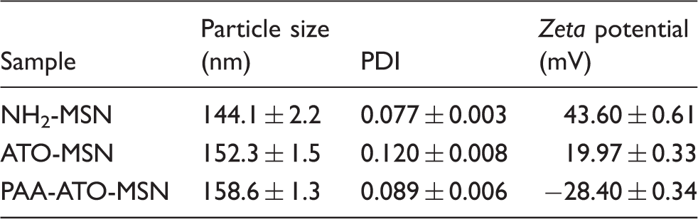
The functionalization of MSN with amino groups by co-condensation method could modify both the internal and external surfaces. This functionalization not only provided positively charged groups to absorb ATO but also afforded functional group for further modification with PAA. Morphology of NH2-MSN and PAA-ATO-MSN was spherical with uniform size, as is shown in Figure 2. The ordered mesoporous on NH2-MSN was shown in the highly magnified TEM image (Figure 2(a), inset). After coated with PAA, the surface of nanoparticles became rough indicating successful functionalization with the organic polymer (Figure 2(b), inset). The dispersion of nanoparticles was not affected after the modification (Figure 2(b)). Mean particle size, PDI and Zeta potential of NH2-MSN, ATO-MSN and PAA-ATO-MSN were displayed in Table 1. The particle size became bigger to about 150 nm after coating with PAA. It was found that microparticles with the size in the range of 100–200 nm tend to accumulate in tumor tissues much more easily than that in normal tissues because of the enhanced permeability and retention (EPR) effect of macromolecules.
39
Size of our prepared nano-systems was within this scope suggested that they might accumulate in tumor sites easily. The main principles of drug loading were Van der Waals Forces and electrostatic interactions between amino group and ionized forms of H3AsO3 in solution. When ATO was loaded by NH2-MSN, the Zeta potential decreased from 43.60 mV to 19.97 mV, indicating a part of amino group was shielded by the drug. Although the positive potential of ATO-MSN had reduced, they still had the possibility for further coated with negatively charged PAA through electrostatic effect.
40
As a result, the Zeta potential of PAA-ATO-MSN changed to −28.40 mV. This further indicated carboxyl terminated PAA had grafted onto the surface of NH2-MSN successfully.
TEM images of NH2-MSN (a) and PAA-ATO-MSN (b). Particle size, PDI, Zeta potential of NH2-MSN, ATO-MSN and PAA-ATO-MSN.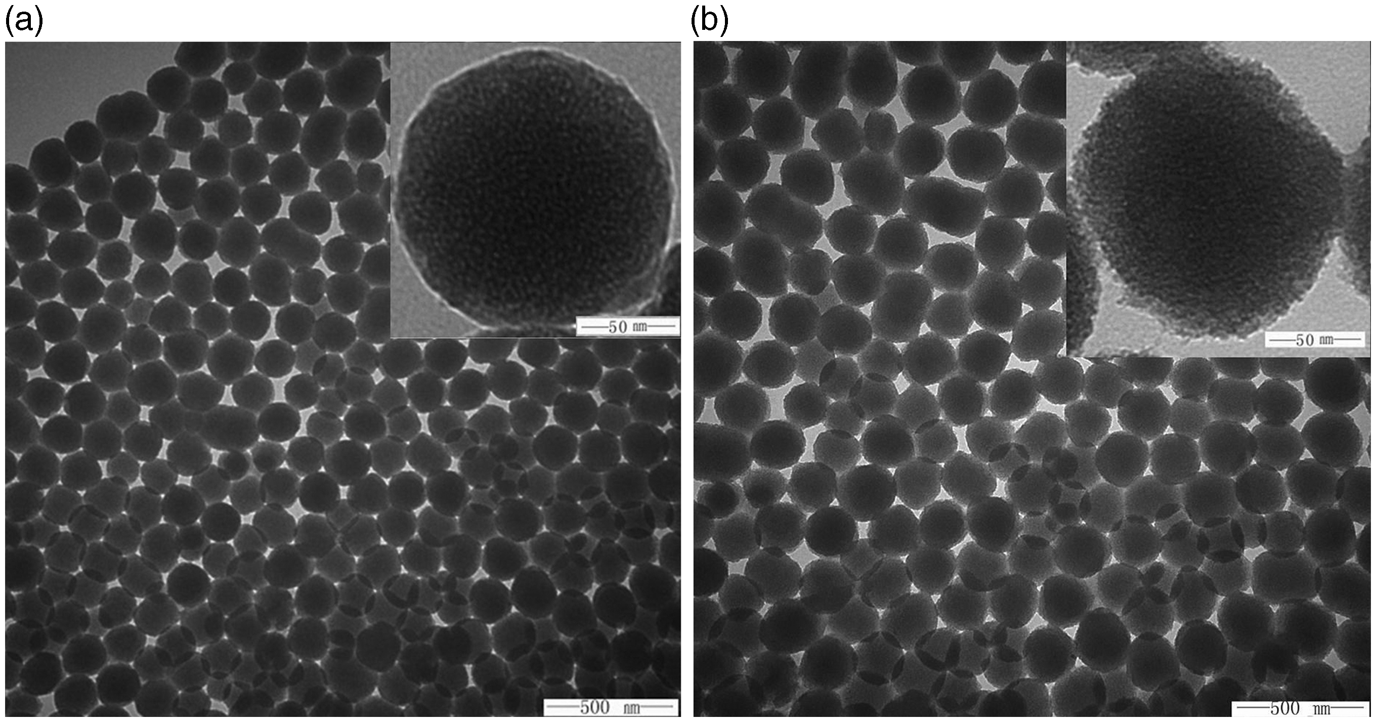
The SAXRD photograph of NH2-MSN and PAA-ATO-MSN is shown in Figure 3(a), a strong diffraction peak occurred around 3° in NH2-MSN, indicating that the internal structure of nanoparticles were in ordered arrangement. While the diffraction peak became very weak in PAA-ATO-MSN because the loaded ATO and grated PAA had filled the pore structure of nanoparticles. As displayed in Figure 3(b) and (c), the N2 adsorption-desorption patterns of NH2-MSN exhibited the characteristic type IV according to the IUPAC classification,
41
and the pore size distribution of the NH2-MSN showed a narrow distribution with a mean value of 3.71 nm. The specific surface area, the pore size and cumulative pore volume of NH2-MSN were 997.33 m2·g−1, 3.71 nm and 1.02 cm3·g−1, respectively. The parallel isotherms of adsorption and desorption assigned to H1 hysteresis loop, further proved that mesoporous were regular and uniform.
42
However, after loading ATO and grafting PAA, the specific surface area, the pore size and cumulative pore volume of the nanocarriers were reduced to 512.28 m2·g−1, 2.56 nm and 0.23 cm3·g−1, respectively. That is to say, the adsorbed nitrogen of PAA-ATO-MSN was reduced and peak of pore size nearly disappeared in pattern of PAA-ATO-MSN. This was due to ATO occupied internal channel and PAA coated external surface. The decrease of pore size may rely on the capping of PAA, making it possible for further sustained release of the drug.
SAXRD patterns of NH2-MSN and PAA-ATO-MSN (a). N2 adsorption-desorption isotherm of NH2-MSN and PAA-ATO-MSN (b). Pore-size distribution of NH2-MSN and PAA-ATO-MSN (c). TGA curves of NH2-MSN, ATO-MSN and PAA-ATO-MSN (d). FT-IR photograph of MSN, NH2-MSN and PAA-MSN (e).
As the FT-IR photograph of MSN, NH2-MSN and PAA-MSN showed in Figure 3(e), the adsorption peak at 1083.82 cm−1 in MSN was assigned to the asymmetric stretching vibration of Si-O-Si and the adsorption peak at 800.33 cm−1, and 462.84 cm−1 were resulted from symmetric stretching vibration and bending vibration of Si-O-Si, respectively. Simultaneously, bending vibration and asymmetric stretching vibration of Si-OH occurred at 956.54 cm−1 and 3446.23 cm−1, respectively, and became weaker after functionalized with amino group or carboxyl of PAA. New adsorption peak appeared at 1508.09 cm−1, due to the bending vibration of N-H, suggested that amino groups had been modified successfully. PAA modified NH2-MSN was confirmed by the strong C=O adsorption of acylamino carboxylic. Compared with NH2-MSN, the appearance of new adsorption peaks at 1652.72 cm−1 and 1720.22 cm−1 indicated the attachment of PAA to NH2-MSN.36,43 The grafted amount of PAA onto NH2-MSN was determined by TGA, shown in Figure 3(d). When heated from 100℃ to 800℃, the weight loss of NH2-MSN, ATO-MSN and PAA-ATO-MSN were 18.5%, 29.6 wt. % and 39.8 wt. %. Since the nanoparticles are mainly composed of SiO2 with high thermal stability, the weight loss of NH2-MSN was due to the organic part of APTES. 44 Drug loading rate was 11.1% and the graft ratio of PAA was about 10.2 wt. %.
Drug loading and in vitro release study
As is mentioned above, the drug loading process was mainly based on the electrostatic interaction. According to the condition of close to neutral, positive charged NH2-MSN naturally absorbed the negative charged ATO. In addition, as the concentration outside the NH2-MSN was much higher than inside, ATO tended to diffuse into the channel of the nanoparticles. Ultracentrifugation combined with ICP method was applied to determine the entrapment efficiency and drug-loading rate of PAA-ATO-MSN. The result exhibited relatively high entrapment efficiency and drug-loading rate with 40.95 ± 3.21% and 11.42 ± 1.75%, respectively. Drug-loading rate calculated by this method was nearly equal to the TGA result. That meant both methods were suitable for measuring drug loading rate of silica-based nanoparticles.
The release profiles of ATO from ATO-Sol, ATO-MSN and PAA-ATO-MSN at pH 5.0, 6.0 and 7.4 showed in Figure 4. The drug release rate of ATO from ATO-Sol was approximate 90% in 2 h. After loaded by MSN, ATO released from ATO-MSN was about 78% in three different pH values within 48 h and an apparently sustained phenomenon was found, but it still showed drug burst release of approximate 50% ATO in the initial 2 h. The rapid initial release of the cargo could be explained by wake bonding between the drug and the surface of the nanoparticles through electrostatic absorption and Van der Waals Forces. However, the burst release rate was significantly suppressed to 42%, 34% and 21% after coated by PAA at pH 7.4, 6.0 and 5.0. This was because PAA sheltered the naked drug on the surface. Meanwhile, slight pH-response release action was observed in ATO-MSN due to amino groups which could either accept or provide protons to undergo pH-dependent changes.27,45 The release rate of ATO from PAA-ATO-MSN displayed obviously pH-dependent and increased with the decrease of pH value. The result showed that the cumulative release amount of ATO from PAA-ATO-MSN could reach up to 76.15% and 59.80% at pH 5.0, 6.0, respectively, in 48 h, while only 40.52% of ATO was measured at pH 7.4. At high pH value, PAA had good solubility and could be combined onto the surface of NH2-MSN by covalent graft and electrostatic interaction to cap the mesoporous structure. With the decrease of pH, the solubility of PAA became weak and dissociated so that more ATO could be released.
46
In a word, PAA-ATO-MSN possesses the pH-responsive and enhanced sustained-release characteristics compared with ATO-Sol and ATO-MSN in vitro study and this DDS could control the drug concentration within a narrow range compared with ATO-Sol. So, the designed nanocarriers are suitable for delivering dose-limited toxic drug.
In vitro release profiles of ATO from ATO-Sol, ATO-MSN and PAA-ATO-MSN at pH 5.0, 6.0 and 7.4 (n = 3) during 48 h.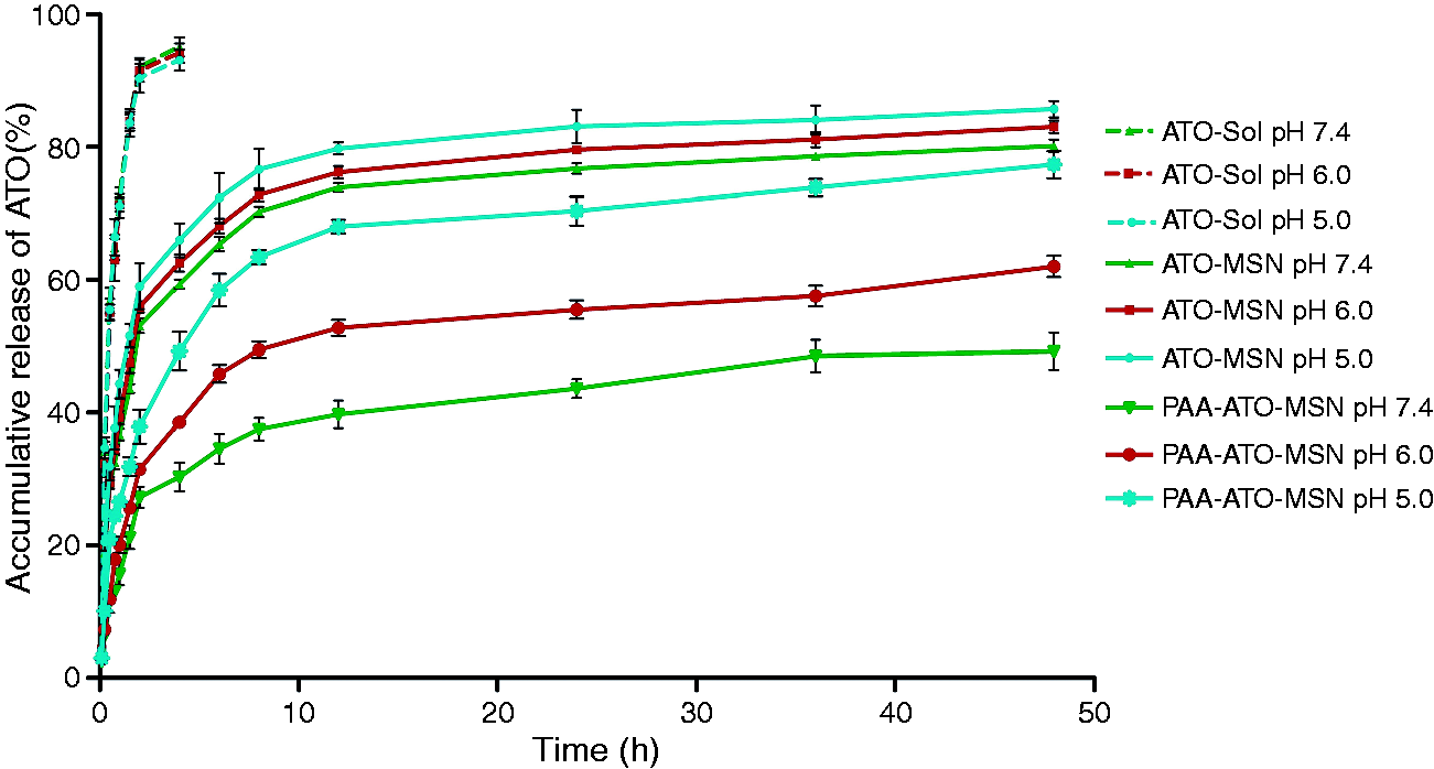
Cytotoxicity and anti-tumor evaluation
The cytotoxicity studies of MSN, NH2-MSN, PAA-MSN on SMMC-7721 cells were carried out at different concentrations of three blank nanocarriers. (Figure 5(a)) Above 85% of cell remained viable with a low concentration of incubated carriers (below 10 µg·mL−1). This meant all drug free nanocarriers exhibited intrinsically low toxicity and good biocompatibility. With the increase of concentration, NH2-MSN revealed higher toxicity compared with MSN, PAA-MSN. It is may be attributed to amine-modified MSN caused positive charge- and concentration-dependent hemolytic activity.
47
The viability of cell treated with PAA-MSN was lower than that of NH2-MSN treatment, which indicated that the modification of PAA could partially conceal the cytotoxicity caused by naked amino on NH2-MSN. The result suggested that organic polymeric grafts could improve the biosecurity of amino-modified nano-silicon biomaterials.
The viability of SMMC-7721 cells after being treated with MSN, NH2-MSN and PAA-MSN at concentrations ranging from 0.1 µg·mL−1 to 100 µg·mL−1 for 48 h (a). The viability of SMMC-7721 cells after being treated with ATO-Sol, ATO-MSN and PAA-ATO-MSN at a concentration ranged from 1 µM to 12 µM for 48 h (b).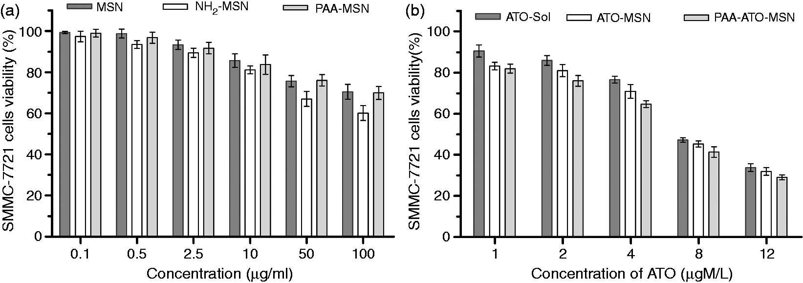
To evaluate the cytotoxicity of the DDS, SMMC-7721 cells were exposed to series of concentrations of ATO-Sol, ATO-MSN and PAA-ATO-MSN for 48 h (Figure 5(b)). The half maximal inhibitory concentration (IC50) values of ATO-Sol, ATO-MSN, and PAA-ATO-MSN were 7.97 ± 0.23 µM, 7.82 ± 0.41 µM, and 6.09 ± 0.37 µM, respectively. The enhanced antitumor activity of PAA-ATO-MSN than both ATO-Sol and ATO-MSN was likely due to the enhanced sustained-release drug in tumor sites.
Cell cycle analysis
SMMC-7721 cells were incubated with ATO-Sol, ATO-MSN, PAA-ATO-MSN at a concentration of 1 μg·mL−1 (ATO) with culture medium as control for 48 h. We used flow cytometry to monitor the cell cycle progression for characterizing the inhibitory effect of ATO on cell proliferation. As shown in Figure 6, ATO-Sol, ATO-MSN, PAA-ATO-MSN could arrest 69.3 ± 2.9%, 71.9 ± 2.1% and 79.5 ± 1.4% of SMMC-7721 cells specifically at G0/G1 phase cell cycle, respectively, much higher than the controlled group (58.8 ± 1.7%), which demonstrated that the drug could inhibit the proliferation of SMMC-7721 by inducing G0/G1 transition. And a significant difference appeared between PAA-ATO-MSN (p < 0.05) and the other two formulation groups. What’s more, PAA-ATO-MSN showed the strongest inhibition of G2-M transition, which further confirmed its best anti-tumor effect among three ATO formulations.
(a) and (b) SMMC-7721 cells cycle perturbations induced by ATO formulations at a concentration of 1 μg·mL−1 for 48 h. (ΔΔp < 0.01, compared with control group and *p < 0.05, compared with ATO-Sol).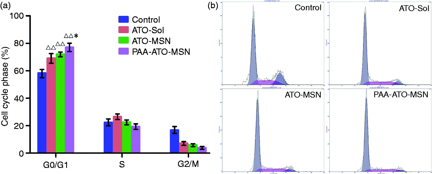
Pharmacokinetic study
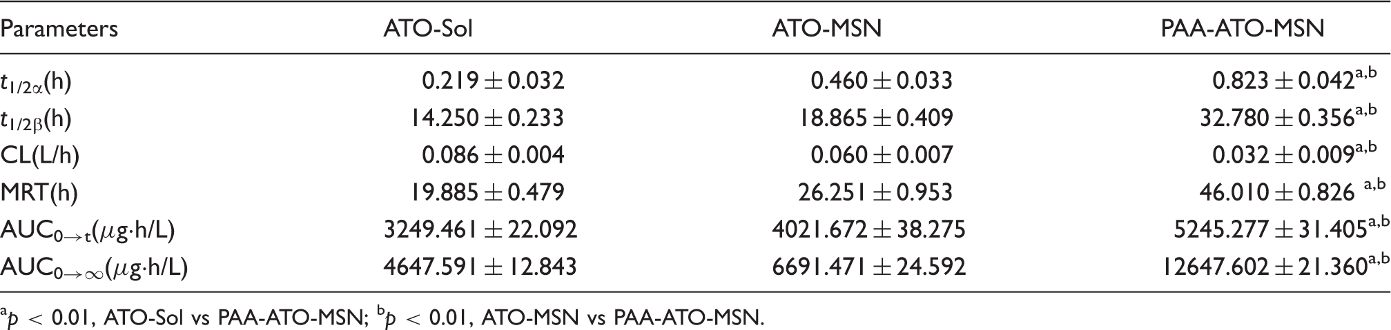
There was a number of pharmacokinetic researches of ATO,48–50 but few studies focused on its PK characteristics after it was loaded by smart nanosized DDS. In this paper, ATO was loaded by pH-triggered MSN and its PK features were studied. The plasma concentration-time profile was depicted in Figure 7. Due to the sustained-release characteristics of MSN, sustained release characteristics could be observed in ATO-MSN and PAA-ATO-MSN, especially PAA-ATO-MSN, which could be attributed to the mesoporous of MSN blocked by PAA. The PK parameters complied with two-compartment model was summarized (Table 2). Compared with ATO-Sol and ATO-MSN groups, sustained release characteristics of PAA-ATO-MSN were observed with the significantly prolonged t1/2β (p < 0.01), reduced clearance (CL) (by 2.7 fold of ATO-Sol) (p < 0.01) and improved AUC (by 2.7 fold of ATO-Sol) (p < 0.01). Remarkably, a prolonged MRT (p < 0.01) indicated the extended circulation time of PAA-ATO-MSN. It implicated that PAA-ATO-MSN possess an enhanced sustained-release characteristics and improved the pharmacokinetic behavior of ATO in rats by prolonging the retained time in circulation.
The profiles of mean drug concentration-time in rats after intravenous injection (n = 6). Main parameters of ATO after vein injection in rats ( p < 0.01, ATO-Sol vs PAA-ATO-MSN; bp < 0.01, ATO-MSN vs PAA-ATO-MSN.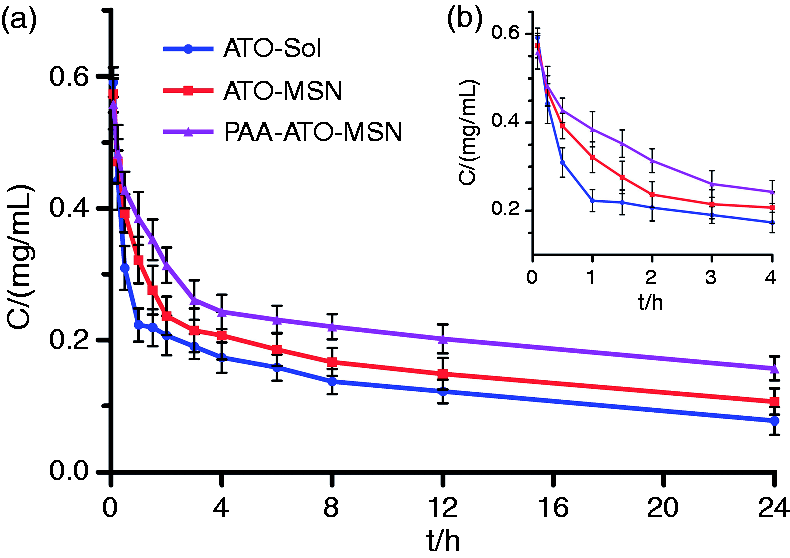
Pharmacodynamic study
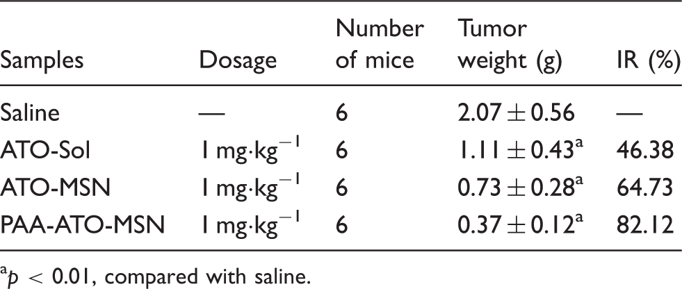
According to the excellent antitumor effect observed in vitro, we further performed an in vivo therapeutic study of PAA-ATO-MSN in H22 tumor bearing mice. During the experiment, six mice of each group were sacrificed on each sample day, respectively. As shown in Figure 8(a), the tumor volume in saline group kept increasing during the treatment process, but volumes in three ATO formulations increased much slowly, which indicated that all drug formulations contained anti-tumor effects. PAA-ATO-MSN showed stronger antitumor effect from the fifth day. The mean tumor volume of PAA-ATO-MSN group on 13th after treatment (477.99 ± 37.64 mm3) was significantly smaller than that of the saline group (2302.67 ± 199.65 mm3, p < 0.01), ATO-Sol group (1405.62 ± 117.20 mm3, p < 0.01), and ATO-MSN group (796.42 ± 104.70 mm3, p < 0.05), Figure 8(a). Consistently, the mean tumor weight of PAA-ATO-MSN (0.37 ± 0.12 g) was considerably less than that of saline group (2.07 ± 0.56 g, p < 0.01), ATO-Sol group (1.11 ± 0.43 g, p < 0.01) and ATO-MSN group (0.73 ± 0.28 g, p < 0.01) on day 13. Furthermore, the tumor inhibition rate (IR) of PAA-ATO-MSN group was 82.12%, 1.77 and 1.27 times higher than that of ATO-Sol (46.38%) and ATO-MSN group (64.73%), indicating the enhanced anti-tumor activity of PAA-ATO-MSN (Table 3). Despite ATO-Sol and ATO-MSN having similar antitumor effects on cancer cells in vitro, the latter was more effective in inhibiting tumor growth in vivo, which may be ascribed to the EPR effect and the sustained release the cargos controlled by porous structure through gradual diffusion from inside to outside of the nanocarriers. Besides, PAA-ATO-MSN induced significantly higher inhibition of tumor growth than other groups. This may be attributed to the two facts: a more obvious sustained release action caused by the coating of mesoporous structure of MSN with PAA, and the pH-sensitive release of ATO trigged by the acidic microenvironment of tumor.
Tumor volume curves after intravenous injection of saline, ATO-Sol, ATO-MSN or PAA-ATO-MSN with ATO 1 mg·kg−1 (*p < 0.05, **p < 0.01, compared with saline; △p < 0.05 △△p < 0.01, compared with ATO-Sol) (a). Mean body weight of the mice in different groups during the treatment (b). The tumors photograph in each group on the thirteenth day (c). Tumor H&E histology images of the mice after administration of saline, ATO-Sol, ATO-MSN and PAA-ATO-MSN for 12 days (black arrows indicate the typical necrotic cells in tumors and scale bar is 200 μm) (d). The data of in vivo anti-tumor activity. p < 0.01, compared with saline.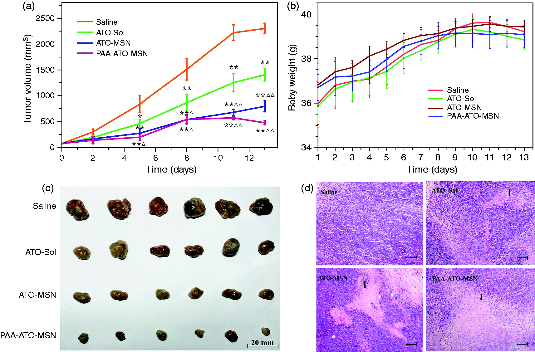
In recent years, with the transformation of medical model, the life quality of animals is seen as a vital evaluation level for pharmacodynamics of an anti-cancer drug. 51 Body weight is one of the most important indicators. During our experiments, body weights of the mice with the treatments were measured per day. As seen in Figure 8(b), weight gain was due to the rapid growth of the solid tumor in the first 10 days. When the tumor grew to a certain size, weight loss was seen from 10th day. PAA-ATO-MSN group contained the minimum rate of weight increase in the first 10 days indicated its best anti-tumor effect. After that, the weight of saline group and ATO-Sol group decreased, while the PAA-ATO-MSN group maintained a consistent trend, suggested that mice in this group had the best life quality.
H&E staining examinations of the tumor tissues after treatment are displayed in Figure 8(d), it appeared that the tumor tissue displayed a typical necrotic response after treatment with ATO-Sol, ATO-MSN and PAA-ATO-MSN. Comparing with ATO-Sol and ATO-MSN, the area of cell necrosis of PAA-ATO-MSN was the largest, which added the evidence that PAA-ATO-MSN provided the best anti-tumor effect.
Conclusions
In summary, ATO-loaded amino functioned MSN coated with PAA were successfully prepared as a pH-responsive and sustained release DDS. The obvious sustained release behavior of ATO was exhibited after loaded by MSN and pronounced by capping with PAA. Importantly, ICP-MS method was capable for pharmacokinetic studies and the results showed that PAA-ATO-MSN really reduced the renal clearance rate, exhibited prolonged circulation time, and significantly increased the bioavailability of ATO. The treatment of PAA-ATO-MSN resulted in the highest suppression of tumor growth in cultured SMMC-7721 cells in vitro and in H22-bearing mice in vivo. This work represented the first trial using a pH-responsive drug delivery system of PAA-MSN as carrier to load the toxic drug ATO, and all the findings proved a novel sense on the aspect of synthesis of pH-triggered biological drug delivery system for poor pharmacokinetics or dose-limited toxicity drugs in solid tumor treatment.
Footnotes
Acknowledgment
We thank Mr. Luo Fang (Zhejiang Cancer Hospital) and Zhenhua Sheng (Zhejiang Chinese Medical University) for their technical assistance.
Declaration of Conflicting Interests
The author(s) declared no potential conflicts of interest with respect to the research, authorship, and/or publication of this article.
Funding
The author(s) disclosed receipt of the following financial support for the research, authorship, and/or publication of this article: This study was financially supported by the National Science Foundation of China (Nos.81274089 and 81473361), the Natural Science Foundation of Zhejiang Province (LZ13H280001 and LY12H28004) and the Science and Technology InnovationTeam Project of Zhejiang Province (2015R410051).