
Abstract
Nanofibrous materials produced by electrospinning have attracted considerable attention from researchers in regenerative medicine. A combination of nanofibrous scaffold and chondrocytes is considered promising for repair of cartilage defect or damage. In the present study, we fabricated a poly(
Introduction
Cartilaginous tissues have limited self-healing ability once damaged by tumor, trauma, inflammation, dysplasia, or aging. This is due to lack of vasculature that might supply progenitor cells to repair defects, and to the poor proliferation and migration abilities of the highly differentiated chondrocytes encapsulated in dense extracellular matrix (ECM). 1 If cartilage injury is not treated properly in a timely manner, this can result in swelling and pain, gradually leading to degenerative lesions, osteoarthritis, or even disability. 2
Severe wear of cartilage at the joints often requires joint replacement,3,4 which greatly improves the quality life of patients with end-stage osteoarthritis. However, it has an unsatisfactory survivorship after implantation due to many factors, such as postoperative infection, wear and corrosion of the prosthesis.5–8 Traditional approaches to treat cartilage injury primarily include physiotherapy, drug therapy, marrow stimulation, and tissue transplantation. However, each of these protocols has its own shortcomings. For example, physiotherapy and drug therapy are mainly suitable for patients as postoperative adjuvant treatment. Marrow stimulation may accelerate cartilage degeneration in the long term, because the repaired tissues lack the characteristics of normal hyaline cartilage. The application of tissue transplantation is limited by the need for grafts. Autograft can cause secondary trauma and increase postoperative pain in patients, and allograft can result in immune rejection and cross infection, and thus surgical failure. In addition, the structure of repaired tissues is not identical to normal cartilage, and thus it fails to restore normal biological functions perfectly. 9
Tissue engineering is a promising and multi-disciplinary technique for fabricating functional biological substitutes for the repair or replacement of damaged tissues. 10 Cartilage tissue engineering is specifically applied to treat cartilage injury, with three key considerations: seeding cells, growth factors, and scaffolds. Generally, an ideal scaffold should have good biocompatibility and biomechanical properties, suitable degradation time, and a three-dimensional (3D) nanoporous structure. 11
Poly(
Electrospinning is a convenient, economic, and effective method recently developed to fabricate composite materials with a nanoporous structure similar to natural ECM.17,18 The technique has been widely applied to regenerate bone,19,20 nerve, 21 cartilage, 22 blood vessels, 23 and cardiac tissue. 24 In the present study, we successfully fabricated a PLLA/SF composite scaffold through electrospinning, and evaluated its surface morphology, wettability, and tensile strength relative to that of a PLLA scaffold without SF. In addition, the chondrogenesis of the composite scaffold was investigated.
Materials and methods
Materials and reagents
The two components for fabrication of the PLLA/SF composite scaffold, PLLA (Mw¼ 300,000) and SF were purchased from Shandong Institute of Medical Appliances, China and Huzhou Heavens Silk Biological Technology, China, respectively. Trifluoroacetic acid (TFA) and 1,1,1,3,3,3-hexafluoro-2-propanol (HFIP), which were used as the solvents of PLLA and SF, were obtained from Tianjin Fuchen Chemical Reagents (China) and Sigma-Aldrich (St. Louis, MO, USA), respectively. Trypsin-ethylenediaminetetraacetic acid (EDTA), collagenase II, and fetal bovine serum (FBS) were purchased from Gibco (Life Technologies, Carlsbad, CA, USA). Dulbecco's modified Eagle's medium (DMEM), Ham's nutrient solution F-12, penicillin, and streptomycin were obtained from Sigma-Aldrich.
Fabrication of PLLA and PLLA/SF scaffolds
PLLA and SF were fully dissolved in HFIP and TFA, respectively, at a concentration of 4% (w/v), and were mixed together at a mass ratio of 60:40. The polymer solutions were placed into a 5-mL syringe with a needle (inner diameter: 0.4 mm), and aluminum foil was used to collect the nanofibers. The electrospinning conditions for the PLLA and PLLA/SF scaffolds were working voltage 15 kV; collection distance 20 cm; and injection rate 0.5 mL/h. The nanofibers were dried in a vacuum oven for two days at 37℃ to remove the solvent completely. Similarly, the PLLA scaffold was fabricated as a reference in the following characterizations.
Characterization of PLLA and PLLA/SF scaffolds
Surface morphology and scaffold thickness
The PLLA and PLLA/SF scaffolds were sputter-coated with gold to increase conductivity, and the surface of the scaffolds was observed by scanning electron microscopy (SEM; Hitachi SU8000, Japan) at an accelerating voltage of 3 kV. The diameter of the nanofibers and the diameter distribution were quantitatively measured using Image-Pro Plus software (Media Cybernetics, Rockville, MD, USA). Approximately 100 nanofibers from the SEM image were analyzed. The thickness of the scaffolds was measured using a micrometer caliper.
Surface wettability and swelling ratio
The surface wettability of the PLLA and PLLA/SF scaffolds was characterized according to the static contact angle, which was measured by the sessile drop method using a contact angle measurement instrument (JC2000C2, Shanghai Zhongchen Digital Technic Apparatus, China) at room temperature, in triplicate for each scaffold.
The swelling ratio of the PLLA and PLLA/SF scaffolds was measured according to water absorption ability, to simulate the swelling process in the in vivo environment (in triplicate for each scaffold). Briefly, the scaffolds were manufactured as specific samples with a size of 1 cm × 1 cm, dried, and the dry molecular weight was determined as Mdry. Subsequently, the scaffolds were immersed in 10 mL of phosphate-buffered saline solution (PBS, pH = 7.4), and incubated at 37℃ for 24 h. The interfacial water in the scaffolds was removed with filter paper, and the wet scaffolds were weighed as Mwet. The swelling ratio was calculated as: swelling ratio, % = (Mwet − Mdry)/Mdry × 100%.
Tensile strength
The tensile strength of the PLLA and PLLA/SF scaffolds was evaluated using a universal material testing machine (Shanghai Instrument, China), in triplicate for each scaffold. The scaffolds were fabricated into specific samples with a dumbbell strip (25 mm × 5 mm), which were attached to the material testing machine. A tensile load was applied at a stretching speed of 10 mm/min until failure was observed, with the stress–strain curve obtained simultaneously. The thickness of the samples was measured using a micrometer caliper at five random sites.
In vitro scaffold–cell interactions
Cell isolation and culture
The Animal Care and Use Committee of Jilin University approved these procedures. Chondrocytes were isolated and cultured in accordance with a reference previously published. 25 Briefly, articular cartilage was isolated from the superficial layer of the knee joints of a two-week-old rabbit under aseptic conditions, and minced into fragments of about 1 mm3. The cartilage fragments were washed with PBS (containing 100 units/mL penicillin and 100 mg/mL streptomycin) three times, and digested with 0.25% trypsin plus 0.02% EDTA at 37℃ for 30 min. The trypsin–EDTA solution was removed, and 0.1% collagenase II in serum-free DMEM/Ham's nutrient solution F-12 (DMEM/F12; 1:1) was added, allowing digestion at 37℃ overnight with shaking.
The cell solution was filtered through a 100-mesh sieve and centrifuged at 1000 r/min for 5 min to obtain a cell pellet after removal of the supernatant. The pellet was resuspended in DMEM/F12 supplemented with 15% FBS, 100 units/mL penicillin, and 100 mg/mL streptomycin, and then seeded in a 25 cm2 culture flask at a density of 5 × 10⁵ cells/mL, which was incubated in an incubator (Thermo 311, Marietta, USA) at 37℃ and 5% CO2. The culture medium was refreshed every two to three days.
An inverted phase contrast microscope was used to observe the morphology, adherence, and growth of the cells. The cells were subcultured upon reaching 85–90% confluence, and cells at the second passage were used for the following experiments.
Cell seeding on scaffolds
The PLLA and PLLA/SF scaffolds were sterilized and wetted prior to seeding of the chondrocytes, as follows. Briefly, the scaffolds were manufactured into disk samples with a diameter that fit the wells of different culture plates. Then, the scaffolds were sterilized under ultraviolet light for 1 h, soaked in 75% ethanol for 30 min, and finally washed with PBS three times, each for 5 min.
For the prewetting procedure, the scaffolds were soaked in DMEM/F12 supplemented with 15% FBS, 100 units/mL penicillin, and 100 mg/mL streptomycin overnight. The culture medium was then absorbed onto sterile blotting paper. Subsequently, the cell suspension was added to the wells of the culture plates (cell seeding density: 2 × 10⁴/scaffold), which were further incubated in an atmosphere of 5% CO2/95% humidified air at 37℃ for 2 h, allowing for cell attachment. After 2 h when the cells had adhered to the scaffolds, an additional 1–2 mL of culture medium was added to immerse the scaffold–cell construct. The culture medium was refreshed every two to three days.
Cell morphology
After culturing for one and seven days, the scaffold–cell constructs were harvested, rinsed thoroughly with PBS to remove unattached cells, and fixed with 2.5% glutaraldehyde solution in PBS overnight at 4℃. Subsequently, the scaffolds were stained with 1% osmium tetroxide for 30 min at 4℃, dehydrated in a graded series of ethanol (30%, 50%, 70%, 80%, 90%, 95%, 100%), and freeze-dried overnight. Finally, the scaffolds were sputter-coated with a gold layer, and examined by the SEM.
After culturing for one and seven days, the scaffolds were washed with PBS and fixed in 4% paraformaldehyde in PBS for 20 min at room temperature, and DAPI solution (5 µg/mL, Roche, Los Angeles, USA) was added to immerse the scaffold. The scaffolds were incubated for 5 min at room temperature and examined under a confocal laser scanning microscopy (CLSM, Olympus FV-1000, Tokyo, Japan).
Biochemical analyses
The biochemical experiments were performed to detect quantitatively the proliferation and ECM secretion of chondrocytes on the scaffolds, as follows. Briefly, a 50 µL cell suspension (5 × 10⁶/mL) was seeded on the PLLA and PLLA/SF scaffolds, which were placed in the wells of 24-well culture plates, in triplicate for each scaffold. After culturing for 3, 7, 14, and 21 days, the scaffold–cell constructs were removed, cut into pieces, and digested with 1 mL of papain solution (papain, cysteine, EDTA, disodium hydrogen phosphate, PBS) for 16 h at 65℃. For the following evaluations, a tissue culture polystyrene surface (TCPS) was used as the positive control, as it was treated with a hydrophilic surface to promote cell attachment. 25 We did not use chondrogenic supplements for 21-day matrix formation studies.
DNA
The papain-digested solution was used to quantify DNA content through a Hoechst 33258 Assay (Sigma, #861405) in accordance with a reference previously published. 26 Briefly, a 100 µL solution was added to 100 µL of Hoechst in a 96-well plate, and measured using a fluorescence spectrophotometer (Fluoroskan Ascent FL, Thermo, Waltham, MA), at excitation 355 nm and emission 460 nm. The measurements were normalized to calf thymus DNA (Sigma, #D0805).
Sulfated glycosaminoglycan (sGAG)
The sGAG content was quantified through a 1, 9-dimethylmethylene blue (DMMB) assay using the papain-digested solution in accordance with a reference previously published. 27 Briefly, a 40-µL solution was mixed with 125 µL DMMB reagent in a 96-well plate, and the optical absorbance was immediately measured using a microplate reader (RT-6000, Lei Du Life Science and Technology, Shenzhen, China) at 595 nm. The measurements were normalized to a standard curve established from chondroitin 6-sulfate (Sigma, C4384).
Collagen
The quantity of collagen was determined by measuring the hydroxyproline content of the constructs in accordance with a reference previously published. 28 Briefly, 50 µL of papain-digested solution was hydrolyzed with 50 µL of HCL (12 N, 37%) at 120℃ for 30 min. Chloramine-T and p-dimethylaminobenzaldehyde (p-DMBA) reagents were added to the hydrolysates, and the optical absorbance was measured at 550 nm using the microplate reader. The hydroxyproline content was expressed as ng/g DNA.
Histological staining
Histological staining was performed to observe the morphology, distribution, and cartilage-specific ECM secretion of chondrocytes on the scaffolds. The procedure for cell seeding was the same as that described for biochemical analyses. After culturing for 3 and 14 days, the scaffold–cell constructs were fixed in 4% paraformaldehyde in PBS overnight at 4℃, dehydrated using a graded series of ethanol, embedded in paraffin, and finally sectioned into 4 µm thick sections. The cross sections and vertical cross sections were stained with hematoxylin and eosin (H&E) to observe the structure of the construct. In addition, Alcian blue-nuclear fast red staining was used to locate sGAG in the construct. The images were recorded using an optical microscope (Nikon Eclipse E600, Melville, NY, USA) with an attached video camera (Sony DXC950P, New York, NY, USA).
Reverse transcription (RT)-polymerase chain reaction (PCR)
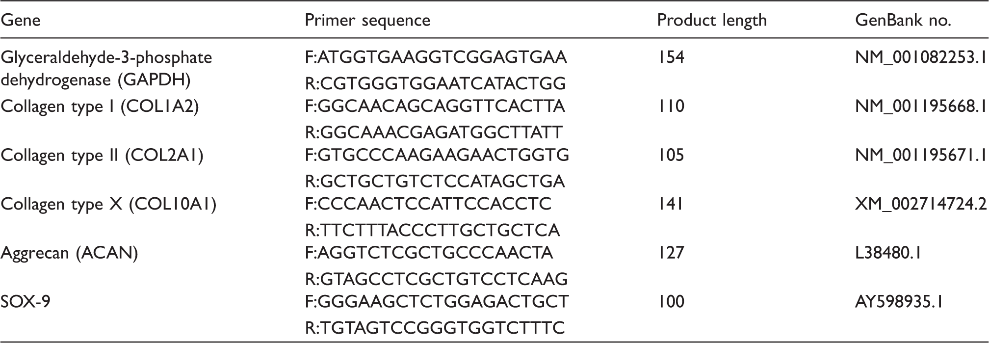
RT-PCR was performed to evaluate the expressions of cartilage-related genes in the scaffolds, specifically collagen type I, collagen type II, collagen type X, sox-9, and aggrecan. Briefly, the PLLA and PLLA/SF scaffolds were placed in the wells of the culture plates, and seeded with 50 µL of cell suspension at a density of 1 × 10⁷ cells/mL, in triplicate for each scaffold. After culturing for 7, 14, and 21 days in an atmosphere of 5% CO2/95% humidified air at 37℃, the scaffolds were removed, and the total RNA of the cells on the scaffolds was extracted by Trizol reagent (Invitrogen, USA).
Forward (F) and reverse (R) primers used for quantitative RT-PCR.
Statistical analyses
All quantitative values are presented as mean ± standard deviation. Statistical analyses were performed using Statistical Product and Service Solutions 19.0 software (SPSS, Chicago, IL, USA). Differences were assessed by one-way analysis of variance (ANOVA), and were considered as statistically significant at P < 0.05. The data were indicated with (*) for P < 0.05, (**) for P < 0.01, and (***) for P < 0.001, respectively. Three scaffolds were used for each experiment and all the experiments were repeated at least three times.
Results
Characterization of PLLA and PLLA/SF scaffolds
Surface morphology and scaffold thickness
TFA and HFIP were good solvents for PLLA and SF, and the two materials could each dissolve equally well in the solvents. The SEM micrographs showed that the nanofibers of both the PLLA and PLLA/SF scaffolds were randomly oriented (Figure 1(a) and (b)) and smooth. No fracture or adhesion between nanofibers was observed. In addition, the scaffolds had an interconnective porous structure. The porosity and pore diameter of PLLA/SF scaffolds were 88.65% and 520 nm, respectively. Besides, both the scaffolds had rough surfaces because of the criss-cross nanofibers, which was different from the flat tissue culture plate surface (TCPS). The mean diameter of the PLLA/SF nanofibers was less than that of the PLLA nanofibers, with a more uniform diameter distribution (Figure 1(c) and (d)). The thicknesses of the PLLA and PLLA/SF scaffolds were 125 ± 0.32 µm and 129 ± 0.41 µm, respectively.
SEM images of (a) PLLA and (b) PLLA/SF nanofibers. Fiber diameter distribution of (c) PLLA and (d) PLLA/SF nanofibers.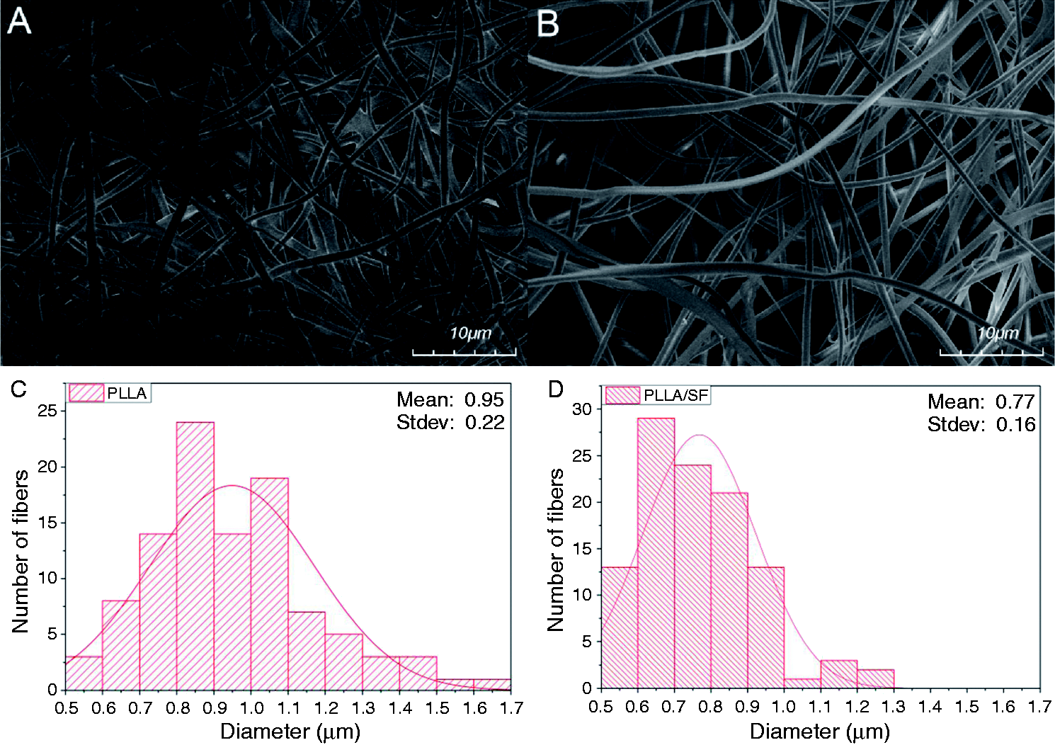
Surface wettability and swelling ratio
The static contact angle of the PLLA/SF scaffold was smaller than that of the PLLA scaffold with dwelling times of 10 s and 60 s, respectively (Figure 2(a)). This result suggests that the hydrophilicity of the PLLA/SF scaffold was better than that of the PLLA scaffold.
Characteristics of the scaffolds. (a) Water contact angle of the scaffolds. (b) Swelling ratio of the scaffolds. *P < 0.05 indicates a significant difference between the scaffolds.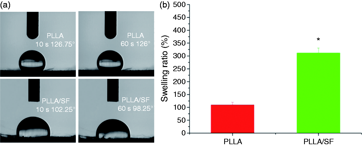
The swelling ratio of the PLLA/SF scaffold (311.56 ± 19.46%) was significantly greater than that of the PLLA scaffold (109.49 ± 10.40%; Figure 2(b)).
Tensile strength
The stress–strain curve of the PLLA and PLLA/SF scaffolds is shown in Figure 3. The tensile strength of the PLLA and PLLA/SF scaffolds was about 6.75 MPa and 5.27 MPa, respectively. In addition, it was indicated from this figure that the elongation at break and Young's modulus of the PLLA/SF scaffold were less than that of the PLLA scaffold (21.57 vs 71.26; 0.59 MPa vs 0.92 MPa).
Stress–strain curve of PLLA and PLLA/SF scaffolds.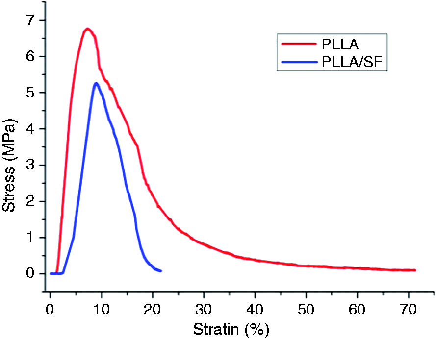
Cell morphology
Based on the SEM micrographs (Figure 4), chondrocytes could attach and proliferate well on both the PLLA and PLLA/SF scaffolds. Chondrocytes were spherical and had a diffused distribution on both scaffolds after one day of culturing. After culturing for seven days, chondrocytes maintained their round shape on the PLLA/SF scaffold, but they were flattened on the PLLA scaffold. A great deal of ECM secretion was observed from chondrocytes on both scaffolds, which became confluent.
SEM images of cell/scaffold constructs of (a, b) PLLA and (c, d) PLLA/SF at time points (a, c) one day and (b, d) seven days.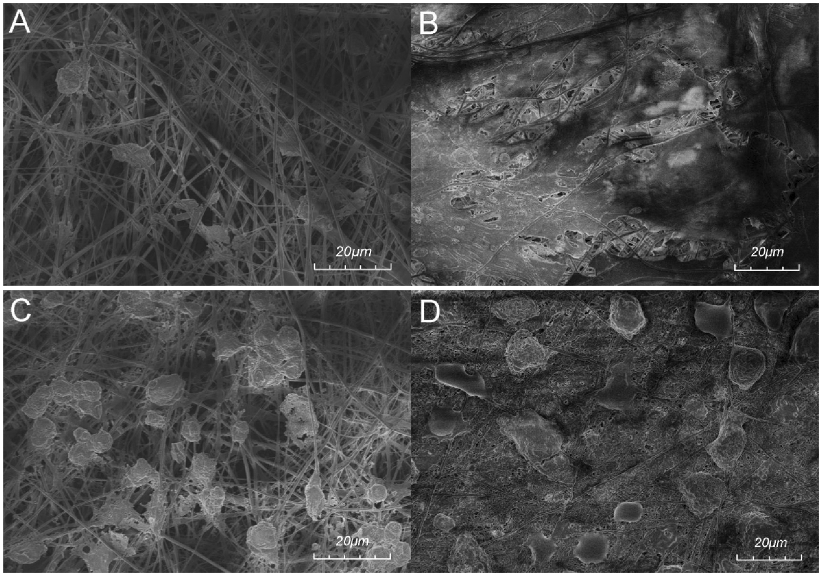
CLSM images showed the cell nuclei stained blue with DAPI fluorochrome (Figure 5). It was clear that the number of chondrocytes increased over time on both scaffolds, but the distribution of chondrocytes on the PLLA/SF scaffold was denser than that of the PLLA scaffolds after culturing for one and seven days. This result indicated that chondrocytes adhered and proliferated better on the PLLA/SF scaffolds than on the PLLA scaffolds.
CLSM images of cell/ scaffold constructs of (a, b) PLLA and (c, d) PLLA/SF at time points (a, c) one day and (b, d) seven days.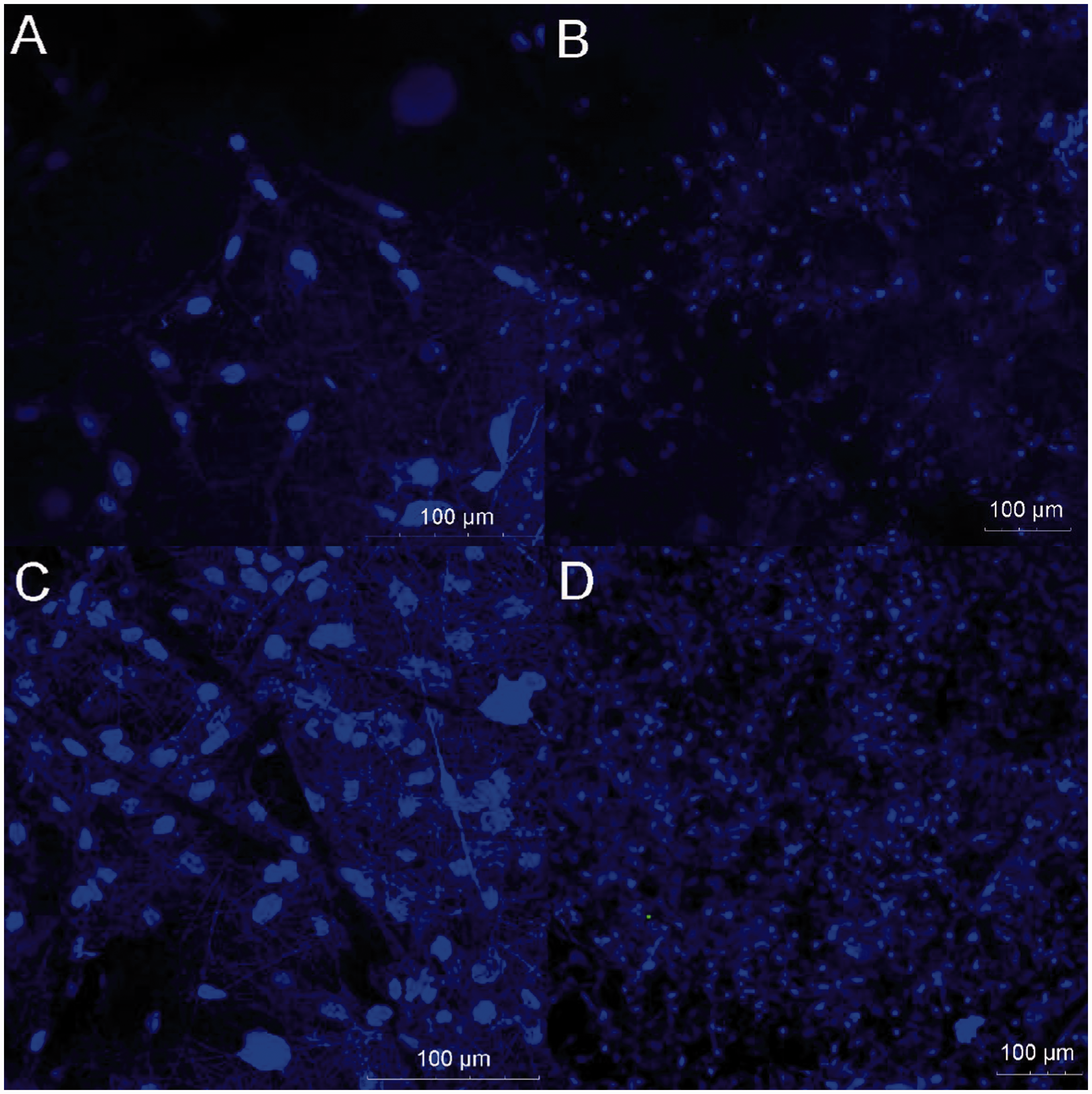
Biochemical analyses
The total DNA of chondrocytes on the PLLA, PLLA/SF, and TCPS scaffolds was measured after culturing for 3, 7, 14, or 21 days (Figure 6(a)). Initially, the growth of chondrocytes on TCPS was the fastest (days 3 and 7), and then slowed after day 14. Proliferation of chondrocytes on the PLLA/SF scaffold was similar to that of TCPS at day 14, and was the highest at day 21. By contrast, the PLLA scaffold showed the slowest proliferation of chondrocytes, with the smallest total DNA observed for all the time points.
Biochemical analyses. The total amount of (a) DNA, (b) GAG, and (c) collagen of the constructs at 3, 7, 14, and 21 days. Total GAG and collagen were normalized with (d, e) total DNA at 3, 7, 14, and 21 days. *P < 0.05, **P < 0.01, and ***P < 0.001.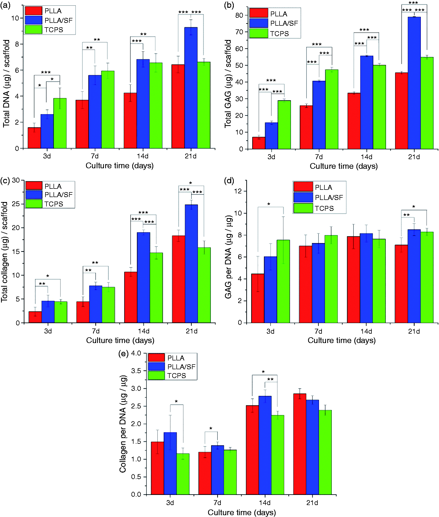
The sGAG deposition of chondrocytes on the PLLA, PLLA/SF, and TCPS scaffolds was measured after culturing for 3, 7, 14, or 21 days (Figure 6(b)). On all the scaffolds, chondrocytes secreted GAG from day 3 to day 21. At days 3 and 7, GAG production on TCPS was the greatest, but on days 14 and 21 GAG was greatest on the PLLA/SF scaffold. The GAG production rate on TCPS slowed after day 14, but increased consistently on the PLLA/SF and PLLA scaffolds. For all the time points, GAG production on the PLLA scaffold was the least.
The total collagen content of chondrocytes on the PLLA, PLLA/SF, and TCPS scaffolds was measured after culturing for 3, 7, 14, or 21 days (Figure 6(c)). All the scaffolds showed continuous increase in the total collagen content, which was greatest value on the PLLA/SF scaffold at all time points. At days 3, 7, and 14, the collagen content for the PLLA scaffold was less than that of TCPS, but exceeded TCPS at day 21.
The total GAG and collagen contents normalized with total DNA (Figure 6(d) and (e), respectively) are indicators of the secretory ability of chondrocytes. The ability of chondrocytes to generate GAG on TCPS was the strongest at days 3 and 7, and slightly less than that of the PLLA/SF scaffold at days 14 and 21, with no significant difference observed between TCPS and the PLLA/SF scaffold. In addition, the ability to synthesize collagen by chondrocytes on PLLA, PLLA/SF, and TCPS was not consistent over time. Generally, collagen synthetic ability was the strongest for the PLLA/SF scaffold at days 3, 7, and 14, and it was exceeded by the PLLA scaffold at day 21. However, the difference was not statistically significant between the PLLA/SF and PLLA scaffolds.
Histological staining
Through H&E staining of the scaffold–cell constructs (cross section, Figure 7(a) to (d)), it was observed that a large number of chondrocytes were distributed on the scaffolds, and some chondrocytes grew into the scaffolds. Chondrocytes appeared with blue nuclei and reddish cytoplasm, and ECM was stained light blue. It was clear that the color of the PLLA/SF scaffold at day 14 was the darkest among the scaffolds, which indicated that more cartilage-specific ECM was secreted. In addition, chondrocytes in the PLLA scaffolds were fusiform or scalene triangle in shape at days 3 and 14, while chondrocytes in the PLLA/SF scaffolds changed from scalene triangle at day 3 to ellipsoidal or round at day 14, which is similar to the morphology of natural cartilage tissue. Cartilage cavities and capsules were also observed in the PLLA/SF scaffolds at day 14. As observed after Alcian blue-nuclear fast red staining, sGAG was scattered on the PLLA and PLLA/SF scaffolds at day 3, while it increased to a much higher level at day 14 (cross section, Figure 7(e) to (h)). From the vertical cross section images (Figure 7(i) and (j)), it was observed that a small number of chondrocytes grew into the PLLA and PLLA/SF scaffolds, and most of cells were arranged in 3–5 layers on the surface of the two scaffolds. Overall, the results of histological staining indicated that the PLLA/SF scaffold was superior to the PLLA scaffold, promoting adhesion, proliferation, and secretion of cartilage-specific ECM by chondrocytes.
Histological staining of the cell–scaffold constructs with (a, b, c, d, i, j) H&E and (e, f, g, h) Alcian blue-nuclear fast red of (a, b, e, f, i) PLLA scaffolds and (c, d, g, h, j) PLLA/SF scaffolds at (a, c, e, g) day 3 and (b, d, f, h, i, j) day 14. The first eight images were cross sections, and the last two images were vertical cross sections. The nucleus were red, the cytoplasm were reddish, and the sGAG secreted by chondrocytes were blue.
RT-PCR
The expression levels of collagen type I, collagen type II, collagen type X, sox-9, and aggrecan for chondrocytes cultured on the PLLA, PLLA/SF, and TCPS scaffolds were measured (Figure 8). The expression of collagen type I was significantly lower for PLLA and PLLA/SF at day 7 and day 14 compared with that of TCPS. This suggested that chondrocytes could maintain phenotype when cultured on PLLA and PLLA/SF scaffolds. The expression of collagen type II was significantly higher on the PLLA/SF scaffold at day 21 than on the PLLA and TCPS. This could be attributed to the advantageous performance of natural SF.
Quantitative gene expression results for chondrocytes cultured on TCPS, PLLA, and PLLA/SF scaffolds at (a) day 7, (b) day 14, (c) day 21. Data are presented as fold change after normalization to GAPDH. Fold changes are shown as mean ± standard deviation for n = 3 from one experiment, although similar trends were observed in three independent experiments. *P < 0.05, **P < 0.01, and ***P < 0.001.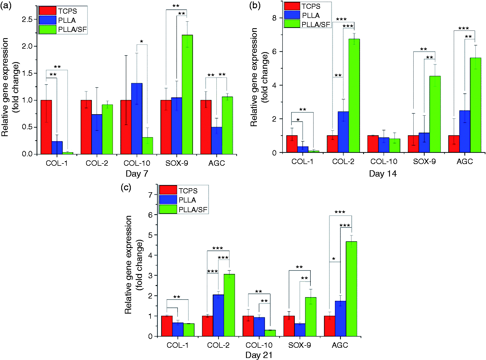
As a factor indicating an enhancement of transcription of collagen type II, Sox-9 showed a significantly higher expression on the PLLA/SF scaffold than the PLLA and TCPS scaffolds at days 7, 14, and 21. The expression of aggrecan was significantly higher on the PLLA/SF scaffold than on the PLLA or TCPS at days 14 and 21. Collagen type X indicates hypertrophy and aging of chondrocytes, which is not desirable for cartilage tissue engineering. The expression of collagen type X was significantly lower on the PLLA/SF scaffolds than on the PLLA or TCPS at days 7 and 14.
Discussion
Nowadays, more and more researchers are paying intensive attention to cartilage tissue engineering due to wear-related issues and postoperative infection associated with total joint replacement. Great progress has been made in insights into the wear mechanism and reducing the generation of wear debris.30–33 The ECM of cartilage has a 3D structure consisting of water (70–80%), collagen (50–75%), and GAGs (15–30%). 34 The scaffold, a crucial factor in tissue engineering, provides the environment for cell seeding and regeneration of new tissue. As a temporary ECM, optimally the engineered scaffold should mimic the composition, structure, function, biocompatibility, biodegradability, and mechanical properties of natural cartilage and promote chondrocyte attachment, proliferation, differentiation, and generation of new tissues.35,36 Currently in cartilage tissue engineering, there is much research interest in biomimetic scaffolds, as an alternative to treat joint disorders besides total joint replacement.37–39 In previous studies, PLLA and SF have been used as scaffold material for cartilage tissue engineering,40,41 but the PLLA/SF composite scaffold prepared by electrospinning has not been investigated in detail, especially in terms of chondrogenesis of chondrocytes. Consequently, in the present study, we manufactured a PLLA/SF composite scaffold and examined its potential application in cartilage tissue engineering.
In the present study, the PLLA/SF scaffold was prepared by electrospinning at a ratio of 60:40, and PLLA was used as a reference. The SEM images showed that the surfaces of both the PLLA and PLLA/SF scaffolds had a porous structure that was similar to the ECM of natural cartilage. A porous structure is necessary for the scaffold to facilitate transportation of nutrient and metabolic waste. The PLLA/SF scaffold has a small nanofiber diameter, which is closer to the size of the biomacromolecule in the ECM of natural cartilage, and contributes to result in a better chondrogenesis effect.42,43 Thickness is an important parameter for an engineered tissue scaffold. For an electrospinning scaffold, if it is too thick, due to fiber layers and close arrangement, this reduces pore diameter and porosity, thus restricting cell growth and the transport of nutrient and metabolic wastes. If it is too thin, the cells could be affected by the property of tissue culture plastics rather than the actual scaffolds. However, so far there has been no report of optimal thickness for an electrospinning scaffold, as the final thickness is material-dependent.
SF is a natural protein with good hydrophilicity. As a result, the swelling ratio of PLLA/SF is larger than that of PLLA. Generally, a scaffold with good hydrophilicity can promote adhesion of cells, which is beneficial to cartilage tissue engineering. However, the mechanical properties of PLLA/SF were slightly weaker than that of PLLA, which was caused by the addition of SF, a natural protein with poor mechanical properties. From the viewpoint of materials science, cartilage is a viscoelastic material, and an ideal scaffold should possess good mechanical properties that match that of natural cartilage. However, so far no materials are known to replicate natural cartilage tissue. 34
The adhesion of cells to the scaffold is the first biological behavior that occurs in the scaffold–cell interaction, and has an important effect on subsequent differentiation, proliferation, gene expression, apoptosis, and migration of cells on the scaffolds. 44 Consequently, adhesion of chondrocytes to the scaffold is preliminary to the construction of tissue-engineered cartilage. In the present study, the SEM and CLSM images both showed that chondrocytes could adhere to the PLLA and PLLA/SF scaffolds. The structure and surface properties of a scaffold can determine the type, number, and conformation of absorbed proteins, causing different gene expressions of the adherent cells and consequently different biological behaviors. 45 Surface properties of a material include topography, wettability, surface energy, surface charge, chemical groups, and bioactive factors. 46 A material with appropriate hydrophilicity, rough surface topography, and positive surface charge can promote the adhesion and spreading of cells. We consider that the surface wettability and surface charge of the PLLA/SF composite scaffold can be adjusted by the chemical groups associated with SF, and therefore the PLLA/SF scaffold is a suitable material with potential application in cartilage tissue engineering.
Cartilage tissues consist of chondrocyte and ECM. Chondrocytes secrete ECM, and in turn ECM provides mechanical and chemical information that chondrocytes require to regulate biological behavior. The interaction between chondrocytes and ECM is a dynamic process, and the secretion of ECM by chondrocytes has an important role in the construction of cartilage and repair of damaged cartilage tissues. In the present study, we used biochemical and histological staining to analyze quantitatively and qualitatively the proliferation and ECM secretion of chondrocytes on the scaffolds. From the histological staining images, it was observed that chondrocytes could proliferate and secrete ECM on the PLLA and PLLA/SF scaffolds, with a homogeneous distribution of blue ECM and dense cells. The results of the biochemical analyses showed that the PLLA/SF scaffold was best for chondrocyte adhesion, proliferation, and growth, among the scaffolds compared. But from the vertical cross section images of H–E staining, we observed that only a small amount of cells grew into the internal structure of both scaffolds. This was considered to be caused by the small pore size of the scaffolds that chondrocytes could hardly grow in.
One obvious disadvantage of electrospinning technology for the fabrication of scaffold is that the scaffold is not thick enough to form a 3D structure. Additionally, it is difficult for the chondrocytes to grow into the relatively smaller pore size of the scaffold, although the pore size may enlarge with the gradual degradation of the scaffolds. Many attempts have been proposed to solve these shortcomings, e.g. increasing the fiber diameter by optimizing the electrospinning parameters. 47 However, it was indicated that, with the increase of the nanofiber diameter, the pore enclosed by the fibers significantly increased from plane view, but the inner pore size of the scaffolds could not increase due to superpose and dense deposit of the nanofibers. 48 The combination of salt leaching and ice crystals as a removable void template to increase pore size during electrospinning was investigated, but the distribution of pore size was found to be extremely uneven.49,50 Recently, various promising and cutting-edge electrospinning techniques have been developed, including multi-layering electrospinning, post-processing after electrospinning, liquid-assisted collection, template-assisted collection, porogen-added electrospinning, and self-assembly, and electrospinning is considered as a powerful tool in fabrication of 3D fibrous macrostructures besides traditional two-dimensional (2D) nanofibrous structures. 51
TCPS was treated to provide a hydrophilic surface to promote cell adhesion and spreading. This resulted in a higher cell number than either the PLLA or PLLA/SF scaffold at days 3, 7, and 14. However, TCPS has a 2D planar and monolayer structure, and chondrocytes stop proliferating due to contact inhibition when they reach complete confluence. The nanofibrious structure of scaffolds has a higher surface area (the nanometer size effect), and the cell number of the PLLA and PLLA/SF scaffolds exceeded that of the TCPS at day 21. As reported earlier, chondrocytes with a flattened morphology grew more rapidly but synthesized less ECM, while those with a round shape proliferated slowly but synthesized more ECM. 52 From the SEM images in the present study, chondrocytes maintained their round shape on the PLLA/SF scaffold. Thus, the amount of sGAG, total collagen content, GAGs per DNA, and collagen per DNA on the PLLA/SF scaffold were higher than that on the TCPS at days 14 and 21. However, there is no significant difference with regard to GAG and collagen activities between the PLLA/SF and PLLA scaffolds, as indicated in Figure 6(d) and (e). One potential reason for this is that the main biological effect of the PLLA/SF scaffold is to better promote cell adhesion, proliferation, differentiation, and synthesis of cartilaginous matrix, compared with the PLLA scaffold. The total DNA, GAG, and collagen on PLLA/SF scaffolds are more than that on the PLLA scaffolds.
Chondrocytes are the only cells approved by the American Food and Drug Administration for clinical applications to repair cartilage damage or defects. However, chondrocytes dedifferentiation is problematic when cultured in a 2D environment in vitro. The dedifferentiated chondrocytes gradually change to a flattened, spindle shape and synthesize collagen type I rather than collagen type II. 53 Consequently, it is very important to maintain the stability of chondrocyte phenotype in order to form normal hyaline cartilage but not fibrocartilage, as fibrocartilage is weaker and can accelerate cartilage degeneration. Collagen type I is a marker of dedifferentiation of chondrocytes, while collagen type II and aggrecan are cartilage-specific and are often used to indicate normal chondrocytes and to evaluate their chondrogenic potential. Sox-9 is the key transcription factor participating in the process of cartilage development and formation, which, combined with an enhancement of the COL2A1 gene, can promote synthesis of collagen type II. Collagen type X is synthesized by hypertrophic chondrocytes and facilitates cartilage ossification. In the present study, our results showed that chondrocytes maintained their phenotype on the PLLA/SF scaffold, with lower levels of collagen type I. In addition, the PLLA/SF scaffold had higher levels of collagen type II, aggrecan, and Sox-9, indicating the best chondrogenesis potential.
In the present study, we prepared the PLLA and PLLA/SF scaffolds by electrospinning and investigated their chondrogenic potential through observing chondrocyte morphology, biochemical analyses, histological staining, and RT-PCR. The results showed that chondrocytes could adhere, proliferate, and secrete cartilage-specific ECM when cultured on PLLA/SF scaffold, and their phenotype remained stable. The PLLA/SF scaffold, as a promising material with good chondrogenic potential, may be applied in cartilage tissue engineering. An in vivo cartilage critical-sized defect model will be established to evaluate the PLLA/SF scaffold in further study.
Footnotes
Declaration of Conflicting Interests
The author(s) declared no potential conflicts of interest with respect to the research, authorship, and/or publication of this article.
Funding
The author(s) disclosed receipt of the following financial support for the research, authorship, and/or publication of this article: This study was financially supported by the Natural Science Foundation of Jilin Province (grant no. 3D512K513431).