
Abstract
Delivery of amphiphobic drugs (insoluble in both water and oil) has been a great challenge in drug delivery. SNX-2112, a novel inhibitor of Hsp90, is a promising drug candidate for treating various types of cancers; however, the insolubility greatly limits its clinical application. This study aimed to build a new type of drug delivery system using single-walled carbon nanotubes (SWNTs) for controllable release of SNX-2112; chitosan (CHI) was non-covalently added to SWNTs to improve their biocompatibility. SWNTs-CHI demonstrated high drug-loading capability; the release of SNX-2112 was pH triggered and time related. The intracellular reactive oxygen species of SWNTs–CHI increased, compared with that of SWNTs, leading to higher mitogen-activated protein kinase and cell apoptosis. The results of western-blotting, lactate dehydrogenase (LDH) release assay, and cell viability assay analyses indicated that apoptosis-related proteins were abundantly expressed in K562 cells and that the drug delivery system significantly inhibited K562 cells. Thus, SWNT–CHI/SNX-2112 shows great potential as a drug delivery system for cancer therapy.
Keywords
Introduction
Heat shock protein 90 (Hsp90) is an adenosine triphosphate (ATP)-dependent molecular chaperone responsible for the stability and function of a diverse range of client proteins, including Akt, Raf, Erk and IKKa, which regulate cell survival and proliferation and are responsible for malignant transformation.1–4 Hsp90 is abundantly expressed in eukaryotes and comprises over 1% of eukaryote total cellular content.5,6 However, the expression levels of Hsp90 in tumour cells are two-fold to 10-fold higher than in normal cells, indicating that Hsp90 may be of great importance to the survival and proliferation of tumour cells. 7
Hsp90 inhibitors reportedly induce apoptosis in tumour tissues through various pathways, such as mitochondria-mediated and death receptor-induced pathways, but have only a modest effect in normal cells.8–11 SNX-2112, a novel inhibitor of Hsp90 that binds to the N-terminal ATP binding site of Hsp90 with high affinity,12–14 is a promising drug candidate for treating various types of cancers, including multiple myeloma, 13 HER-kinase-dependent cancers, 15 and human chronic leukaemia. 16 However, SNX-2112 is only slightly soluble in water and oil and poorly soluble in other lipophilic excipients, which has significantly limited its clinical application. Therefore, an effective carrier for SNX-2112 delivery is needed to improve its bioavailability.
Among the currently available delivery vehicles, single-walled carbon nanotubes (SWNTs) are gaining increasing attention due to their significant advantages. SWNTs are formed from a graphene layer rolled in a seamless cylinder, the hollow construction and large surface area of SWNTs enables them to combine with aromatic drugs through π–π stacking, as well as hydrophobic and van der Waals interactions,17–20 making them ideal for drug loading. However, upon delivery and drug accumulation in tumour tissues, the nanostructure of SWNTs allows them to leave the tumour area via abnormally leaky tumour blood vessels. 18 Several studies have shown that short (< 1 µm), well-functionalised SWNTs are rarely retained in the reticuloendothelial systems (RES) and could be cleared via renal pathways, thereby minimising the likelihood of toxicity in vivo.18,21,22 However, poor water solubility and potential toxicity limit SWNT development. 23 It is particularly encouraging that SWNTs can be modified readily using physical and chemical methods. Moreover, in vitro results have shown that multifunctional SWNTs greatly improve the therapeutic efficiency of the drug while reducing toxicity. Additionally, SWNTs can be modified with many biological species, such as poly (ethylene glycol) 24 and polysaccharides 25 to improve biocompatibility.
Chitosan (CHI), a type of polysaccharide, is often used for biocompatible modification, due to its favourable characteristics including biocompatibility, biodegradability and non-toxicity. 26 Moreover, the amino group of CHI contributes to its cellular uptake and endosomal escape, which gives it a great advantage for drug delivery applications. 27 As such, there have been numerous studies of CHI-modified drug carriers in the form of nanoparticles, liposomes and micelles.28–31
The purpose of this study was to explore the feasibility of SWNTs for the controllable delivery of SNX-2112; SWNTs were non-covalently modified by CHI to improve their biocompatibility. Subsequently, SNX-2112 in aqueous solution was attached to modified nanotubes by mere mixing. The pharmaceutical efficiency was further examined for K562 cells through cell viability assay in vitro and LDH release assay. Intracellular reactive oxygen species (ROS) and western-blotting analyses were used to monitor cell apoptosis.
Materials and methods
Materials
The following materials were used in this research: carboxyl SWNTs (purity > 90%; carboxyl content: 2.73%; length: 5–30 µm; diameter: 1–2 nm; Chengdu Organic Chemistry Institution, Chengdu, Sichuan, China); CHI (MW: < 5000 Da; Jinan Haidebei Co., Ltd., Jinan City, Shandong, China); hyaluronan (MW: 6000 Da, Shandong Furuida Co., Ltd., Dezhou, Shandong, China); SNX-2112 (Guangzhou Jinan Biomedicine Research and Development Center, Guangzhou, China); WST-1 reagent (Beyotime Biotechnology Institution, Haimen, China); RPMI 1640, foetal bovine serum, penicillin, streptomycin and trypsin (Gibco, Grand Island, NY, USA); and dichlorofluorescein diacetate (DCFH-DA), lysis buffer and TOX7 Kit (Sigma-Aldrich, St. Louis, MO, USA). All other chemicals were of analytical grade and used as received.
Synthesis of SWNT–CHI
SWNTs (20 mg) were sonicated in CHI solution (60 mg in 0.1 M NaCl and 0.02 M acetic acid, 40 mL) for 30 min and stirred at room temperature for 24 h. The SWNT–CHI was dialysed (dialysis bags, MWCO = 10 kDa) for 3 d in ultrapure water. After drying at 37℃ under vacuum, the modified SWNT–CHI was collected.
Loading of SNX-2112 onto nanotubes
SWNTs and SWNT-CHI were sonicated in SNX-2112 solution (15 mg in 15 mL pH 7.4 PBS) for 30 min and stirred at room temperature for 24 h. The products were washed with pH 7.4 PBS buffer and centrifuged several times until the supernatant became colourless. The nanotubes, loaded with SNX-2112, were collected after drying at 37℃ under vacuum. Then, 1-mg nanotubes (SWNTs, SWNTs–CHI, SWNTs/SNX-2112 and SWNTs–CHI/SNX-2112) were dispersed in 5 mL of ethanol. Next, the dispersal solution was dropped onto a copper mesh. The morphology of the nanotubes was then observed by transmission electron microscopy (TEM; JEM-2100F; JEOL, Tokyo, Japan).
Drug loading efficiency and embedding ratio measurement
After collecting free SNX-2112 solution, the amount of free SNX-2112 unbound on nanotubes was determined by high-performance liquid chromatography (HPLC) using a Dionex Ultimate 3000 HPLC system (Thermo Scientific, Waltham, MA, USA). SNX-2112 was separated against a C18 column (Syncronis C18, 5 µm, 4.6 × 250 mm), guarded with a precolumn at 40℃ and monitored at 251 nm with an injection volume of 20 µL. The mobile phase consisted of 70% methanol and 30% water pumped at a flow rate of 1.0 mL/min.
In vitro drug release
First, 1-mg nanotubes (SWNTs/SNX-2112 and SWNTs–CHI/SNX-2112) were dispersed in 0.5 mL of pH 7.4 and pH 5.5 PBS in dialysis bags (MWCO = 10 kDa) and mixed in 20 mL of pH 7.4 and pH 5.5 PBS at 37℃ at 60 rpm, respectively. After 2, 6, 12, 24, 48 and 72 h, 2-mL aliquots were taken and free SNX-2112 was measured by HPLC, as described before.
In vitro cytotoxicity assay
K562 cells were maintained in RPMI 1640 medium, supplemented with 10% FBS and 1% antibiotics (penicillin–streptomycin, 10,000 U mL−1) at 37℃ in a humidified atmosphere containing 5% CO2. Exponential-phase cells were collected and seeded in a 96-well plate. After reaching confluence, the cells were treated with SWNTs/SNX-2112, SWNTs–CHI/SNX-2112 and free SNX-2112 at a concentration of 80 mg/mL. Before adding WST-1, the background absorbance of each well at a wavelength of 450 nm was measured. The cell viability was determined using a microplate reader (Thermo, Multiskan MK3) at 450 nm after 2 h.
Estimation of intracellular reactive oxygen species (ROS)
Due to oxidation by a ROS, non-fluorescent dichlorofluorescein diacetate (DCFH-DA) converts to a fluorescent molecule; in this research, this was used to measure the intracellular ROS in K562 cells. Briefly, 5 × 10³ cells were treated with 80 µg/mL of SWNTs/SNX-2112 and SWNTs–CHI/SNX-2112 in serum-free medium for 4 h, washed with 1 × PBS, and then incubated with 100 µL serum-free medium supplemented with 50 μM of DCFH-DA for 3 h at 37℃. The medium was then aspirated and the product was harvested in 150 µL of lysis buffer. Fluorescence was recorded using a Perkin Elmer (Norwalk, CT, USA) fluorescence spectrometer at λex = 505 nm and λem = 523 nm. The final value of intracellular ROS concentration was normalised and represented as a percentage with respect to an untreated control.
Lactate dehydrogenase (LDH) release
Released LDH was quantified using a TOX7 kit. Briefly, 5 × 10⁴ K562 cells were seeded per well of a 24-well plate and were treated with SWNTs/SNX-2112, SWNTs–CHI/SNX-2112 and SNX-2112. The assay was performed according to the manufacturer's protocol. First, 100 µL of assay mixture was added to 50 µL of supernatant from all samples. The samples were incubated for 30 min at room temperature, and the reaction was terminated by the addition of 15 µL of 1 N HCl. The absorbance was then recorded using an MRX microplate reader (Dynex Technologies, Chantilly, VA, USA) at 490 nm, with a reference wavelength of 650 nm.
Western blotting
K562 cells, grown to 70% confluence in a flask, were cultured with a medium containing 20 mg/mL SWNTs/SNX-2112, SWNTs–CHI/SNX-2112 and SNX-2112. After 48 h, cells were washed with PBS, harvested with a scraper, and centrifuged at 2000 × g for 10 min at 4℃. After discarding the supernatant, cells were lysed to collect the cellular proteins. Equal amounts (50 µg) of proteins were separated on 12% SDS-PAGE gels and transferred to nitrocellulose membranes. The membranes were blocked with 5% non-fat milk in tris-buffered saline and Tween 20 (TBST) at room temperature. The specific antibodies used for this experiment were anti-Bax primary antibody (Abcam, Cambridge, UK), anti-Bcl-2 primary antibody (Abcam) and anti-caspase-3 primary antibody (Abcam). Blots were developed using enhanced chemiluminescence.
Statistical analysis
All results are expressed as means ± standard deviation (SD). Statistical analyses were performed using SPSS for Windows software (ver. 16.0; SPSS Inc., Chicago, IL, USA). Continuous data were analysed by one-way analysis of variance (ANOVA) and the Kruskal–Wallis test. Categorical data were analysed using the χ2 test. A p value < 0.05 indicated statistical significance.
Results and discussion
TEM images
Figure 1 shows TEM images of the SWNT, SWNTs–CHI, SWNTs/SNX-2112 and SWNTs–CHI/SNX-2112 structures; the unmodified SWNTs were straight tubes with a diameter of ∼20 nm with hollow construction and a smooth, compact single wall. Modification with CHI enhanced the diameter of the nanotubes; although the nanotubes were smooth and uniformly coated, the clarity of the sidewalls was reduced due to screening by CHI. Through π–π stacking interactions, hydrophobic interactions and van der Waals interactions, SNX-2112 was largely absorbed onto the nanotube sidewalls; the wall surfaces were no longer smooth. In contrast, nanotubes loaded with SNX-2112 were thicker, and the loading of SNX-2112 was clearly visible.
Transmission electron microscopy (TEM) images of (a) single-walled nanotubes (SWNTs), (b) chitosan (CHI)-modified SWNTs (SWNTs–CHI), (c) SWNTs loaded with SNX-2112 (SWNTs/SNX-2112) and (d) CHI-modified SWNTs loaded with SNX2112 (SWNTs–CHI/SNX-2112).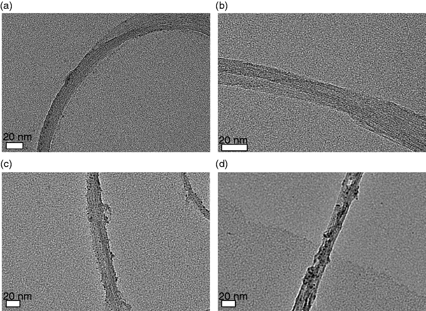
SWNTs are filamentous; thus, SNX-2112 attached to the SWNT surface; note that this attachment is reversible. Therefore, when the organism absorbed the nanotubes, SNX-2112 could be easily dissociated to take effect. 32 The diameter of SWNT-CHI/SNX-2112, ≤ 30 nm, prevented its elimination by the RES, prolonging the retention time in the blood33,34; in addition, their small size facilitates their penetration of cancer cells.19,35
Drug loading
The drug-loading efficiencies and embedding ratios of SWNTs and SWNTs–CHI are shown in Figure 2. The drug-loading efficiencies of both nanotube types were above 110%; the drug embedding ratios all exceeded 70%. The drug-loading capacity of SWNTs–CHI was nearly the same as that of SWNTs, indicating that the wrapping of CHI did not have any effect on SNX-2112 loading. Thus, the loading of the drug depended mainly on the hollow structure and the large surface area of SWNTs, which benefit from the trapping and containment of SNX-2112 via π–π stacking and electrostatic interactions18,27,36; CHI did not have much of an effect in this regard. Therefore, SWNTs-based materials have a high drug-carrying capacity and show great potential in drug delivery.
(a) Drug-loading efficiency and (b) drug-embedding ratio of modified SWNTs.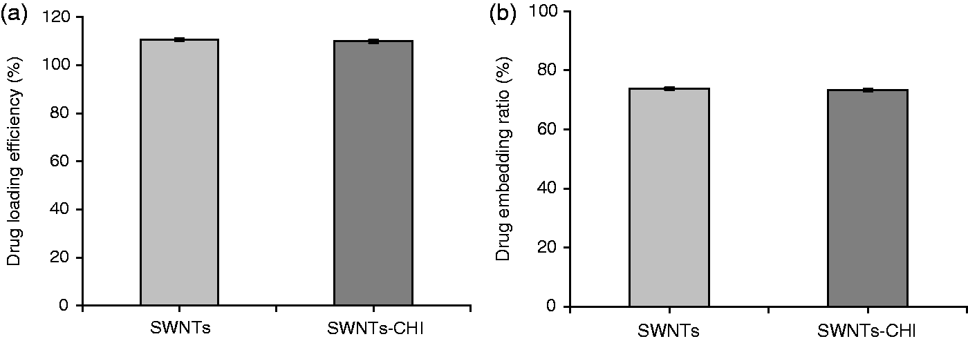
Drug release
The release of SNX-2112 from SNX-2112-loaded nanotubes was pH-triggered, as shown in Figure 3; SNX-2112 on both the nanotubes was stable in PBS buffer at pH 7.4 and 37℃. SNX-2112 was released quickly within 10 h; the release ratio tended to be flat over the following 62 h. The total amount of released drugs was no more than 30%. This demonstrated the stability of SNX-2112 loading on both modified and unmodified SWNTs for the given physiological pH values. In slightly acidic solutions such as pH 5.5 PBS, corresponding to lysosomal pH, the cumulative release amount was considerable over a 72-h period, at nearly 80%, and the release was time-related. After a quick rise to about 50% within 10 h, the curve slowed down over the following 62 h, during which another 30% of the drug was released. The release behaviours of both nanotubes were fairly similar, as SWNTs are the dominant factor in SNX-2112 loading; CHI modification did not appear to have much of an effect on drug loading. The pH-triggered drug release from nanotubes, in which the SNX-2112 stably bound to SWNTs under normal physiological conditions, is released at a reduced pH typical of micro-environments of intracellular lysosomes, endosomes or cancerous tissue, provides a built-in mechanism for selective drug release.
36
SNX-2112 release at 37℃ in (a) pH 7.4 and (b) pH 5.5 PBS.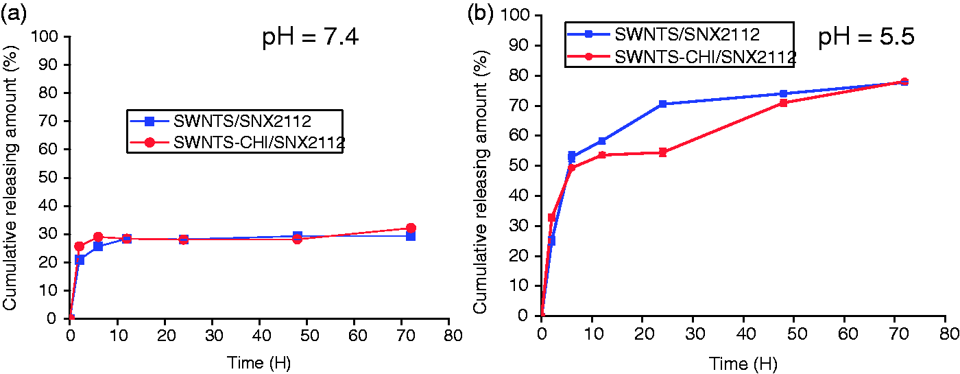
Inhibition effects
The cell viability of the various drug-loaded nanotubes and free SNX-2112 was determined in K562 cells using the WST-1 assay to evaluate their inhibition effects; the results are shown in Figure 4. The SWNTs–CHI/SNX-2112 had the highest inhibition ratios compared with groups treated with SWNTs/SNX-2112 or free SNX-2112 at the same concentration. The inhibition ratio of the three groups at the concentration of 10 µg/mL towards K562 was less than 20%. The inhibition ratio increased with the concentration of drug-loaded nanotubes. At the concentration of 80 µg/mL, the inhibition ratios exceeded 80%.
Inhibition ratio of K562 cells after treatment with modified SWNTs and SNX-2112 for 48 h. The results represent the means ± SD (n = 6). Asterisks indicate statistical significance (p < 0.05).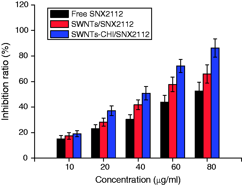
Effects of nanotubes on ROS production
ROS reflects the intracellular oxidative stress status, an important indicator of cell health, and can induce intracellular protein inactivation (through oxidation and nitration), lipid peroxidation, dysfunction of the mitochondria and, eventually, apoptosis or necrosis.37,38 In this work, the effects of SWNTs and SWNTs–CHI at the concentration of 80 µg/mL on intracellular ROS were investigated in K562 cells, as shown in Figure 5. An increase of approximately 100% intracellular ROS percentage in cells treated with SWNTs–CHI was observed over unmodified SWNTs, which could enhance mitogen-associated protein kinase (MAPK) activation. Specifically, MAPK can induce G2/M cell cycle arrest, thereby pushing the cancer cells into a new mitotic cycle that makes them vulnerable to apoptosis.
39
Effect of nanotubes (80 µg/mL) on intracellular reactive oxygen species (ROS) (percentage of control) in K562 cells at the end of 4 h. The results represent the means ± SD (n = 6). Asterisks mark statistical significance (p < 0.05).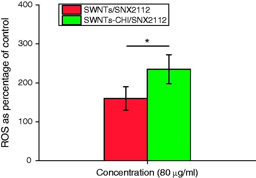
LDH levels
Lactate dehydrogenase (LDH) is a type of enzyme that is released by the cell if the cell membrane becomes damaged;
40
thus, the estimation of LDH release provides a quantitative basis for the loss of cell viability. The LDH release of K562 cells after treatment with SNX-2112 and SNX-2112-loaded nanotubes was examined to evaluate their therapeutic effect; the results are shown in Figure 6. In contrast to the control, the LDH release levels of the other three groups were much higher. The absorbance of the drug-loaded nanotube groups was higher than the free drug group, and the absorbance of SWNTs–CHI/SNX-2112 was the highest, almost three-fold that of the control, indicating the most serious cell injury of K562 cells. The LDH release results demonstrate that both nanotube types were capable of destroying cell membrane integrity, with SWNTs–CHI/SNX-2112 showing the strongest effect, in agreement with the previous results of the highest inhibition ratio of K562 cells.
Lactate dehydrogenase (LDH) release from K562 cells following 24-h treatment with SNX-2112 and SNX-2112-loaded nanotubes.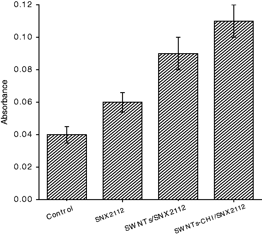
Western blot analysis
Western blot analysis was conducted towards K562 cells to directly detect caspase 3, Bax and Bcl-2. Caspase-3 is an important evectional caspase of apoptosis.
41
Generally, caspase-3 is activated in apoptotic cells by both extrinsic (death ligand) and intrinsic (mitochondrial) pathways40,42,43 and is considered as one of the most important execution caspases. The Bcl-2 family acts as a critical regulator of mitochondrial permeability and consists of pro- and anti-apoptotic members that form heterodimers to inhibit or activate each other.
44
The anti-apoptotic member, Bcl-2, protects against apoptotic stimuli. Many anticancer drugs trigger mitochondria-mediated apoptosis in cancer cells via downregulation of Bcl-2 and/or upregulation of Bax
9
; thus, the ratio of Bcl-2 to Bax is important in determining apoptosis susceptibility.
45
The results of Western blotting (Figure 7) showed that the Bax band of the SWNTs–CHI/SNX-2112 group was much darker than the others, indicating that Bax was significantly upregulated. The Caspase-3 bands of the three groups were nearly the same, and the band of SWNTs–CHI/SNX-2112 group was slightly darker and broader. As far as the Bcl-2 band was concerned, the band of SWNTs–CHI/SNX-2112 group had the least expression. The Western blotting results were consistent with ROS results, all suggesting the strongest apoptosis induced by SWNT–CHI/SNX-2112.
Western blotting of Bax, Bcl-2 and Caspase-3 in K562 cells treated with (a) SNX2112, (b) SWNTs/SNX2112 and (c) SWNTs–CHI/SNX2112.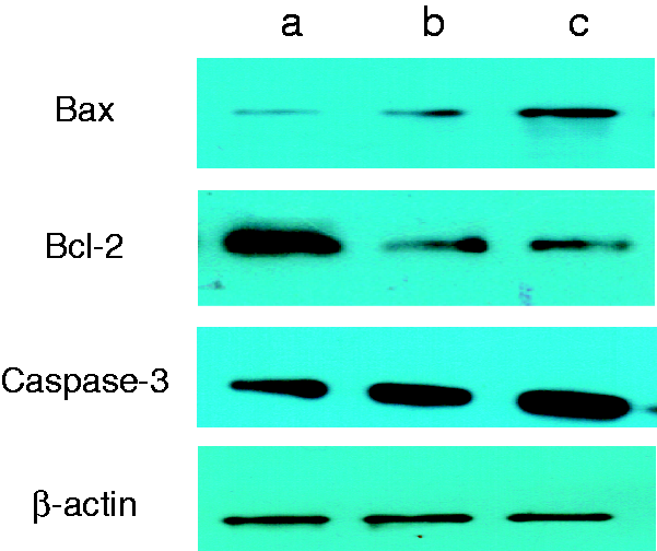
Conclusion
In this study, a novel drug delivery system was established for the controlled release of SNX-2112. SWNTs, used as the core for drug loading, were wrapped by CHI to improve the biocompatibility. SWNTs–CHI nanotubes showed exceptional drug-loading capability. According to ROS measurements, cell inhibition assay, LDH release assay and western-blotting analyses, the SWNT-CHI/SNX-2112 had a significant therapeutic effect on K562 cells. Therefore, SWNT-CHI/SNX-2112 shows great potential as a drug delivery system for anti-tumour therapy.
Footnotes
Declaration of Conflicting Interests
The author(s) declared no potential conflicts of interest with respect to the research, authorship, and/or publication of this article.
Funding
The author(s) received no financial support for the research, authorship, and/or publication of this article.