
Abstract
The bioactivity of yttrium and cerium are investigated when substituted for Sodium (Na) in a 0.52SiO2-0.24SrO-0.24-xNa2O-xMO glass-ceramics (where x = 0.08 and MO = Y2O3 or CeO2). Bioactivity is monitored through pH and inductively coupled plasma-optical emission spectrometry where pH of simulated body fluid ranged from 7.5 to 7.6 and increased between 8.2 and 10.0 after 14-day incubation with the glass-ceramic disks. Calcium (Ca) and phosphorus (P) levels in simulated body fluid after incubation with yttrium and cerium containing disks show a continual decline over the 14-day period. In contrast, Con disks (not containing yttrium or cerium) caused the elimination of Ca in solution after 1 day and throughout the incubation period, and initially showed a decline in P levels followed by an increase at 14 days. Scanning electron microscopy and energy dispersive spectroscopy confirmed the presence of Ca and P on the surface of the simulated body fluid-incubated disks and showed precipitates on Con and HCe (8 mol% cerium) samples. Cell viability of MC3T3 osteoblasts was not significantly affected at a 9% extract concentration. Optical microscopy after 24 h cell incubation with disks showed that Con samples do not support osteoblast or Schwann cell growth, while all yttrium and cerium containing disks have direct contact with osteoblasts spread across the wells. Schwann cells attached in all wells, but only showed spreading with the HY-S (8 mol% yttrium, heated to sintering temperature) and YCe (4 mol% yttrium and cerium) disks. Scanning electron microscopy of the compatible disks shows osteoblast and sNF96.2 Schwann cells attachment and spreading directly on the disk surfaces.
Introduction
Bioactive glasses are defined by their ability to become securely bonded with host tissues in vivo. This characteristic arises from a reaction cascade beginning with the exchange of hydrogen ions (H+ or H3O+) from physiological fluids with R+ and R2+ (commonly Na+ and Ca2+) modifying ions in the glass network. Subsequently, dissolution of the Si-O glass network creates a surface consisting of silanol (Si-OH) groups which proceeds to condensate to Si-O-Si and facilitates the deposition of an amorphous hydroxycarbonated apatite (HCA) layer.1–3 The formation of the HCA layer on the biomaterial surface is essential for bonding with host tissues. As such, bioactivity is evaluated based on the formation of a HCA surface layer after incubation in physiological fluids, commonly simulated body fluid (SBF). SBF replicates the pH and ion content of blood plasma, of particular interest is the change in Ca and phosphorus (P) content of SBF with initial concentrations at 2.5 mM and 1.0 mM, respectively. 4 The fluctuation of these ions in solution can be indicative of dissolution from the glass network, if the glass contains either element, or precipitation, potentially for the formation of an HCA layer. In addition to indicating bioactive potential for typical hard tissue bonding applications, Ca and P deposition also indicate glass dissolution which has implications relating to cell attachment and proliferation. Cell interaction with material surfaces in vitro determine the potential for tissue integration, where cell attachment controls cell morphology which in turn affects cell activity.5,6 Specifically relevant is the relation between dissolution and surface chemistry, charge and polarity, which affects both Ca and P deposition and the adsorption of molecules and protein cells use to facilitate binding to the biomaterial surface.5,6 Therefore, dissolution of bioactive glasses and deposition of Ca and P may indicate a potential for proteins enabling cell attachment of the commonly studied osteoblast line as well as the Schwann line of most interest in this work. 6
Aside from Ca and P, other dissolution products from silicate-based bioactive glasses have been found to encourage the bonding of bioactive glasses with host tissues. These ions commonly activate or upregulate the cellular response to the HCA layer, and encourage osteogenesis; one example is Si, which is essential for metabolic processes relating to formation and calcification of the bone matrix and can improve bone mineral density.7,8 Of particular interest in this work is Sr, which has been shown to perform similarly to calcium (Ca) in both the glass network and the body.7,9–11 As such, it makes Sr an ideal candidate to replace the Ca component of typical bioactive glass compositions. One drawback to the replacement of Ca with Sr is the reduction of glass network solubility and changes in the formation and morphology of the HCA layer resulting in reduced bioactivity.12–14 The replacement of Ca is considered due to the excessive Ca accumulation at the site of nerve injury and the dissolution behavior of bioactive glasses. Aside from the physical damage associated with nerve injury, loss of ionic homeostasis in the extracellular fluid occurs due to the influx of calcium and loss of Na. This Ca accumulation alone can cause excitotoxic necrosis in adjacent cells and tissue and limit the regeneration potential of nerve tissue, which already has limited ability to reconnect over gaps of 5 mm but also stimulates the production of radical oxygen species (ROS).15–22 The presence of ROS and accompanied oxidative stress on neighboring cells and tissue, in addition to the impediments created by the inherent biological response further reduce the chance at successful regeneration and reconnection for functional recovery.20,23–27
Current techniques employed to aid in the regeneration and reconnection of nerves focus on methylprednisolone sodium succinate to reduce the inflammatory response, or nerve guide conduits.15,27 Conduits provide physical stabilization and axonal guidance, as well as having the capability to deliver and maintain growth factors at the site of regenerating axons.28–30 Recently, bioactive glass fiber wraps have been considered for use as nerve conduits where a study by Bunting et al. 19 found rat schwann cells grew on Bioglass® and, in vivo, conduits populated with Bioglass® fibers produced axonal regrowth (of the sciatic nerve in rats) equivalent to that of an autograft over a 0.5 cm gap after a month. Another study by Jeans et al. 20 using conduits formed from Corglaes® mesh bonded with a biodegradable polymer found axonal regeneration of the median nerve in the forelimb of sheep to be equivalent to that seen in the epineurial repair group. Finally, a study on neurite outgrowth of dorsal root ganglion cells (closely associated with the central nervous system) by Marquardt et al. 25 found borate-based bioactive glass fibers in fibrin scaffolds-guided neurite extension and did not exhibit significant toxicity to neuronal cells. The promising results obtained in these studies show that bioactive glasses can be successfully incorporated into composite nerve conduits in fiber form, and thus is a potential delivery mechanism for the glasses studied herein. Improvements on bioactive glasses for nerve conduit applications have been suggested through the removal of traditional ions such as Ca while maintaining Na to supplement the ionic fluctuations seen after injury. Additionally, for the purpose of encouraging functional recovery of nerves, the oxidative stress may also be addressed. Elimination of ROS is proposed through the addition of yttrium (Y) and cerium (Ce) as dopants, where studies have found that 12 nm yttria and ceria nanoparticles (non-toxic up to 200 µg/mL) offered neuroprotection via direct dose-dependent ROS scavenging.19,31 Therefore, upon inclusion in the glass composition, one or both elements may create a local coordination environment in the glass network that will allow it to act as an antioxidant. One consideration with the introduction of trivalent and tetravalent ions into the glass network is the effect on bioactivity. Studies by Leonelli et al. 32 and Cacaina et al. 33 revealed that Y and Ce have been successfully incorporated into bioactive glasses; however, bioactivity decreased with increasing Y and Ce content. The deposition of a HCA layer was delayed in these glasses forming after 7–14 days incubation in SBF.32,33
Previous work evaluating the effect of Y2O3 and CeO2 on SiO2-SrO-Na2O glass structure and solubility found Y and Ce to perform as modifier ions within the glass network, though they produce an increase in rigidity and decrease the solubility due to ionic differences with Na for which they are being substituted.34,35 Despite the reduced solubility, the dissolution products still posed toxicity in osteoblast viability studies, so glasses were thermally treated to produce partially crystalline glass-ceramics and further reduce the solubility.34,36 While partial crystallization reduces the solubility, it has also been shown, through numerous studies, to reduce the bioactivity of bioactive glasses.37–39 This work uses pH, inductively couples plasma-optical emission spectroscopy (ICP-OES), scanning electron microscopy (SEM), and energy dispersive spectroscopy (EDS) to evaluate the bioactivity in terms of Ca and P presence on the surface of Y and Ce containing glass-ceramics heat treated at a high and low temperature, namely the first crystallization temperature (Tc1) or 50℃ above the glass transition (Tg).
Materials and methods
Disk synthesis
Glass compositions in mol% and characteristic temperatures (℃) and disk size.
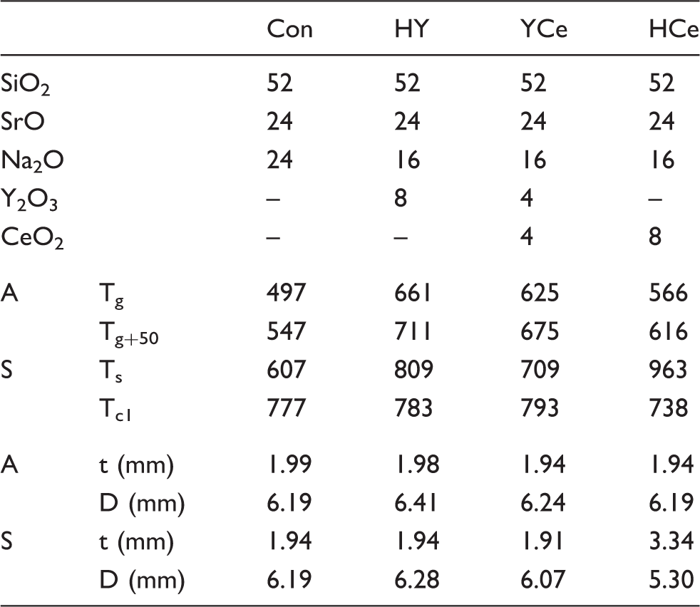
Glass powders were prepared by weighing out appropriate amounts of analytical grade reagents (SiO2 99.5%, SrCO3 99%, Na2CO3 99.5%, Y2O3 99.99%, CeO2 99.9%, Fisher Scientific, PA, USA) and ball milling (1 h). The mix was then oven dried (100℃, 1 h), fired (1500℃, 1 h, Lindberg Blue M 1700, Fisher Scientific, PA, USA) in a platinum crucible and shock quenched in water. The resulting frit was dried, ground (Gy-Ro Mill, Glen Creston Ltd, London, UK) and sieved to retrieve glass particles <20 µm. Glass powders were ball milled for 24 h with <75 µm polyvinyl alcohol (100 k MW, 97.5%, Fisher Scientific, PA, USA) added at 10 wt% to act as a binder. Glasses were thermally analyzed prior to this work using thermal gravimetric analyzer-differential scanning calorimetry and hot stage microscopy to obtain characteristic temperatures which are summarized in Table 1.34,40
Glass powders (0.15 g/disk, n = 3) were pressed (Carver Laboratory Press M, Carver, WI, USA) in a stainless steel die at a pressure of 4 tons and subsequently heat treated at two processing temperatures (Tp1 and Tp2), where the processing temperatures are included in Table 1. Disks were heated to Tp1 at a rate of 10℃/min and held for 6 h, then heated at a rate of 1℃/min to Tp2, followed by an additional 6 h hold and cool down at 5℃/min. Processing temperatures were selected at the glass transition (Tg), 50℃ above Tg (Tg+50), or sintering (Ts) and first crystallization temperatures (Tc1) for each glass individually and are denoted by annealed disks (A) and sintered disks (S). Final disk dimensions measured with digital callipers (n = 15) are included in Table 1 (where t represents disk thickness and D represents disk diameter, ±0.06 mm) yielding a surface area of 100 mm2 ± 6 mm2 between disk types.
pH analysis
Sterile liquid extracts were prepared by incubating glass-ceramic disks (n = 3/time period) in sterile 15 mL centrifuge tubes with 5 mL SBF prepared as described by Koboku et al. 4 The tubes, including disk-free control samples, were sealed and incubated for 1, 7, and 14 days. Upon removal, each sample was filtered (Amicon Ultra-4 Centrifugal Filters, Fisher Scientific, PA, USA) to ensure particle-free extracts. A 1.5 mL aliquot from each sample extract was removed into a sterile vial to be used for cytocompatibility studies.
The pH of each sample extract was measured using an Accumet® Excel XL 15 pH meter (Fisher Scientific, PA, USA). Prior to testing, the pH meter was calibrated using pH buffer standards 4.00 ± 0.02, 7.00 ± 0.02, and 10.00 ± 0.02. The probe was rinsed with deionized water (DI) water and allowed to stabilize in the pH 7 buffer between readings which were taken after the pH value stabilized for 10 s.
ICP-OES
The Ca and P ion contents were measured axially using ICP-OES on a Perkin-Elmer Optima 8000 (Perkin Elmer, MA, USA) with a detection limit of 0.1 µg/mL. ICP-OES calibration standards for Ca and P were prepared from stock solutions (Perkin-Elmer, MA and Fisher Scientific, PA, USA) at 0.1, 1, 10 µg/mL for samples diluted 1:10 after Ca and P concentrations in the SBF-only samples were found to be 93.4 ± 1.5 µg/mL and 38.0 ± 0.7 µg/mL, respectively. Ca and P precipitation from SBF solution after incubation with disks is reported based on the percent remaining in solution relative to that of the SBF-only controls using equation (1)

Osteoblast viability
MC-3T3-E1 osteoblasts (ATCC CRL-2593) were maintained on a regular feeding regime with minimum essential medium (MEM) alpha media (w/L-glutamine, ribonucleosides, and deoxyribonucleosides), supplemented with 10% fetal bovine serum (FBS) in a cell culture incubator at 37℃/5%CO2/95% air atmosphere (Fisher Scientific 3530 Incubator, Fisher Scientific, PA, USA). Viability studies were conducted using cells seeded into 96-well plates at a density of 104 cells/well where after 24 h incubation cells were confluent across the well surface. Confluency was not achieved with Schwann cells and therefore not used for viability studies. Glass/ceramic extracts and SBF controls were added to the 100 µL wells, 10 µL/well, producing a final concentration by volume of 9%. After addition of the extracts, the plates were incubated for an additional 24 h.
Cytocompatibility was tested using the methyl thiazolyl tetrazolium (MTT) assay. MTT (10 µL) reagent was added to each well and incubated for 4 hr (37℃/5%CO2). After incubation each well was aspirated and 100 µL of MTT solubilization solution (10% Triton X-100 in acidic (0.1 M HCl) Isopropanol) was added, and mixed by gently pipetting at half the well volume (50 µL). Once the crystals were fully dissolved, the absorbance was measured at 570 nm using a µQuant Microplate Spectrophotometer (Bio-tek Instruments Inc., VT, USA). Media wells with the SBF control samples were used to determine the background effect, and a control cell population was assumed to represent 100% viability to normalize the readings. One-way analysis of variance was employed to compare the difference in cell viability relative to the control population and between time periods for each sample. Comparison of relevant means was performed using the post hoc Bonferroni test. Differences between groups were deemed significant when P < 0.05.
Bioactivity
After SBF incubation, disks were removed, rinsed gently with deionized water (DI) water, and stored in a desiccator. Any HCA deposition on the disk surface (n = 1) was observed with a Quanta 200 F Environmental SEM (FEI, OR, USA) under a vacuum at a pressure of 0.60 torr. The electron beam was used at an accelerating voltage of 20 kV and a spot size of 3.0. EDS was carried out using an FEI EDAX system equipped with a silicon-drift detector (FEI, OR, USA).
Cell adhesion
MC-3T3-E1 osteoblasts (ATCC CRL-2593) were maintained as described in the osteoblast viability section. sNF96.2 Schwann (ATCC CRL-2884) cells were maintained on a regular feeding regime with Dulbecco's modified Eagle medium (DMEM) low media supplemented with 10% FBS in a cell culture incubator at 37℃/5%CO2/95% air atmosphere. Adhesion studies were conducted after allowing the glass-ceramic disks (n = 1) to buffer for 24 h in Media with three exchanges. Osteoblast and Schwann cells were seeded onto individual disks in 24-well plates at a density of 105 cells/well and incubated for 24 h. Images of each disk/well were taken at 4 × magnification using an Olympus IX81 Optical Microscope (Olympus America Inc., NY, USA). Osteoblasts were subsequently fixed using the procedure from Wang et al. 28 An additional milliliter of DMEM low media was added to each well for the Schwann cell samples which were then incubated for an additional 72 h and fixed. SEM images were collected using the parameters given in the bioactivity section.
Results
pH analysis
pH (presented in Figure 1) of the SBF was found to be 7.5 ± 0.1 with a statistically insignificant increase to 7.6 ± 0.1 after 14 days. All disks produced an initial increase in pH after 1 day incubation and continued to increase over the 14-day time period. HCe-A and HCe-S disks produced an initial pH increase of 8.2 ± 0.1 and 7.8 ± 0.05 increasing to 8.7 ± 0.1 and 8.2 ± 0.05 after 14 days. The HY and YCe disk extracts fell within the HCe-A/S pH range, while for Con extracts pH was found to range from 9.0 to 10.0 ± 0.1, with Con-A at a higher pH than Con-S at each time period, respectively.
pH of liquid extracts from Y/Ce disk series over 1, 7 and 14 days incubation.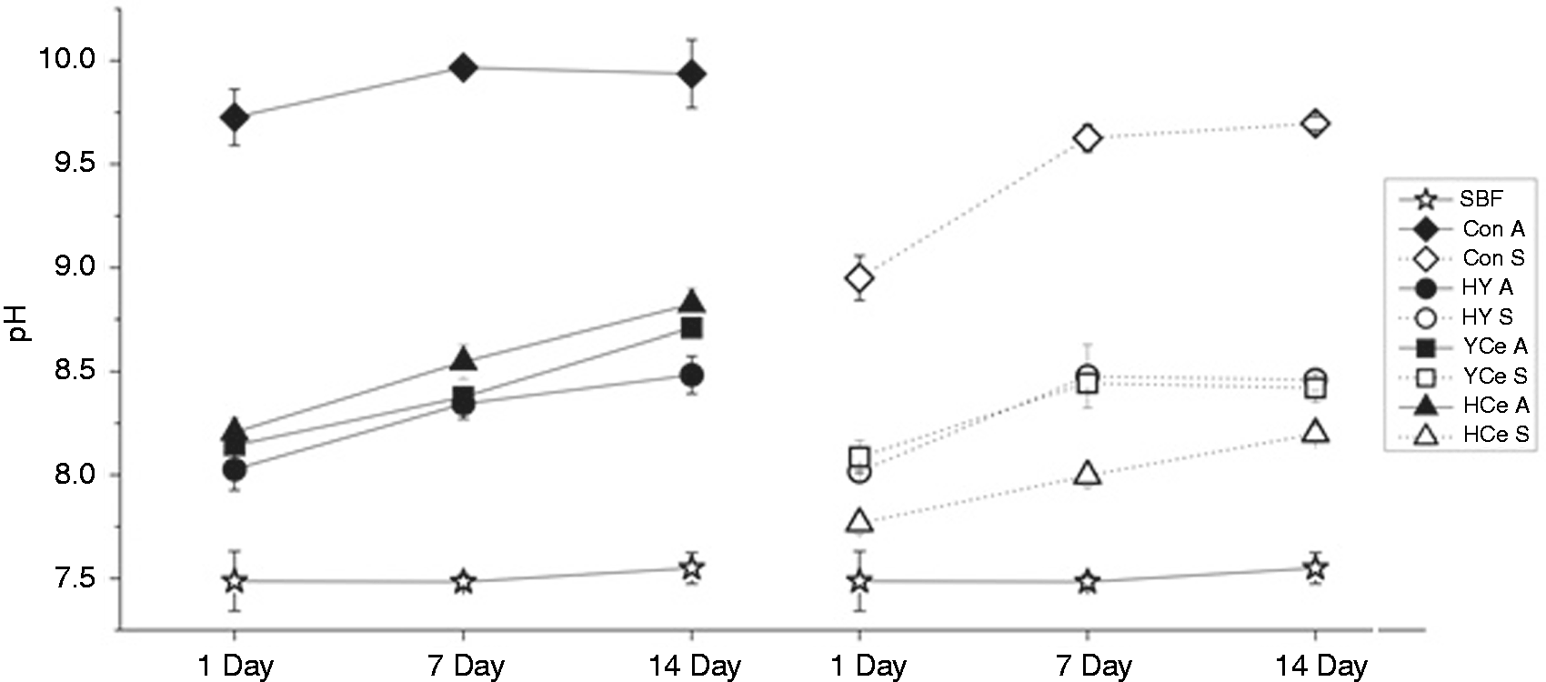
ICP-OES
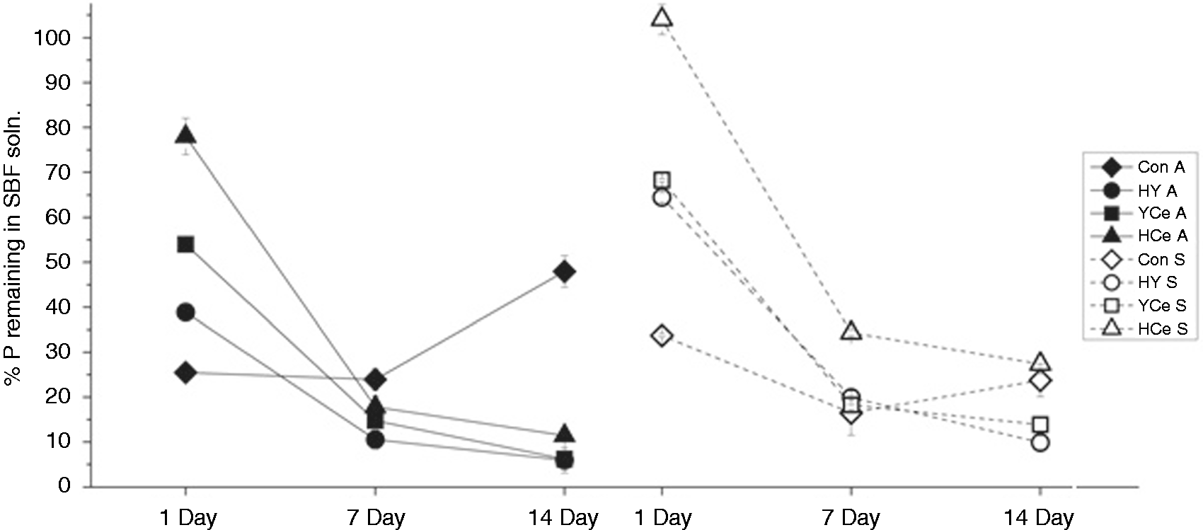
The percent of Ca and P content remaining in solution after disk incubation in SBF is shown in Figures 2 and 3, respectively. It can be seen that over the 14-day incubation, the Ca in solution decreases for the Y and Ce containing disks, with the exception of HY-S which reaches a plateau after 7 days. An initial drop of 27 ± 3%, 24 ± 4% and 2 ± 3% after 1 day occurs for HY-S, YCe-S and HCe-S, further decreasing to 49 ± 11%, 51 ± 6% and 27 ± 16% after 14 days, respectively. The A-disks produce a greater Ca reduction in comparison with the S-disks, of 29 ± 5%, 36 ± 3% and 38 ± 5% after 1 day to 92 ± 9%, 64 ± 10% and 100% after 14 days for HY-A, YCe-A and HCe-A, respectively. Con disks cause 100% decrease of Ca in solution after 1 day; however with P, they cause a 75 ± 1% and 66 ± 1% decrease after one day, with a further decrease at 7 days, and a subsequent increase to 52 ± 4% and 56 ± 4% for Con-A and Con-S at 14 days. The Y and Ce containing disks do not follow this behavior but show continued decrease of P in solution over the 14-day incubation. Similar to Ca, HCe-S causes minimal change in P concentration at 1 day, while the remaining samples cause decrease between 22 ± 4% and 69 ± 4%. After 14 days, a 73 ± 3% to 94 ± 3% decrease in P content is seen for all Y and Ce containing disks.
% Ca ions remaining in solution after 1-, 7-, and 14-day disk incubation relative to SBF-only samples. % P ions remaining in solution after 1-, 7-, and 14-day disk incubation relative to SBF-only samples.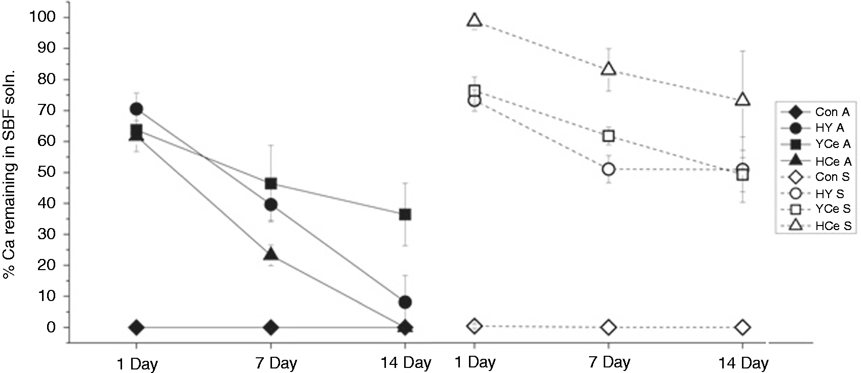
Osteoblast viability
Osteoblast cell viability was tested using liquid extract concentrations of 9% (a concentration previously found to pose toxicity using parent glass extracts
34
), and the results are presented in Figure 4. This shows that viability does not differ significantly from the control population for any sample at any time. While statistical analysis did not find a significant increase associated with the HY-A 7 day (131 ± 12%) or HCe-A 1 (126 ± 20%) and 7 day (131 ± 8%) samples, these samples showed viability higher than that of the control population.
Osteoblast viability after incubation with disk extracts at 9%, 20%, and 33% concentrations by volume over 1, 7, and 14 days.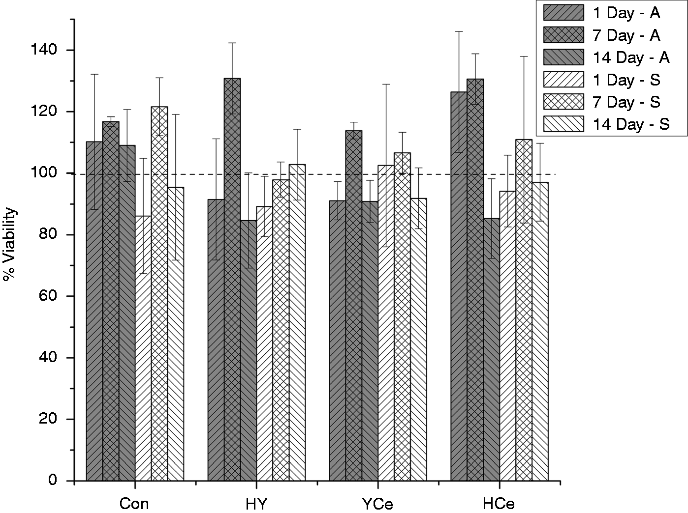
Bioactivity
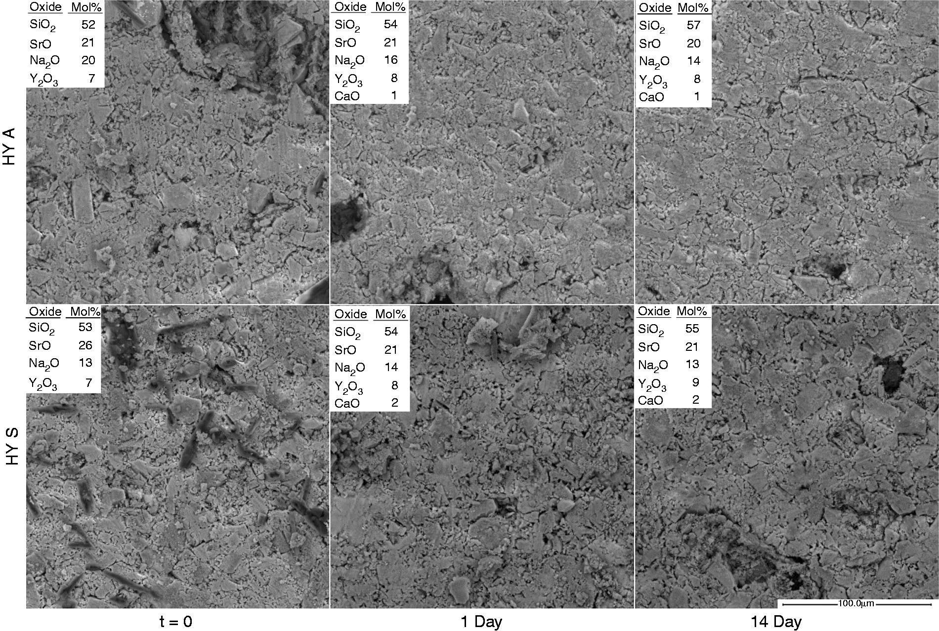
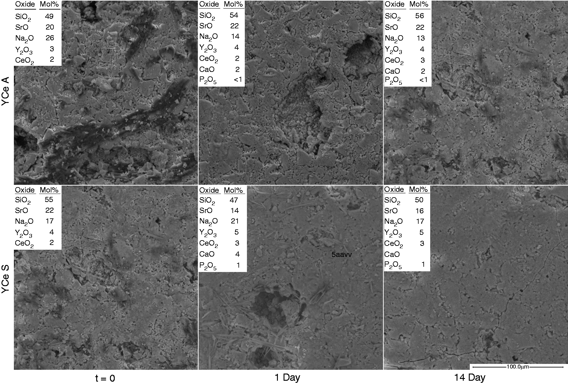
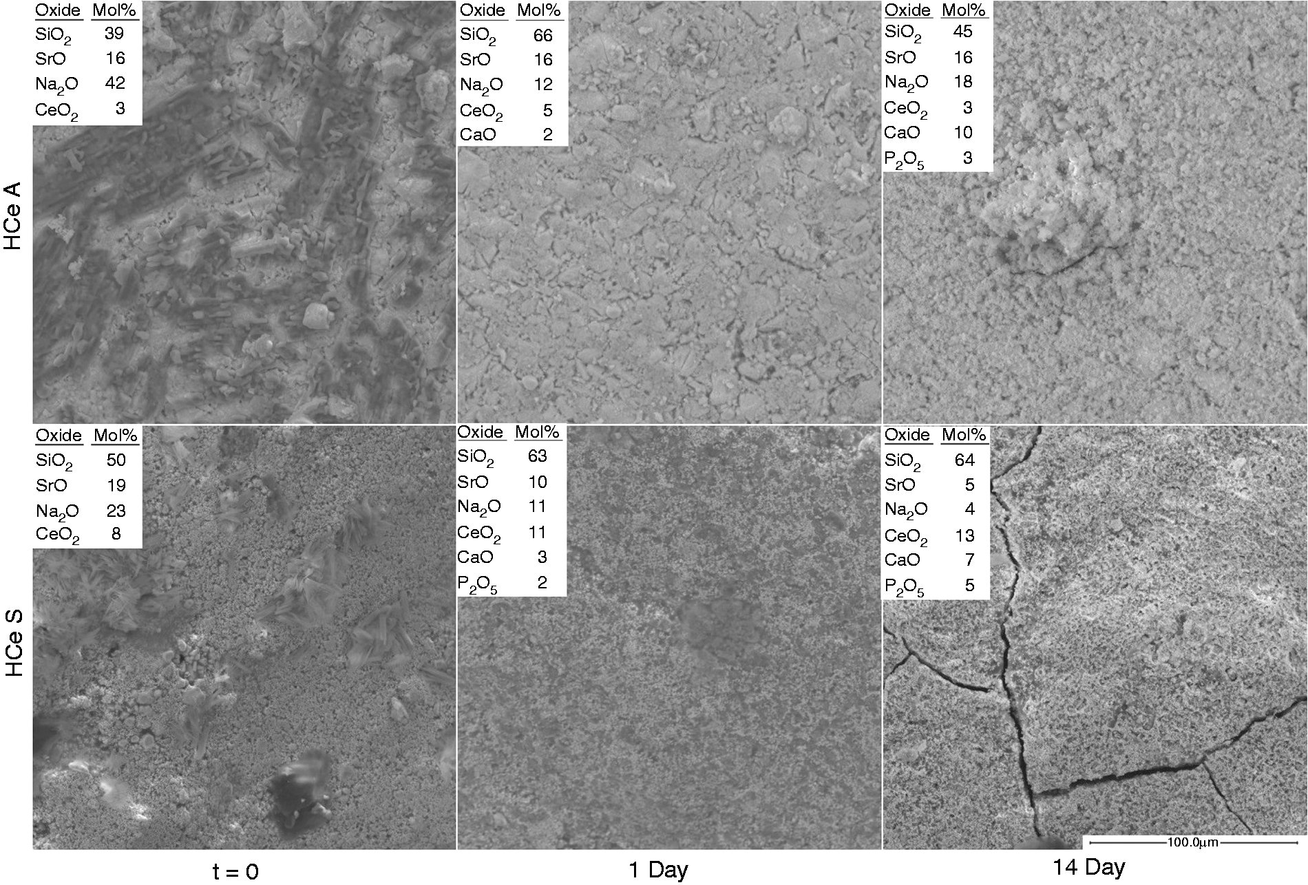
SEM images of the surface of each disk before (t = 0) and after 1- and 14-day incubation in SBF are presented in Figures 5 to 8. Additionally, the EDS-generated oxides are included for each disk. The t = 0 Con disks both exhibit what appears to be a partially reacted surface and correspondingly a high concentration of Na2O relative to SiO2. After incubation, the ratio becomes comparable to the initial glass composition, and the presence of quantities of CaO and P2O5 is detected on the surface. With the exception of the Con-A 14-day sample SrO was not detected at the surface of the disks. The HY disks initially and after 14-day incubation each show a packed and sintered powder surface with SiO2, SrO, and Na2O content comparable to the original glass composition. EDS also detected small quantities of Ca on the surface of the HY disks, though no precipitates are observable. The YCe disk surfaces appear as a compacted/sintered powder surface without visible precipitates over the 14-day time period similar to those of the HY disks. EDS shows surface composition is comparable to that of the original glass, though for YCe-A, Na-rich crystals present at t = 0 skews the ratio between Na2O and SiO2, and presence of Ca and P. HCe-A, t = 0, also shows significant presence of Na-rich surface crystals which are reflected in the oxide ratios. After 1- and 14-day incubation precipitates form on the surface with Ca identified after 1 day and quantities of both Ca and P after 14 days. Similar trends are seen with the HCe-S disks though there is a localization of Ce at the surface, opposite that observed with HCe-A disks. In addition to increasing Ca and P precipitates, after 14 days, cracks appear on the surface similar to those appearing on the Con-A disks over the same incubation period.
SEM images of t = 0-, 1-, and 14-day SBF-incubated disks at 500 × and EDX generated oxides list for Con. SEM images of t = 0-, 1-, and 14-day SBF-incubated disks at 500 × and EDX generated oxides list for HY. SEM images of t = 0-, 1-, and 14-day SBF-incubated disks at 500 × and EDX generated oxides list for YCe. SEM images of t = 0-, 1-, and 14-day SBF-incubated disks at 500 × and EDX generated oxides list for HCe.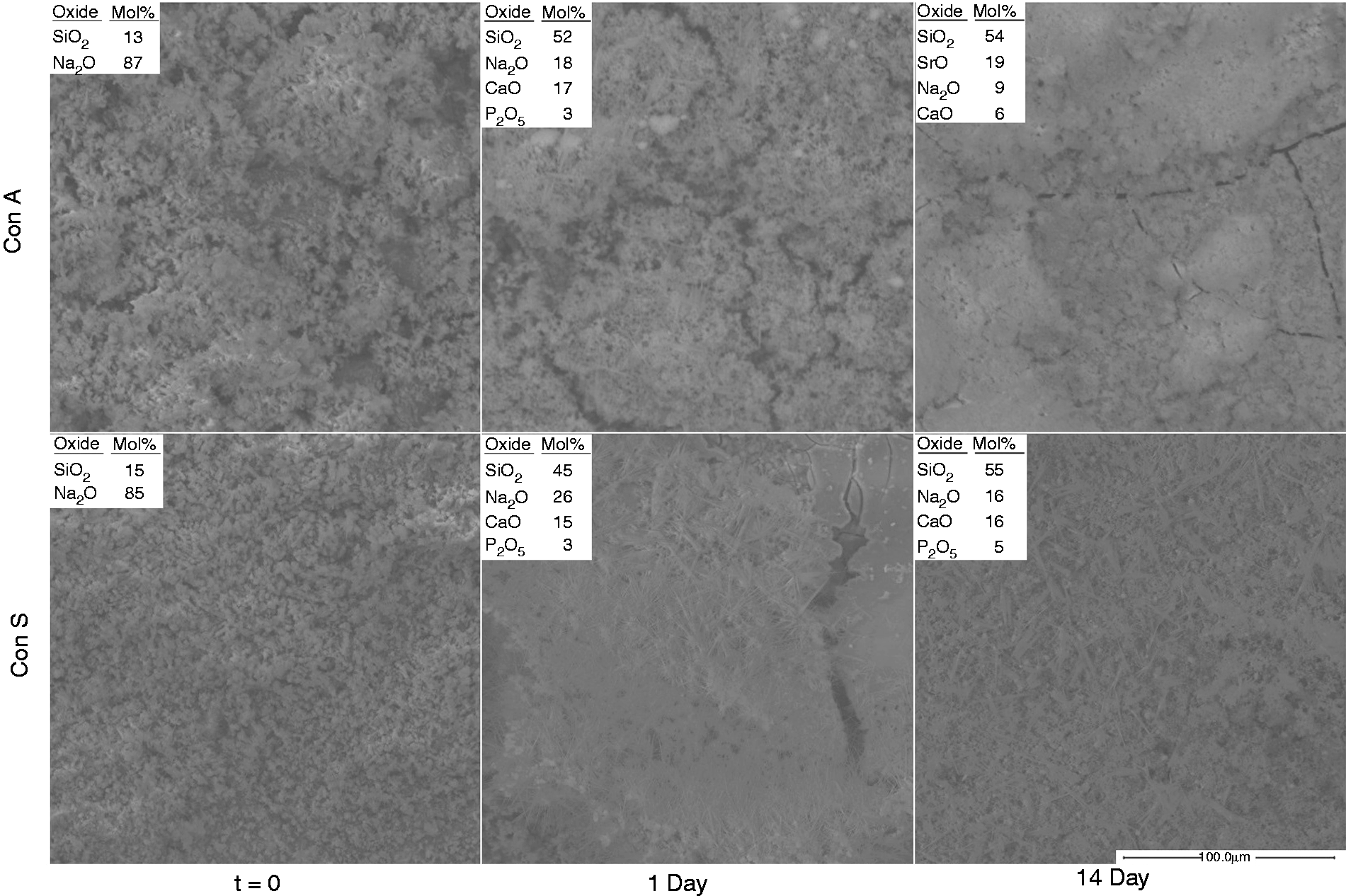
Cell adhesion
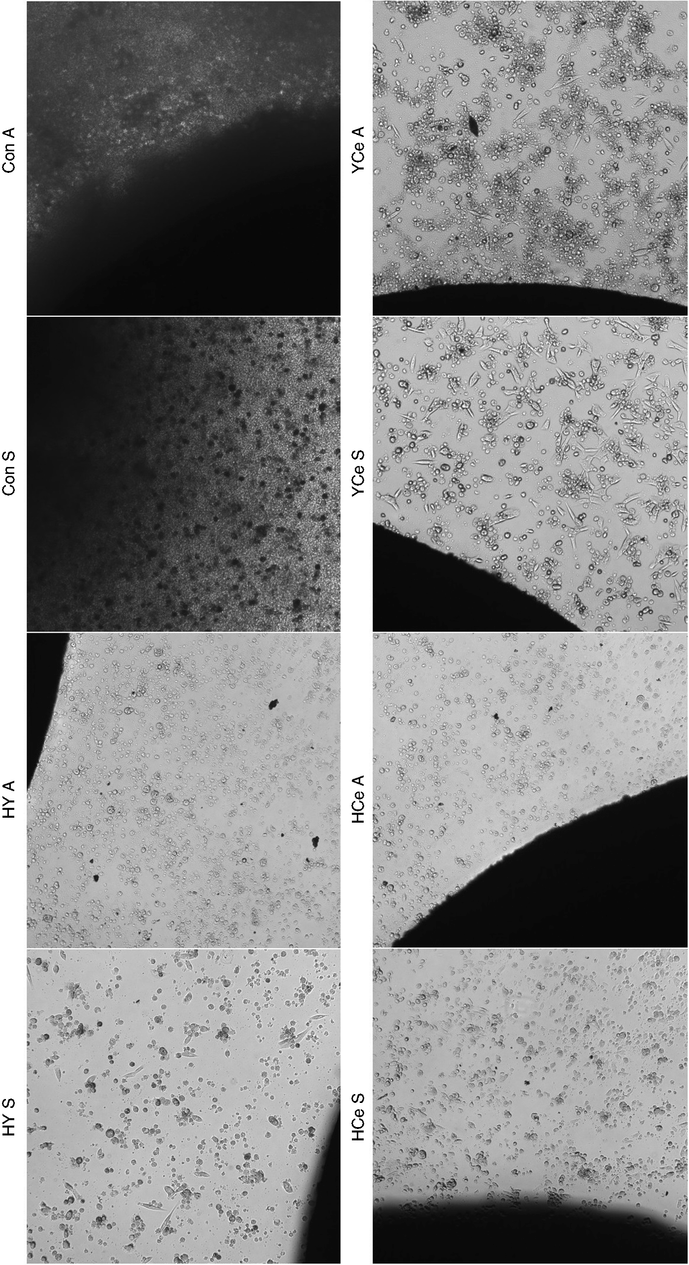
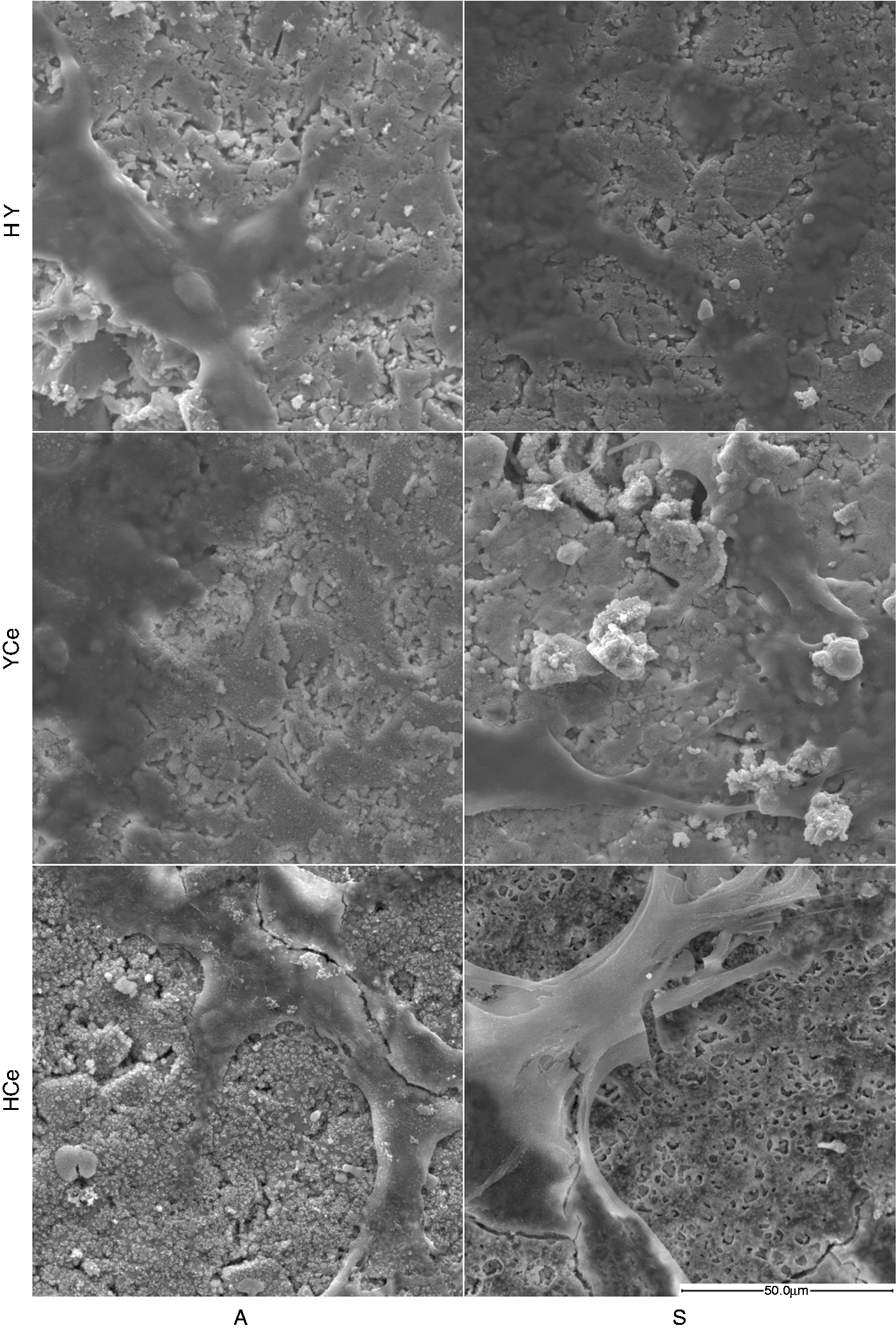
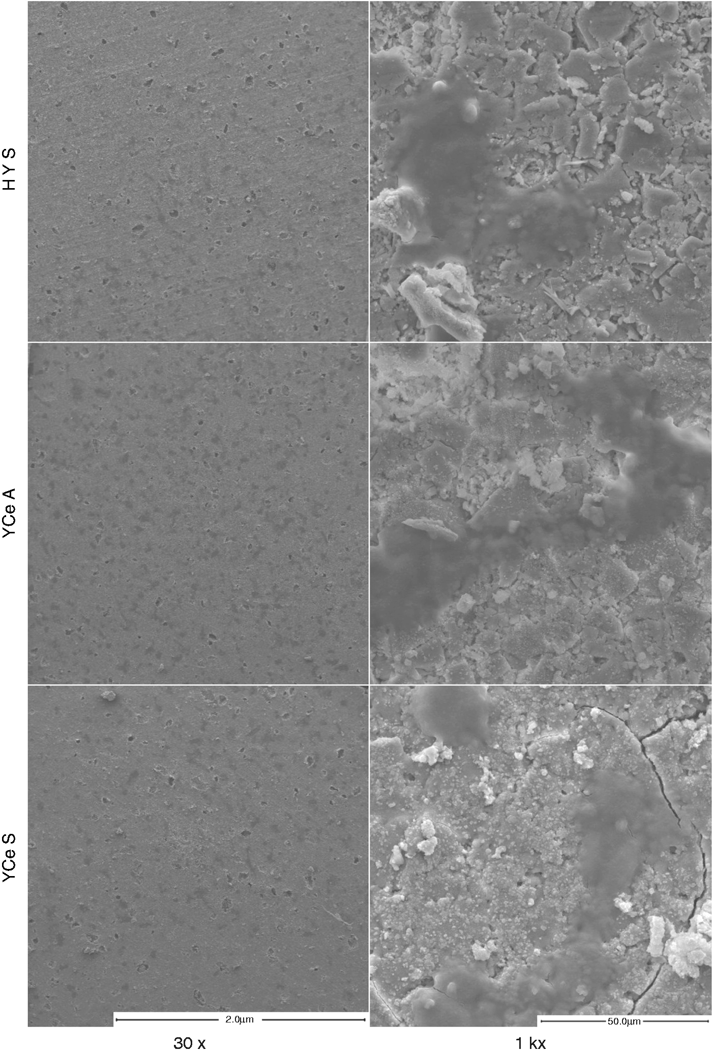
The optical images of the disk-cell interface after 24 h incubation are shown in Figures 9 and 10. It can be seen that the 14 day incubated Con disks continue reacting when in contact with cell media causing it to gel and creating an inhospitable environment for cell attachment and survival. Y and Ce containing disks, while they may continue to react in cell media, allow the media to maintain its viscosity and clarity, and support osteoblast attachment and spreading with many cells directly contacting the disks. Schwann cells show reduced viability when incubated with the Y and Ce containing disks in comparison with the osteoblasts, though it can be seen that cell attachment has occurred. Despite the cell attachment, the majority of the cells adopt a spherical shape and in some cases appear to be ruptured. However, HCe-S, YCe-A and YCe-S show a small population of Schwann cells attached and spreading. Of the samples with attached and spreading osteoblast and Schwann cells SEM images were collected after fixation and are presented in Figures 6 and 7. In Figure 11, it can be seen that the disks containing Y and Ce all support osteoblast attachment and spreading. Schwann cells, at high magnification, are not as easily visible as osteoblasts; therefore, Figure 12 includes 30 × images showing the presence of Schwann cells (the darker spotting) across the HY-S and YCe disk surfaces.
Optical images at 4 × of 14-day incubated disk-osteoblast interface after 24 h incubation. Optical images at 4 × of 14-day incubated disk-Schwann interface after 24 h incubation. SEM images at 1k × after disk incubation with osteoblasts and fixation. SEM images at after disk incubation with Schwann cells and fixation.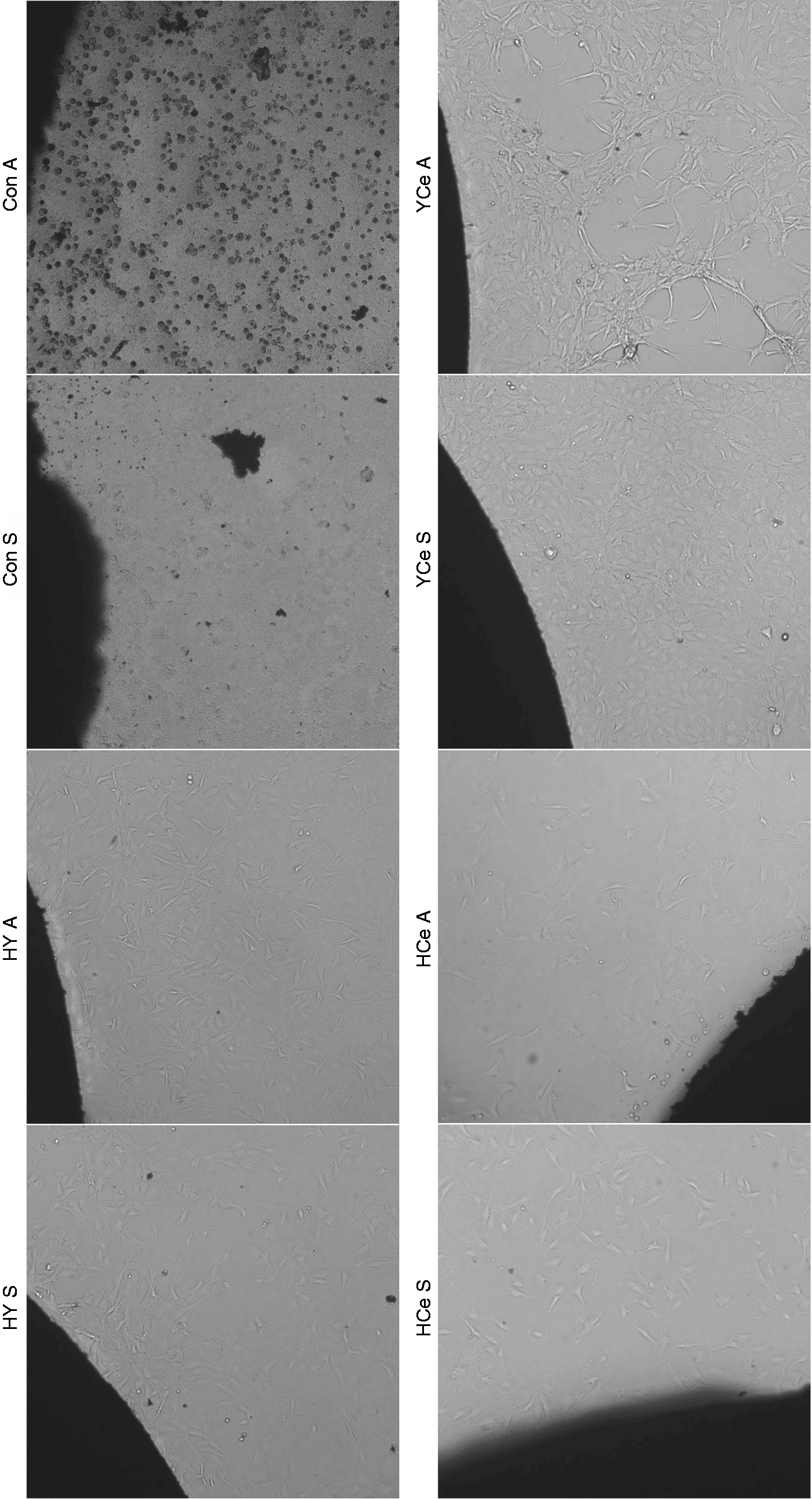
Discussion
The bioactivity of Y and Ce inclusive glass-ceramics for the reduction of oxidative stress generated by excess calcium and ROS upon nerve injury was evaluated after incubation in SBF for 1, 7, and 14 days. The increase in pH seen is similar to that observed in typical bioactive glasses, 41 where the Con-A glass saw the greatest spike from 9.7 after 1 day to 10.0 after 14 days. There is a direct relationship between the increase in pH and the dissolution of the glass network, relating to the initial exchange of hydrogen ions (H+) with Na+. This exchange is most pronounced in the Con glass-ceramics as they contain the highest Na content. In addition, they lack the trivalent/tetravalent Y and Ce ions which reduce the mobility of Na through the network as a result of steric hindrance or charge compensation requirements, thereby reducing the Na content available for exchange upon glass dissolution.33,34,42,43 As such, glass-ceramics containing Y and Ce show a maximum increase in pH of 8.7 which is below that observed for the Con glass-ceramics at any time period. The lower shift in pH observed with the Y and Ce containing glass ceramics may also influence cell viability studies and compatibility. Biological environments maintain pH for proper functioning of cells, and while moderate fluctuations in pH may be buffered within physiological fluids, greater shifts can lead to apoptosis in adjacent cells. Therefore, lower shifts in pH are desired with any biomaterial to minimize adverse effects on surrounding tissues. As such and regardless of thermal processing, the Y and Ce containing glass-ceramics elicit a lower pH than the Con glass-ceramics. In addition, Con glass-ceramics will likely have greater initial ion exchange, consistent with work on the solubility of these glass-ceramics which found the overall network solubility was significantly higher than that of the Y and Ce containing glass-ceramics. 35 Another trend is observed in the pH for Con and HCe glass-ceramics where samples processed at lower temperatures (A) show higher pH than those processed at higher temperatures (S). This is due to the degree of crystallinity imparted on the structures after thermal processing. Glass quenching often freezes molecules with bond lengths that are not optimum and therefore stressed; thermal treatment at and above the Tg allows these bonds to relax and at higher temperatures reform in crystalline structures.37,44 The presence of periodic atomic arrangement increases the rigidity of the material and decreases the solubility, and, in general, the greater the degree of crystallinity, the lower the dissolution potential of a material.39,45 HY and YCe glass-ceramics do not show a significant difference in pH increase at any time period throughout the SBF incubation. This is suggestive that their reactivity is inherently lower than that observed for Con and HCe, where initial studies evaluating the glass solubility in DI water found these glass compositions to have reduced solubility. 35 The high Y content (8 mol%) in the HY composition and the additive effects of 4 mol% of both Y and Ce in the YCe glasses, produce an increase in network rigidity attributed to the higher field strength of Y and Ce in comparison with Na. It was also found that while both Y and Ce have increased, field strength Y produces greater network rigidity due to the presence of a single valance state and ability to partially stabilize the multiple valences adopted by Ce in the glass network. As a result of the multiple valances and coordination environments associated with the Ce ions, charge compensation, and field strength, local network rigidity around the Ce ions varies within the glass network. This results in slightly higher dissolution potential for Ce containing glass-ceramics in comparison with those containing Y.33,34,42,43 Thus, while thermal treatments inducing crystallinity in the glasses should cause reduced reactivity in the S processed glass-ceramics, the rigidity of Y containing A samples must be at a level comparable to that observed for the crystalline phases generated through S temperature processing. While pH changes based on glass-ceramic dissolution do not show a significant difference between the A and S processed Y containing disks, there are differences in surface conditions indicated by Ca and P fluctuations in SBF over the incubation period.
The reduction in Ca content over the 14-day SBF incubation suggests adequate reactivity to produce surface conditions, specifically the proportion of negatively charged silanol groups, and encourage the association and chemisorption of Ca onto the surface, followed by PO4 for charge neutralization.46,47 Con, with the highest pH, suggesting the greatest reactivity, shows elimination of Ca in solution for both A and S samples, suggesting significant calcium precipitation on the sample surface. Since the reacted surface dictates the association with Ca ions and thus facilitates deposition, glass-ceramics processed at lower temperatures, with higher reactivity, would be expected to produce greater Ca reduction from solution, as seen for each of the A vs. S, Y and Ce containing samples. However, considering the trends in pH and Ca content for the Y and Ce containing glasses and the effects of Y in comparison with Ce on rigidity, the reduced pH and minimal Ca deposition seen for the HCe-S glass-ceramic samples relates to a lower reactivity than expected. This is likely due to the presence of Ce crystallites at the surface of these samples, which is not observed in other Ce containing samples and is attributed to the high sintering temperature of the HCe glass composition. The concentration of Ce crystallites likely impedes surface reactions creating silanols (Si-OH), thereby reducing the potential for Ca deposition. P follows identical trends to that of Ca with the Y and Ce containing glass-ceramics where all samples cause a decrease in P content throughout the 14-day incubation period. A samples cause a greater P decrease than S, and HCe-S demonstrates the lowest propensity to cause P precipitation, all of which can be accounted for through the same mechanisms. One difference of note is the precipitation rates of Ca and P, where Ca shows a linear decrease over the 14 days and P shows greater precipitation between the 1 and 7 days than between 7 and 14 days. The variation in rates may be accounted for by the ratio of Ca:P typically found in the amorphous HCA layer on bioactive materials which ranges from 1.54 to 1.73 with the optimum of 1.67. 48 As such, after seven days, it is likely that this ratio is approached and deposition rate is reduced. Con glass-ceramics do not show similar P trends to that of Ca, although it does show an initial decrease greater than that of any Y or Ce containing sample. However, after 14 days, the P content increases from levels seen after seven days suggesting re-release into solution. This may be indicative of continued degradation of the glass-ceramic as release of Na was found to continue throughout the 14-day incubation period. 35 EDS complements ICP-OES through relative comparison of surface species on the glass-ceramic disks, and may provide further insight into Ca and P deposition trends.
SEM images of the Con glass-ceramics correlate with the high reactivity suggested by pH and ICP-OES studies where the t = 0 disks show a reacted Na-rich surface layer. Once incubated, the surface Na decreases, likely dissolving into the SBF and promoting the deposition of Ca and P which are significant after 1 day in correlation with the levels detected in SBF. In addition, after 14 days, the Con-A samples show reduction of Ca on the surface and elimination of P, also consistent with the significant re-release of P observed through ICP-OES. This effect is less pronounced in the Con-S samples where 1 and 14 day Ca and P levels were comparable as listed by EDS. Visually, the Ca and P deposition are presented as small meshed needle-like crystallites at higher Ca content and a porous amorphous deposition at lower concentrations. In contrast, visual depositions on the surfaces of HY, YCe and HCe-A glass-ceramics are nearly undetectable in the SEM images, however consistent with the reduction in both Ca and P in SBF, EDS detected small quantities of Ca, and in the case of YCe samples, P on the surfaces. Previous studies with Y and Ce containing bioactive glasses saw the delay of HCA formation up to 14 days based on the glass composition.32,33 In addition to comparable quantities of Y and Ce in compositions used in this study, the resulting glasses were thermally treated to produce glass-ceramics, which can further reduce the bioactivity. Therefore, it may require additional time for Ca and P deposition to form a visual HCA layer. This is supported by the visual deposits on the 14-day HCe-A samples, which have been shown to have reactivity between Con and other Y and Ce glass-ceramics through pH and ICP-OES. These deposits and relative Ca content appear to fall, visually, between the amorphous character of Con-A, 14-day deposits, and the crystallites seen on samples with higher Ca content. HCe-S samples show incremental increase in the Ca and P content detected on the surface where deposition appears in a thin distribution across the disk surface. The presence of Ca and P detected on the surface of each disk indicates a degree of bioactivity for each and potential for adherence of cells to these surfaces.
Cell attachment and morphology give insight into the biocompatibility of a material. Optical microscopy of the Con glass-ceramic disks incubated for 24 hr with cells shows reduced clarity of the media associated with gelation and unattached spherical cells. The gelation of the liquid media upon incubation is suggestive of continued reactivity and significant silica release. When approaching the saturation limit, silica can polymerize potentially causing an increase in media viscosity and an inhospitable environment for osteoblast and Schwann cell lines.49–51 Y and Ce containing glasses with reduced reactivity do not cause this phenomenon and, as such, cell attachment occurs in each well for these samples. In addition to attachment, osteoblasts exhibit cell spreading and growth across the well, uninhibited by proximity to the disks. This correlates to the results obtained from osteoblast viability analysis conducted with the SBF-incubated disk extracts. Statistical analysis revealed that there was no significant difference between any of the sample incubated populations and the control population. These two observations show that the Y and Ce containing glass-ceramics are not toxic to osteoblasts after processing at either temperature and therefore may have the potential for cell adhesion directly to the sample surfaces. Therefore, SEM was employed and shows the presence of osteoblast cells attached and spreading across each Y and Ce containing glass-ceramic. While there was no toxicity and favorable cell morphologies with osteoblasts, they tend to be robust compared to Schwann cells. This is illustrated in the optical images of disk incubation with Schwann cells where cell attachment of spherical cells is observed in each well, but growth and spreading only occur in the HY-S and YCe disk sample wells and are limited to a fraction of the population. This indicates adverse effects on Schwann cells in proximity with the disks, possible toxicity over time, and may eliminate the potential for cell growth and attachment on the disk surface. However, when the best performing disks HY-S, YCe-A, and YCe-S were observed through SEM, low magnification images revealed the presence of cells that exhibited spreading across the surfaces though did not form a connected network across the disks and are singular or in small groups. Schwann cells function in close proximity in vivo and therefore achieving a network as observed with the osteoblasts would be beneficial and presents a direction for future research.
Conclusion
The ability of bioactive glasses to interact with physiological fluids and induce the deposition of a HCA layer is essential to the formation of a bond with host tissues. This work has shown Con glass-ceramics to have the greatest reactivity with the highest increase in pH and the greatest reduction in Ca content from SBF. In addition, Ca and P deposition appear as a mesh of needle-like crystallites, however, despite the lack of osteoblast toxicity observed through viability analysis when directly incubated with cells Con samples cause complete toxicity to both cell populations. Y and Ce containing glasses show reduced reactivity with moderate pH increases in SBF upon incubation and reduction of Ca and P content and corresponding deposition. However, only HCe-A shows significant Ca and P deposition suggesting the bioactivity may be delayed by more than 14 days for the remaining Y and Ce containing glass-ceramics. Observation of cells in direct contact with Y and Ce containing glass ceramics suggests only HY-S, YCe-A and YCe-S are suitable surfaces for cell attachment of both osteoblasts and Schwann cell types. While bioactivity was demonstrated for Con and HCe-A glass-ceramics, the combination of bioactivity and direct cell compatibility suggests HY-S, YCe-A and YCe-S may prove better candidates considering the sensitive nature of regenerating nerve tissues.
Footnotes
Declaration of Conflicting Interests
The author(s) declared no potential conflicts of interest with respect to the research, authorship, and/or publication of this article.
Funding
The author(s) received no financial support for the research, authorship, and/or publication of this article.