
Abstract
In the present study, polyurethane materials were obtained from castor oil, polycaprolactone and isophorone diisocyanate by incorporating different concentrations of chitosan (0.5, 1.0 and 2.0% w/w) as an additive to improve the mechanical properties and the biological activity of polyurethanes. The polyurethanes were characterized by Fourier transform infrared spectroscopy, thermogravimetric analysis, scanning electron microscopy, stress/strain fracture tests and swelling analysis, and the hydrophilic character of the surface was determined by contact angle trials. The objectives of the study were to evaluate the effect of the incorporation of chitosan on the changes of the physico-chemical and mechanical properties and the in vitro biological activity of the polyurethanes. It was found that the incorporation of chitosan enhances the ultimate tensile strength of the polyurethanes and does not affect the strain at fracture in polyurethanes with 5% w/w of polycaprolactone and concentrations of chitosan ranging from 0 to 2% w/w. In addition, PCL5-Q-PU formulations and their degradation products did not affect cell viability of L929 mouse fibroblast and 3T3, respectively. Polyurethane formulations showed antibacterial activities against Staphylococcus aureus and Escherichia coli bacteria. The results of this study have highlighted the potential biomedical application of this polyurethanes related to soft and cardiovascular tissues.
Introduction
Polyurethanes (PUs) are polymeric materials that are widely applied in medicine as biostable implants and cardiovascular medical devices such as catheters and pacemakers because their properties imitate the behavior of different tissues and are biocompatible. 1 Additionally, it has been demonstrated that PUs can favor tissue growth and restoration, making them candidates for use in tissue engineering.2,3 Hence, different scaffolds have been fabricated for the growth of soft and bone tissues.4,5
Despite the various scientific breakthroughs within the field, certain challenges remain, for example, (i) avoiding the generation of toxic decomposition products, 6 such as aromatic diamines, specifically 2,4-toluenediamine (TDA) and 4,4′-methylenedianiline, which are generated as degradation products of the hard segments of the PUs synthesized from aromatic diisocyanates such as toluene diisocyanate (TDI) and methylene diphenyl diisocyanate (MDI), 7 respectively. Many studies have demonstrated the carcinogenic potential of aromatic diamines under in vivo conditions. Therefore, the use of aliphatic diisocyanates, which do not generate highly toxic decomposition products when incorporated into the hard segments of PU, has drawn significant interest. Nevertheless, the PUs synthesized from aliphatic diisocyanates present lower tensile strengths and melting points with respect to PUs synthesized with aromatic diisocyanates, where the latter also present higher reactivity, lower cost and higher commercial availability. Thus, aromatic diisocyanates represent one of the most widely used class of raw materials for PU synthesis. 8 With respect to the latter, (ii) it is highly important to select the most appropriate monomers and additives for the synthesis of biomedical PUs to favor the biocompatibility of the material and its degradation products without affecting the material’s physico-chemical or mechanical properties and thereby achieve the proper balance between the PU structure and its properties.
Another challenge associated with biomedical PUs is (iii) the prevention of bacterial infection risks at the material surface, 9 which could lead to the formation of pathogenic biolayers and later complications due to infection. The primary method for avoiding bacterial adhesion is surface modification using antibacterial agents, which include heavy metals, antibiotics and phenols, among others. The antibacterial agents are gradually released and, depending on the type of agent, can also present toxic effects for the organism.10,11 Therefore, highly biocompatible antibacterial agents should be used; examples include quaternary ammonium salts and chitosan (Ch). Finally, the PUs generally should (iv) be produced using low-cost monomers derived from renewable sources, whereas at present, most PUs are produced from non-renewable sources. 12
PUs are a class of polymers that contain urethane bonds (–NH–CO–O–) in their structures. PUs are obtained from an exothermic polymerization stepwise growth reaction (polyaddition) of one diol and one diisocyanate. 13 The physico-chemical properties of the resulting PUs are directly related to the chemical composition, molecular weight and ratio between the soft and hard segments that form their structure. 4 Usually, the soft segments of a PU are composed of polyol chains that can be polyol-polyether or polyol-polyester. In turn, the hard segments are formed by isocyanate groups and chain extenders. 14 The thermodynamic incompatibility between the soft and hard segments of a PU generates a phase separation effect, giving rise to a two-phase morphology. 15
Castor oil has been used in the synthesis of PUs, because it is a renewable source. Approximately 90% of its composition by weight is ricinoleic acid, which presents a trihydroxylic structure that allows the synthesis of cross-linked urethanes. Furthermore, castor oil has low toxicity, and the presence of ester groups in its structure favors the hydrolytic degradation of the resulting PU material when it contains the soft segments of this polymer. 16 Different PU formulations have been synthesized with castor oil and aliphatic diisocyanates, such as hexamethylene diisocyanate, lysine diisocyanate and isophorone diisocyanate (IPDI). These diisocyanate monomers are preferred for the synthesis of biomedical PUs, because they do not promote the generation of carcinogenic decomposition products such as the aromatic diamines in in vivo conditions, in contrast to the case of aromatic diisocyanates such as TDI and MDI.2,17
Moreover, the ease with which the properties of PUs can be changed by altering their chemical structure and combining them with other polymers allows their properties to be easily improved and new characteristics, such as antimicrobial activity and biodegradability, to be readily incorporated. Polycaprolactone (PCL) is a widely used polymer in biomedical applications as a result of its biocompatibility and degradability.18,19 PUs with different fractions of incorporated PCL have proved to be candidate biomaterials for use in tissue engineering. 20 In turn, Ch is one of the most abundant polymers in nature, presenting biocompatibility and antimicrobial activity. 15 However, Ch and PCL have weak mechanical properties, limiting their use in different medical applications. One possible approach for improving the use of Ch is in combination with PU materials, which have similar mechanical properties to various tissues and have an average biocompatibility.14,17–20
In the present study, PU materials were synthesized from castor oil, IPDI and mixed with PCL, incorporating Ch as an additive. The objective of this study consisted of evaluating the effect of the incorporation of different Ch concentrations (0.5, 1.0 and 2.0% w/w) on the changes of the physico-chemical and physico-mechanical properties and the biological activity of the PUs. The percentage of water absorption was obtained by swelling analysis, and the surface hydrophilic character was determined by contact angle assays. Finally, the in vitro biocompatibility of the PUs and their degradation products was evaluated by the 3-[4,5-dimethylthiazol-2-yl]-2,5-diphenyl-2 H-tetrazoliumbromide (MTT) assay with fibroblast cell strains L929 and NIH/3T3, respectively. The anti-Staphylococcus aureus and anti-Escherichia coli antibacterial activity was evaluated. Additionally, the level of acceptance as a material for biomedical applications was determined by analyzing these various properties.
Experimental section
Materials
Castor oil was obtained from Ciacomeq S.A.S (Colombia), with a hydroxyl number of 160 mg KOH/mg. PCL diol with an average molecular weight of 2000 g mol−1 and low-molecular weight Ch with a degree of deacetylation higher than 75% were obtained from Sigma-Aldrich (USA). IPDI was purchased from Merck (USA), and potassium bromide (KBr) was purchased from Thermo Scientific (USA). Phosphate-buffered saline solution was prepared by dissolving NaCl (5.85 g), KH2PO4 (0.6 g) and Na2HPO4 (6.4 g), all from Merck (USA), in 1 L of distilled water. The pH of the solution was adjusted to 7.4 by adding HCl and NaOH solutions (0.2 M). 21 MTT, (0.5 mg/ml), trypsin 2.5% (10×), penicillin–streptomycin (contains 10,000 units of penicillin and 10,000 µg of streptomycin per milliliter) for application in cell culture, and RPMI 1640 and Dulbecco’s Modified Eagle medium (DMEM) were purchased from Gibco/Invitrogen (UK). Fetal bovine serum (FBS) was obtained from Eurobio (France). Trypticase soy broth (TSB) and trypticase soy agar (TSA) were purchased from Scharlau Co (Spain), as well as rat embryonic fibroblasts NIH/3T3 (ATCC CRL-1658) and L929 (ATCC CCL-1). E. coli (ATCC 2469) and P. aeruginosa (ATCC 27853) were obtained from the strain library of the University of La Sabana (Universidad de La Sabana).
PUs synthesis
Formulation and mechanical properties of PUs.

Note: All PUs were synthesized with the ratio NCO/OH = 1 – According to the analyses of variances. Average (n = 3) ± standard deviation.
Statistically significant differences (p ≤ 0.05) for the data of each column.
PU characterization
Fourier transform infrared spectroscopy
The PU synthesis and the incorporation of Ch were confirmed by Fourier transform infrared spectroscopy (FTIR) with an FT-IR Nicolet™ iS™10 spectrometer (Thermo Scientific, USA) in the wavenumber range of 4000 to 400 cm−1. 24 The PU samples had been previously processed (scraping) until small particles were obtained, and then were blended with KBr in a ratio of 1:100 until a homogeneous mixture was obtained.
Mechanical properties
Stress–strain analyses were performed to estimate the mechanical properties of the PUs, using a universal testing machine model EZ-LX (Shimadzu, Japan) with a load cell of 5 kN. Test tubes with dimensions of 40 mm × 6 mm × 3 mm (length × width × thickness) were employed, and a vise displacement speed of 10 mm min−1 was utilized, according to ASTM D638-10. Three samples of each formulation were evaluated.
Scanning electron microscopy
The effect of the incorporation of Ch on the surface texture changes of the PUs was evaluated by scanning electron microscopy (SEM) in a Pro Desktop microscope (Phenom, the Netherlands). The samples were evaluated at two magnifications, 1500× and 3500×.
Thermal degradation
Thermogravimetric analysis (TGA) were performed using TGA equipment (TGA/DSC1; Mettler Toledo, USA). The weight of each sample was 15 ± 1 mg. The tests were performed under air atmosphere with a heating rate of 25℃ min−1, in the temperature range of 25–600℃, based on ASTM D6370.
Water absorption and contact angle
The hydrophilic behaviors of the PUs and their surfaces were determined as a function of the percent water absorption and the contact angle, respectively. Absorption tests were performed with distilled water at 37℃ for 48 h, based on ASTM D 570-98. The samples (diameter of 8 ± 1 mm and thickness of 3 mm) were dried and weighed to determine the percent water absorption according to equation (1).
The contact angle trials were performed with a drop shape analysis system (GH11, Kruss, Germany), using the sessile drop technique with distilled water at room temperature, according to Grzesiak et al. 25 The reported value is the average of 10 positions inside the PU slide, specifically in the formulations of 0 and 2% w/w of Ch.
In vitro biocompatibility
Cell culture
NIH/3T3 and L929 rat fibroblasts are widely used in cytotoxicity tests in vitro. The cells were cultured in 25-cm 2 culture flasks with DMEM or RPMI medium, respectively. The medium was supplemented with FBS (10%) and penicillin–streptomycin (1%) in a CO2 atmosphere at 5% and 37℃. The cells were trypsinized with 0.25% trypsin and then were re-suspended in fresh medium with FBS and antibiotics.16,26
PU biocompatibility in vitro
The in vitro cell viability of the cell strain was evaluated with respect to direct contact with the PU samples using the MTT colorimetric method defined in ISO/CD 10993-5.
10
Cylindrical samples of the PUs with a diameter of 4 ± 0.2 mm and a thickness of 3 ± 0.1 mm were exposed to UV radiation for 20 min per side to maintain sterility.
27
Next, a pre-culture of cells was performed in 96-well plates with RPMI medium supplemented with FBS at 37℃ and 5% CO2 for 24 h. Then, the PU samples were added, allowing for 24 h of contact under the same culture conditions. For quantification, the supernatant was removed, and 100 µL of MTT solution (0.5 mg/mL) was added to sterile PBS in each well and then incubated at 37℃ for 4 h. The solution was removed, and dimethyl sulfoxide (DMSO) was added to each well and incubated at 37℃ for 15 min. The optical density reading was performed in a microplate reader model iMark™ Microplate Reader (BIO-Rad, USA) at the wavelength of 595 nm. Cells seeded without the sample were used in medium that was free of FBS and DMSO, as positive and negative controls, respectively. All tests were performed in triplicate. The percent cell viability of the L929 rat fibroblast cell strain was calculated with equation (2), where DO denotes the optical density
10
In vitro biocompatibility of PU degradation products
Three samples of each PU formulation were placed in micro-centrifuge tubes with 100 µL of DMEM medium at 37℃ and 200 r/min for 30 days according to the methodology proposed by Bakhshi et al. 10 The resulting supernatant (100 µL) (extract) was used to determine the cytotoxicity of the degradation products by the MTT assay according to the methodology of the previous reference. The cytotoxicity of the degradation products was evaluated with NIH/3T3 rat cell fibroblasts seeded in 96-well plates. Cells seeded without the experimental sample were used in FBS-free medium as a positive control.
Antibacterial activity tests
The antibacterial activity of the PUs was evaluated based on the method defined by Kara et al. for anti-E. coli (ATCC 2469) and anti-S. aureus (ATCC 6538); these two pathogenic microorganisms are found in infections that result from various surgical procedures. 11 Bacterial suspensions were obtained by growing the cultures in TSB at 37℃ for 16 h. Aliquots were collected from the bacterial suspensions to inoculate new TSB at 37℃ for 6 h. Each bacterial suspension was centrifuged at 3000 r/min for 10 min. The cells were washed twice with phosphate-buffered saline after the supernatants had been removed. The bacteria were suspended in PBS at the concentration of 4.2E + 06 Colony Forming Unit (CFU mL−1). To determine the antibacterial activity, the polymers were placed in 96-well microplates with 100 µL of bacterial suspension and incubated at 37℃ for 24 h. The content of each well was transferred to micro-centrifuge tubes containing 900 µL of sterile PBS. By counting colonies, the number of viable cells was determined for each suspension, using serial dilutions seeded in TSA at 37℃ for 18 h. A polymer-free bacterial suspension was used as a control.
Statistical analysis
The statistical significance of the differences of mean values obtained in the different tests was evaluated using analysis of variance with the significance level p > 0.05. The significant differences between groups were evaluated using Student’s t-test and the Software SPSS Statistics. 23 All results are reported as the value of the mean ± standard deviation.
Results and discussion
PU characterization
FTIR spectroscopy
The FTIR spectra of PCL, Ch and the PUs are represented in Figure 1(a). The spectrum of pure Ch, Figure 1(a), presents peaks at approximately 3490 cm−1, which are related to the vibration of the –NH and –OH bonds. Furthermore, the two characteristic bands of Ch at 1646 cm−1 (amide I: stretching of the C = O bond) and at 1594 cm−1 (amide II: –N–H amino groups) were identified.23,28 The peaks observed in the PCL spectrum, Figure 1(a), were located at 3440 cm−1 (stretching of –OH bond), 2939 cm−1 (asymmetric stretching of –CH2), 2860 cm−1 (symmetric stretching –CH2), 1720 cm−1 (stretching C = O), 1470 cm−1 (flexion –CH2), and 1188 cm−1 (stretching C–O–C).
29
The PU-PCL and PU-PLC-Ch spectra are shown in Figure 1(b).
FTIR spectra: (a) Ch and PCL, (b) PU-PCL and PU-PCL-Ch.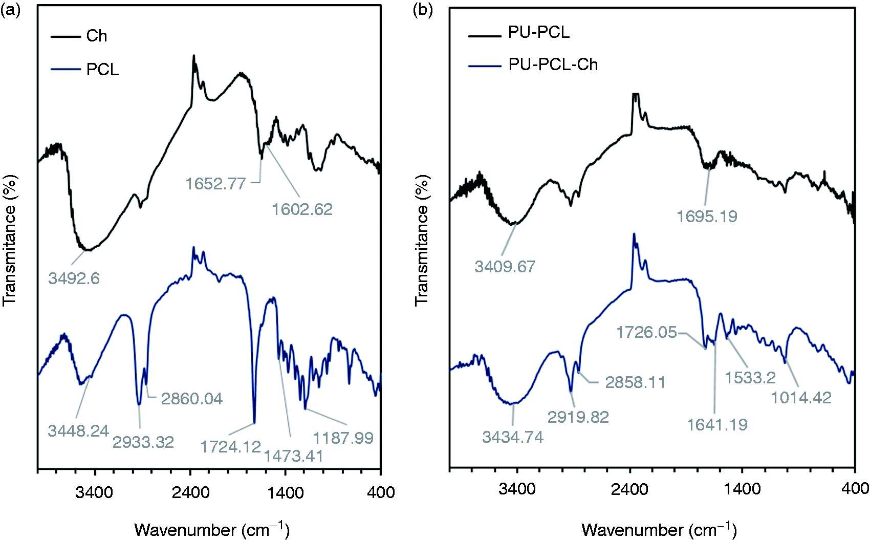
The absence of the peak at 2220 cm−1 (N = C = O group of IPDI) and the presence of peaks at 3400 cm−1 (stretching of urea and urethane bond –N–H) and 1741 cm−1 (combination of NH–CO–O– and –CO–O–) confirm the production of the PUs. In turn, slight displacements of characteristic Ch and PCL peaks are observed in the PU-PCL-Ch spectrum, Figure 1(b). Two original bands of Ch at 1652 cm−1 and 1602 cm−1 were displaced to 1654 cm−1 and 1532 cm−1, respectively. In turn, the characteristic PCL peak at 1724 cm−1 was displaced to 1726 cm−1, reducing its intensity. This phenomenon is likely due to the presence of hydrogen bonds in the amino and hydroxyl groups present in the molecules of the additives and the PU matrix because there is no formation of new covalent bonds expressed in the appearance or disappearance of the characteristic peaks of the molecules.23,29 The interactions identified for the PU-PCL-Ch mixtures are consistent with the formation of hydrogen bonds identified among the different components of PU-Ch-nylon and PCL-Ch mixtures, which were prepared by Jayakumar and Sudha 23 and Zia et al., 29 respectively.
Mechanical properties of PUs
The synthesized PUs presented ultimate tensile strength σ and strain ɛ intervals of 0.47–0.87 MPa and 42.11–76.90%, respectively, Table 1.
The incorporation of Ch on PUs with 5% PCL has a significant effect on the value of the ultimate tensile strength of PUs (p < 0.05) and does not affect the ultimate tensile strain for the PUs. The mechanical properties obtained are characteristic of PUs synthesized from CO and IPDI, 22 a fact that could be related to the low reactivity of castor oil and the low yield of the polymerization reaction when aliphatic diisocyanates, such as IPDI, are used, leaving some terminal groups of –NCO. 30 The PUs containing 10% w/w of PCL have a lower ultimate tensile strength with respect to the PUs with 5% w/w of PCL. In turn, the value of the ultimate tensile strain increases with increasing PCL concentration. This result could be related to the high temperature of the vitreous transition and the low mechanical properties of PCL 31 as well as the incorporation of PCL chains in the soft segments of PU. 32
It was found that the incorporation of Ch significantly increases the σ values of the PUs containing 5% PCL (p < 0.05), specifically for the samples with 0% w/w and 2% w/w of Ch (p < 0.05), while samples with 0.5% w/w and 1% w/w of Ch did not exhibit significant differences (p > 0.05). The previous result could be related to hydrogen bond interactions between either the functional groups of the Ch and PCL molecules or the Ch and the PU matrix, according to the results found in the FTIR analysis. The hydrogen bonds could favor interfacial adhesion and the physical cross-linking of the mixture, resulting in improved mechanical properties of the PUs.19,33,34 The previous result is consistent with that reported by Fu et al., 35 who incorporated different quantities of halloysite nanotubes modified with Ch into PU matrices. It was observed that Ch acts as a reinforcing agent (until concentration values of 2% w/w) and favors interfacial interactions between the nanotubes and the PU matrix.35,36
The formulations of the PUs synthesized in this investigation have favorable mechanical properties for applications in the biomedical engineering fields, especially with respect to soft and cardiovascular tissue engineering because the mechanical resistance of these PUs is similar to that of the aorta (0.5 MPa) and skin (1.0 MPa).37,38 The previous conclusion is based on the study developed by Guan et al., who prepared biodegradable scaffolds of PUs synthesized from PCL, obtaining ultimate tensile strength values ranging from 0.97 MPa to 1.64 MPa, which were comparable to the canine thoracic aorta vein (0.9 ± 0.1 MPa). After determining the degradable character of the PUs and the viability of vascular smooth muscle cells in contact with the PUs, it was concluded that the scaffolds have a significant potential for use in soft and cardiovascular tissue engineering. 37 Among the various applications of cardiovascular tissue engineering, the synthesized PUs could have the potential for application in the regeneration or substitution of infarcted myocardium and, therefore, could be used to improve cardiac function, according to the study performed by Chen et al. 38 In that study, the mechanical behavior of poly (glycerol sebacate) elastomers was evaluated, yielding mechanical properties of 0.5 MPa, 0.05 MPa and 4.5% for tensile strength, elasticity modulus and strain at fracture, respectively. Because the synthesized elastomers presented similar mechanical properties in relation to the heart muscle, the resulting material was suggested for myocardium regeneration. 38
Because bone tissue engineering requires biomaterials to present ultimate tensile strength values ranging from 40 to 140 MPa, the synthesized PUs cannot be used for bone regeneration. 39 The ultimate tensile strain of the PUs was found to be in accordance with the physiological parameters required for cardiac applications, which is on average 60% of that for heart tissues, according to Hidalgo-Bastida et al. 40
Morphology
Figures 2 and 3 show the SEM micrographs of the synthesized PUs as a function of the changes in Ch concentration, maintaining a constant quantity of PCL (5% w/w of PCL). With the magnification of 1500×, a rough and irregular surface was observed in all PU samples, as well as the separation of microphases. The surfaces revealed the presence of phase domains (a disperse phase) of Ch grains on a continuous PU–PCL phase, as pointed by the arrows in Figure 2(b) to (d). A clear separation between the phases, Ch granules and PU matrix was observed, indicating that it is not possible to achieve good incorporation through a simple physical mixture. The Ch granules appear intact and apparently did not suffer alterations during mixing processes and chemical reaction.
SEM micrographs at 1500×. (a) PU-PCL5-Ch0, (b) PU-PCL5-Ch0.5, (c) PU-PCL5.0-Ch1, and (d) PU-PCL5.0-Ch2. SEM micrographs at 3500×. (a) PU-PCL5-Ch0.5, (b) PU-PCL5-Ch1, and (c) PU-PCL5-Ch2.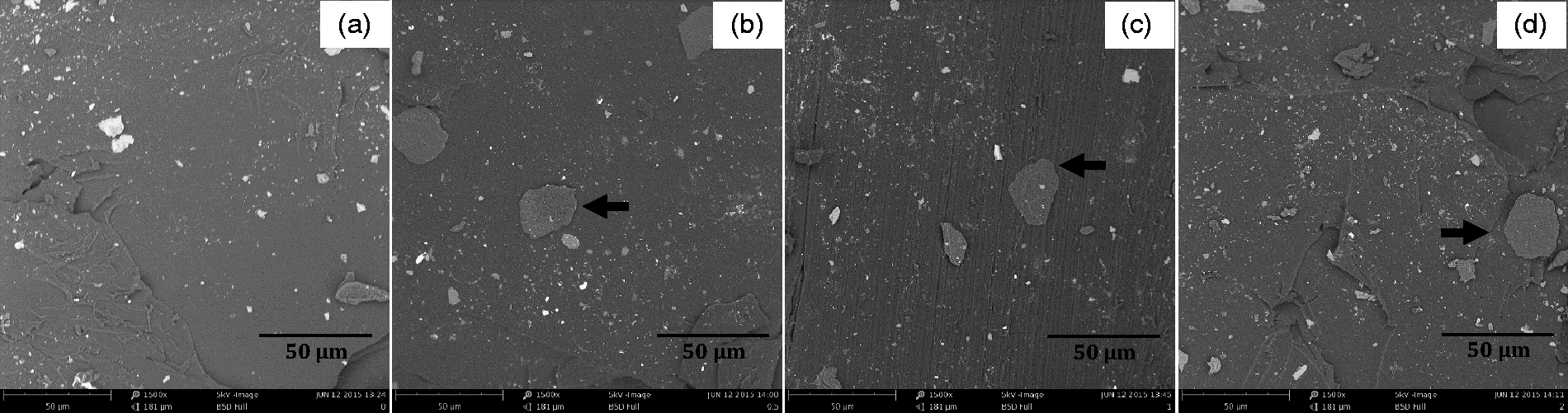
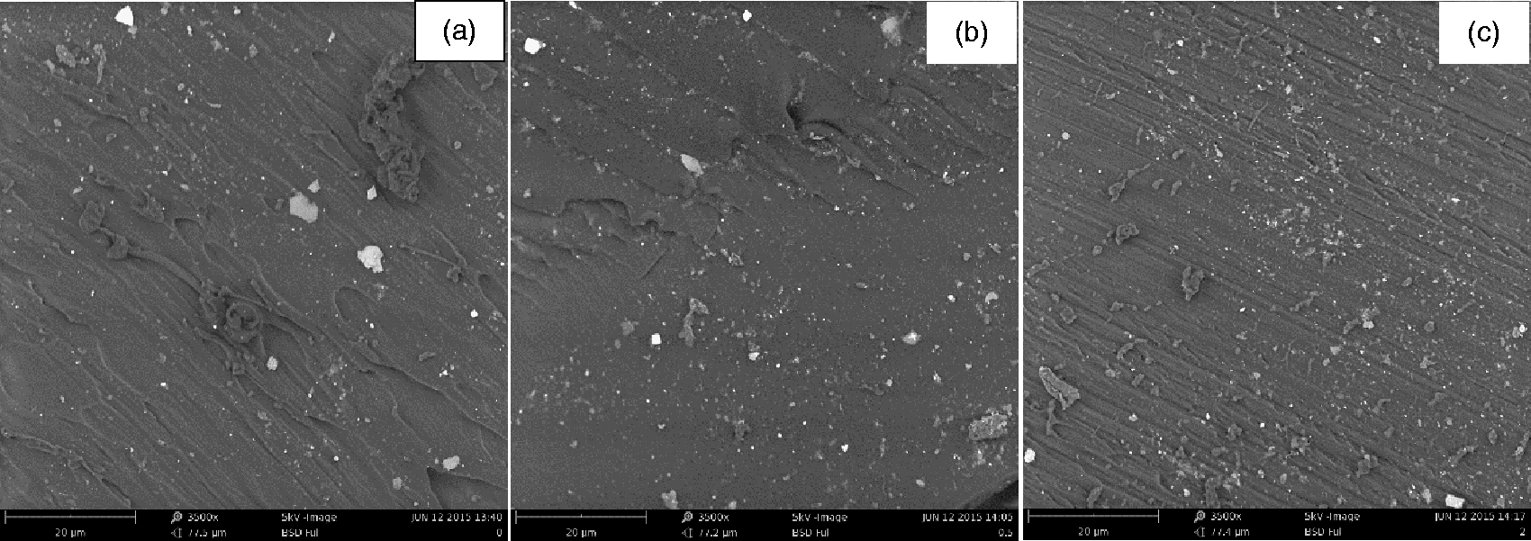
With a magnification of 3500×, a reduction was observed in the roughness and in the heterogeneous character of the PU surface with the increase of the concentration of Ch. 34 The mechanical resistance of PUs can be favored by employing the reinforcing ability of Ch, which conserves the chemical structure of the PU and favors the formation of hydrogen bonds between the component of the mixture, as observed by the FTIR spectroscopic analysis.23,41
Thermal degradation
The results of the TGA tests are shown in Figure 4. The thermograms indicate that all PUs are thermally stable at temperatures less than 220℃ and are completely degraded at approximately 600℃. Four degradation regions were observed. The first region (250–370℃) is related to the degradation of the minor components of castor oil (aliphatic hydrocarbons)
42
and the degradation of Ch molecules.
23
The second region (375–430℃) comprises the rupture of urethane bonds connecting the hard segments of PU.
22
The third region (425–480℃) corresponds to the degradation of PCL.
43
Finally, the decomposition region of the chains of the castor oil fatty acids belonging to the soft segments of PU is located at 450–500℃, according to Anaya et al.
44
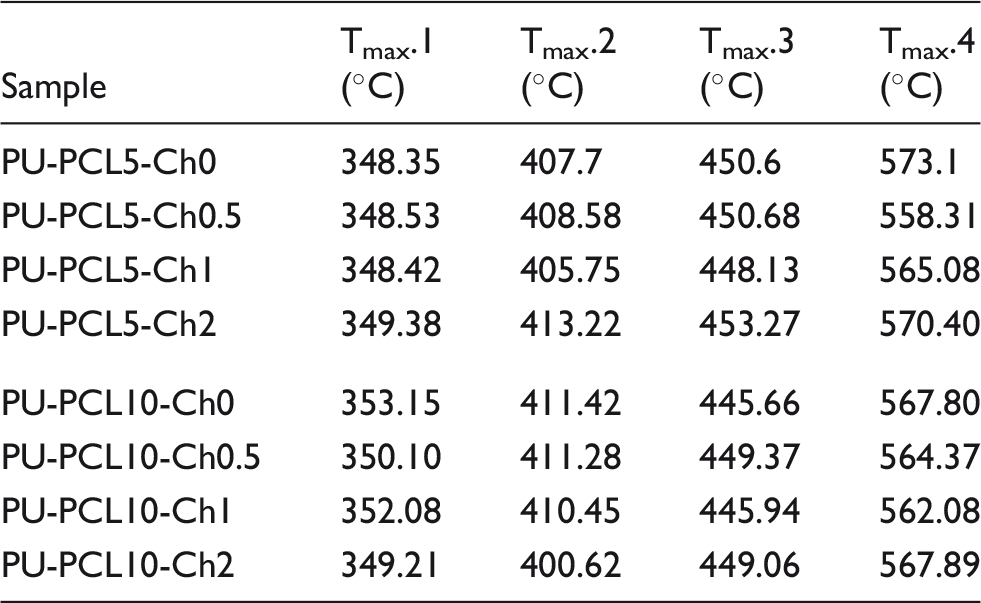
Thermograms of the PUs. (a) PU-5PCL-Ch; (b) PU-10PCL-Ch.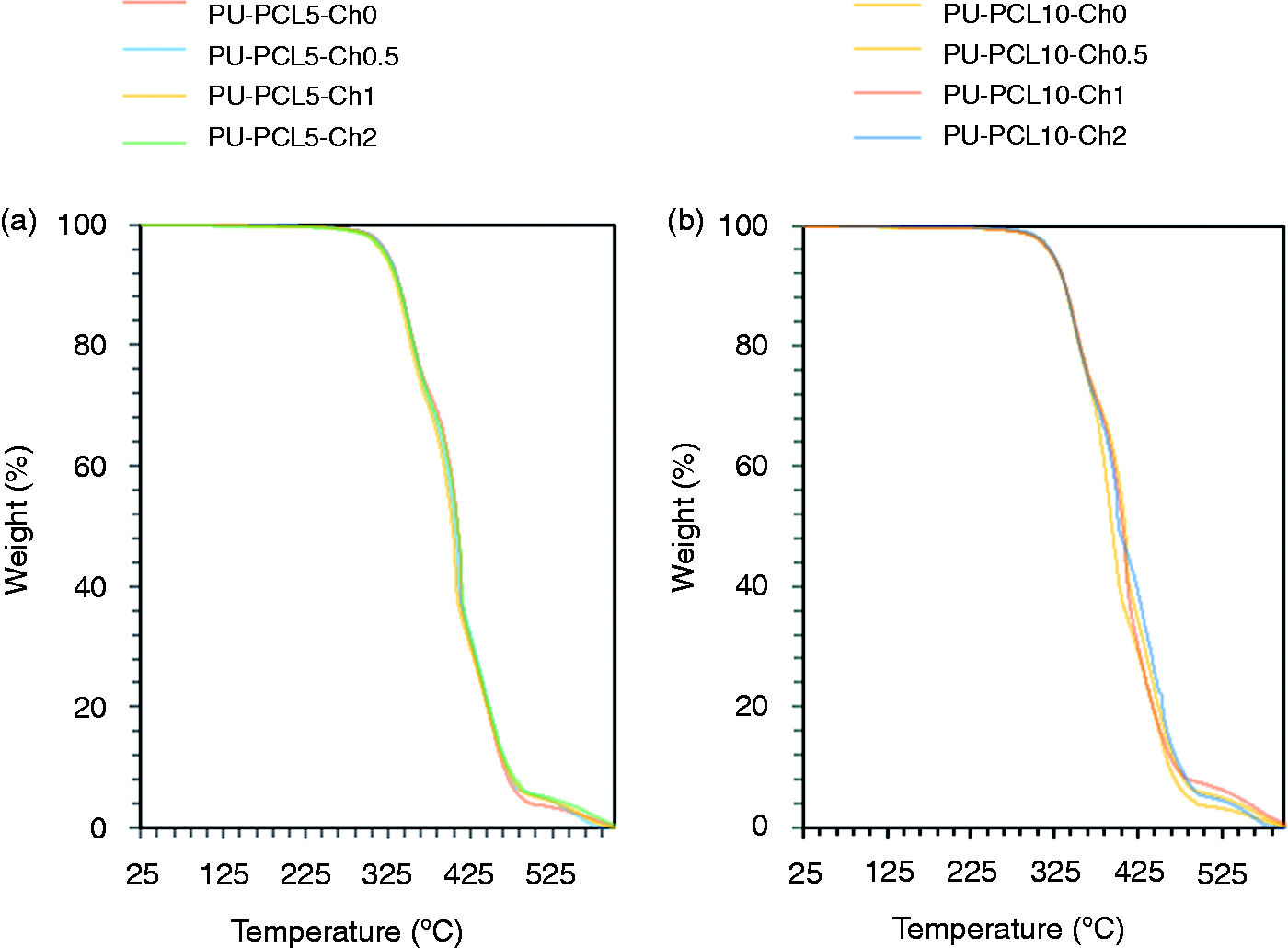
Maximum temperatures of PU degradation.
Water absorption
The water absorption of the PUs is shown in Figure 5. As shown, the water absorption of the samples increased significantly with the increase of the concentration of Ch for PUs with 5% w/w and 10% w/w of PCL (p < 0.05), specifically, for the PU samples containing 0% w/w and 2% w/w of Ch. The previous result can be related to the result of the hydrophilic character of Ch, which is incorporated into the mixture of polymers of hydrophobic character, such as PU and PCL.
46
In a previous study, Wu
47
reported that the increase in the water absorption of PCL-Ch and Acrylic Acid-Ch mixtures is not only caused by the hydrophilic character of Ch but also by the structural arrangements of the polymeric chains generated during the synthesis because the Ch prevents the movement of the chains and avoids the formation of arrangements with enhanced crosslinking, preventing water molecules from penetrating into the polymer network, in comparison to PCL and acrylic acid.21,47
Water absorption of PUs 48 h. (Mean (n = 3) ± standard deviation). According to the analyses of variances, (*) indicates a statistically significant differences (p ≤ 0.05) in water absorption for PUs with 5% and 10% w/w PCL, incorporating different concentrations of Ch.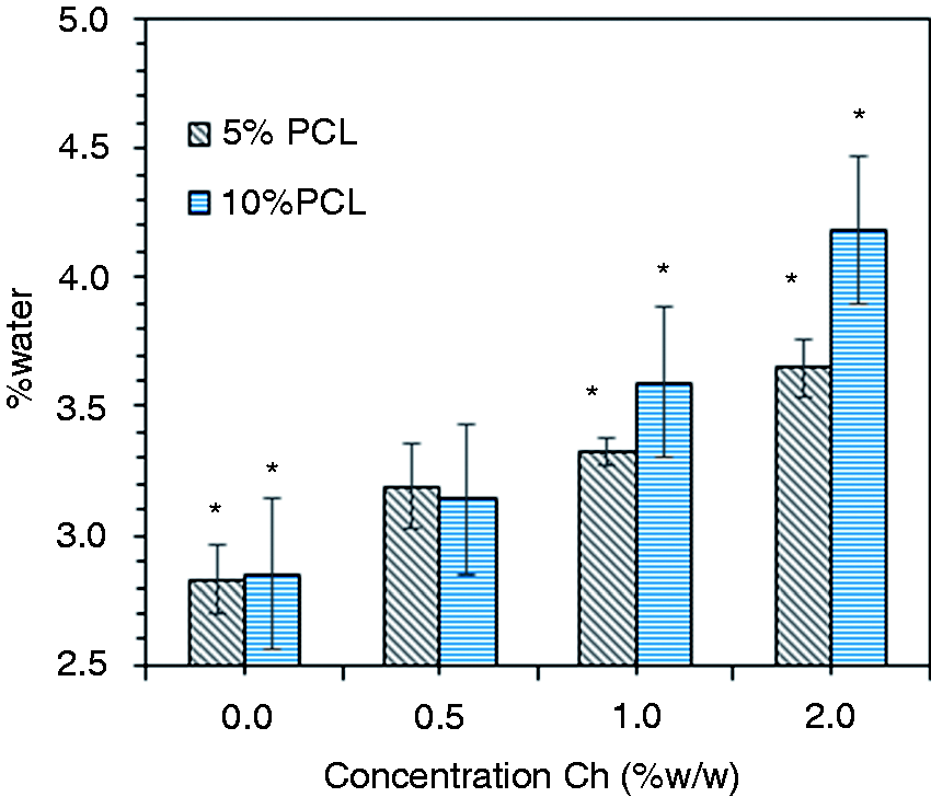
Contact angle values of PUs.
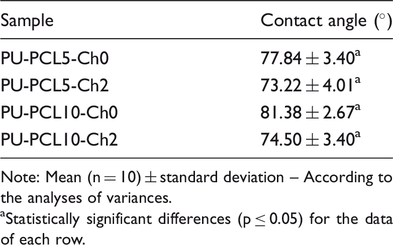
Note: Mean (n = 10) ± standard deviation – According to the analyses of variances.
Statistically significant differences (p ≤ 0.05) for the data of each row.
In vitro biocompatibility
In vitro biocompatibility of the PUs
L929 and 3T3 rat fibroblasts are widely used in the study of the cytotoxicity of different biomaterials, because these cell strains exhibit high sensitivity to toxic effects, similar to human fibroblasts.48,49 The viability of the L929 rat fibroblast cell strain exposed to the PU samples is presented in Figure 6(a). The incorporation of Ch did not have a negative effect on the viability of L929 fibroblasts that remained in incubation with the PUs for 24 h, specifically with the PUs containing PCL at 5% w/w. Additionally, sample PU-PCL5-Ch was found to favor cell viability. The latter is related to the bioactivity of Ch, which is recognized as a biocompatible material.24,50 Moreover, the corresponding increase observed in the viability of L929 rat fibroblasts with the increase in the concentration of Ch, specifically for sample PU-PCL5-Ch2, could be related to the increased hydrophilic character of the surface of the PU. Due to the incorporation of a hydrophilic and biocompatible polymer, the latter favors the interaction between cells and the PUs and, consequently, cell viability and proliferation, as reported by Xu et al.,
51
who evaluated the in vitro biocompatibility of PU nanoparticles coated with Ch on human umbilical vein endothelial cells.
(a) Cell viability of L929 fibroblasts in contact with PU samples at 24 h. (b) Cell viability of NIH/3T3 fibroblasts in contact with the UP degradation products. (Average (n = 3) ± standard deviation). According to the analysis of variance, (*) indicates a statistically significant differences (p ≤ 0.05) in cell viability compared to the control.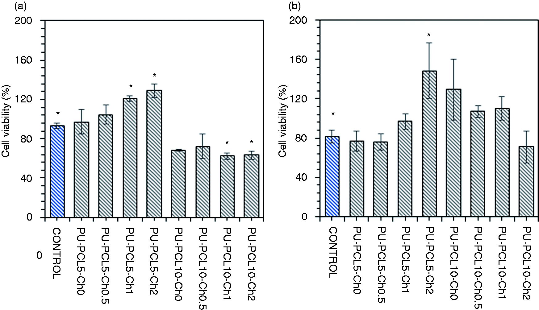
Despite the results reported in Figure 6(a), the statistical analysis for the viability values for L929 fibroblasts in contact with the PU-PCL-Ch samples indicate that there are no significant differences between the groups of viability values obtained (p > 0.05), suggesting that cell viability is maintained after contact with the PU samples. Based on a previous discussion, the PUs with PCL contents of 5% w/w and Ch contents of up to 2% w/w present in vitro biocompatibility with L929 rat fibroblasts at 24 h of incubation, according to the requirements established by ISO/CD 10993-5. Therefore, it could be suggested that the previously mentioned PUs do not present risks to cell viability and can be used in biomedical applications.
In contrast, the PUs containing 10% PCL significantly affect cell viability compared with the PU samples with 5% PCL (p < 0.05). The decrease in cell viability could be explained in terms of the chemical structure of PU, specifically with respect to the number of terminal isocyanate groups of an aliphatic diisocyanate, which have low toxicity, as is the case of its degradation products.2,52 Additionally, the IPDI monomer residues, due to their reactivity, could interact chemically with the cells 53 because they can bond to the carrier proteins through the reaction of the NCO group with the –SH, –NH2 and –OH groups present in the proteins, forming covalent bonds. The latter promotes the increase of cytotoxicity in the materials, as reported by Clour et al., 30 who evaluated the effect of the chemical composition of PUs on the in vitro biocompatibility of a fibroblast cell strain by varying the type of aliphatic and aromatic diisocyanates.
Based on the results of the in vitro biocompatibility of the PU-PCL5-Ch formulations, it is suggested that the materials have high potential for use in biomedical applications, specifically applications that include soft and cardiovascular tissues. This is because these materials are non-toxic and have similar mechanical properties to tissues such as the skin and the aorta.
In vitro biocompatibility of the PU degradation products
The PU extracts were used to determine the in vitro biocompatibility of the PU degradation products. After the extraction in DMEM medium, the PU samples were washed with distilled water, dried until the mass no longer changed, and then weighed. All PUs presented degradation percentages below 1%. The viability of the NIH/3T3 fibroblast cell strain exposed to the PU degradation products is presented in Figure 6(b). As shown, the cell viability of the NIH/3T3 line is maintained after being in contact with the PU degradation products. Furthermore, the statistical analysis corroborated the finding that there were no significant differences between the viability values for each of the tests with respect to the control (p > 0.05); hence, the incorporation of Ch does not affect the cytocompatibility of the PU degradation products, except for sample PU-PCL5-Ch2, which favors cell viability (p < 0.05), Figure 6(b). The cell viability results quantified by the MTT assay suggest that the synthesized PU degradation products, and the incorporation of Ch do not affect the biocompatibility of the materials; therefore, they have potential for use in biomedical applications.
The great interest for the use of Ch in biomedical applications has allowed for the development of different studies focused on determining the cytotoxic effect of different natural and synthetic materials such as Ch and PU, respectively. Wang et al. 54 evaluated the effect of the Ch degradation products (3–120 days in saline solution) on the cell proliferation of endothelial cells during four days of incubation. It was determined that the Ch degradation products at 30 days did not have a negative effect on cell proliferation. In fact, in some cases, the degradation products increased the proliferation of the endothelial cells. 54 Nevertheless, throughout the 90 days over which the test was carried out, inhibited cell proliferation was observed, which could be related to possible changes in the molecular structure undergone by the degradation products over time and under certain conditions, as described by Wang et al. 54 The molecular structure of the substances influences their biological activity. Thus, it is very important to continue to study the effect of the incorporation of Ch on the biological activity of the degradation products of PU-PCL-Ch mixtures.
Moreover, it has been demonstrated that the degradation products of polymers such as PU and PCL do not affect cell viability, according to the reports by Bakhshi et al. 10 and da Silva et al., 55 who evaluated the cytotoxic effect of PU degradation products on ARPE-19 human retina pigment epithelial cells and L929 rat fibroblasts, respectively. In both studies performed by Bakhshi et al. 10 and da Silva et al., 55 no significant differences were observed between the cell viability values. Therefore, it was suggested that the PU degradation products did not present cytotoxic risks for the cultured cells, such as the case of the PU-PCL-Ch mixtures prepared in this study.10,55
Antibacterial activity tests
It was possible to promote antibacterial activity in the polymers by using antibacterial agents applied to the surface of the materials. Antibacterial activity is related either to the release of an antibacterial agent to the medium or to the direct contact of bacteria with the agents on the surface of the material. The latter is known as the active contact method, which is preferable because it does not release agents that could have toxic effects on the organism.56,57
The surface changes induced to biomedical materials with the use of agents with antimicrobial activity, such as silver, quaternary ammonium, and Ch, among others, make it possible to avoid bacterial adhesion that could occur during different medical procedures.10,11 In this study, the antibacterial activity of the PUs was determined against E.coli (Gram negative) and S. aureus (Gram positive) bacteria. Furthermore, the formation of biofilms is undesirable in biomaterials because these multicellular structures allow the microorganisms to persist in hospital environments and afford them more resistance against the antibiotics used routinely in their treatment.
58
The antibacterial effect of PUs against S. aureus and E. coli (after 24 h of exposure) is shown in Figure 7(a) and (b), respectively. For the case of S. aureus and E. coli, the number of colony-forming units (CFUs) decreased after being in contact with the PUs. According to the statistical analysis, the PU formulation with 5% PCL (PU-PCL5-Ch) presented statistically significant differences in the CFU value of S. aureus and E. coli with the increased incorporation of Ch (p < 0.05). The formulations with 10% PCL (PU-PCL10-Ch) were found to inhibit bacterial growth. Nevertheless, no statistically significant differences were observed with the incorporation of Ch (p > 0.05). The results obtained indicate that the PUs have antibacterial activities against S. aureus and E. coli and, therefore, could be useful in biomedical applications.
Antibacterial activity of PUs against (a) Staphylococcus aureus and (b) Escherichia coli. (Average (n = 3) ± standard deviation). According to the analysis of variance, (*) indicates a statistically significant differences (p ≤ 0.05) in cell viability compared to the control.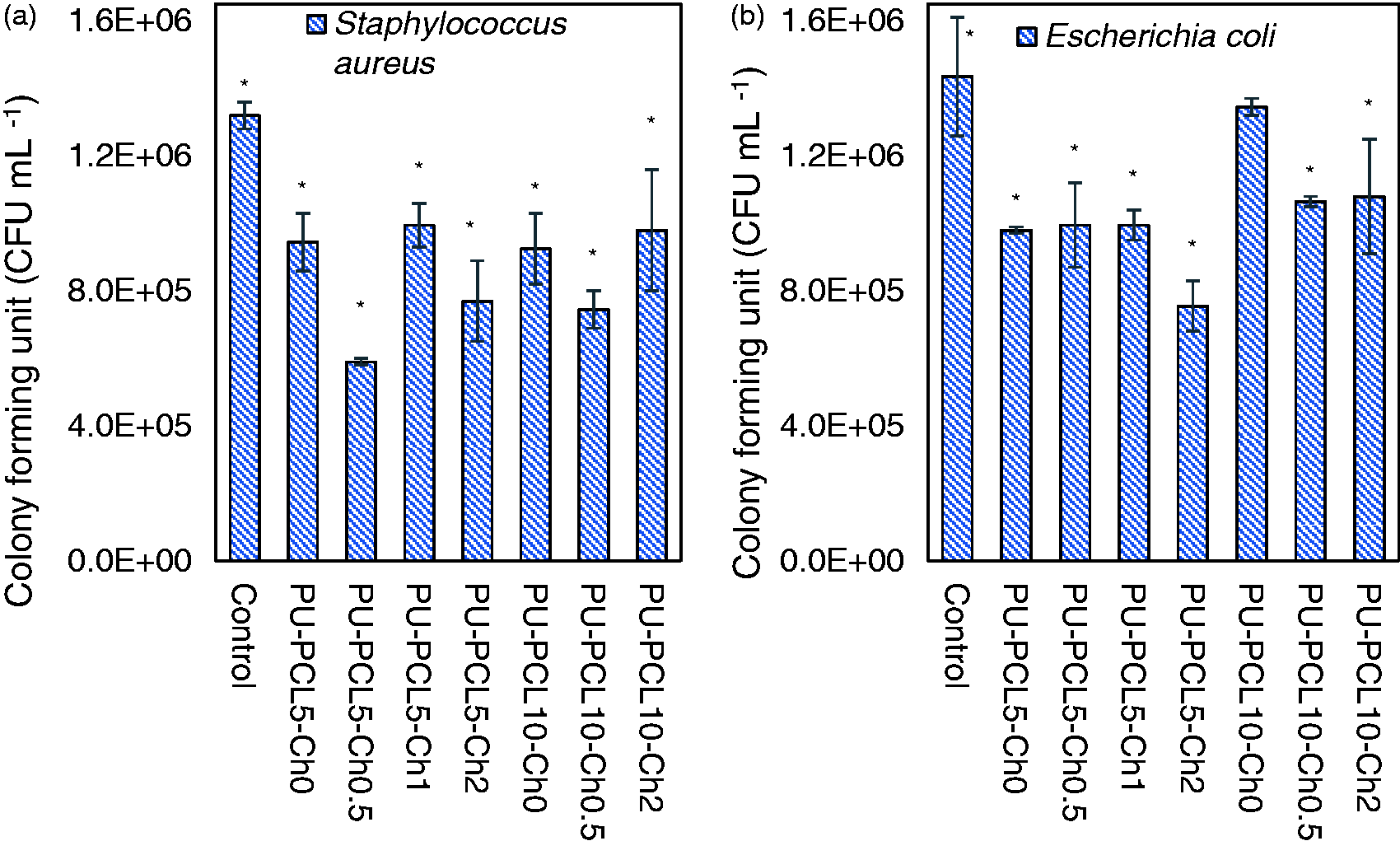
The antibacterial activity of the formulations PU-PCL5-Ch can be explained in terms of the antibacterial action of Ch (which is incorporated into the PU-PCL matrices) against Gram-positive and Gram-negative bacteria such as S. aureus and E. coli, respectively.11,59 The most accepted model for the explanation of the antibacterial activity of Ch is based on the interaction of the negatively charged cell membrane of the bacteria with the Ch molecules, which are positively charged. Such interactions promote changes in membrane permeability, causing internal osmotic imbalances and the leakage of components, which could inhibit microorganism growth.15,60 Additionally, the incorporation of Ch improves the hydrophilic character of the PU surface, generating an increase in the number of polar groups, which could significantly affect bacterial adhesion. The latter was reported by Kara et al. 11 for PU films synthesized with TDI and ethylene glycol, incorporating Ch on the surface by covalent immobilization. The authors also improved the antibacterial activity of the PUs by adding different concentrations of Ch.
By analyzing the results, it can be observed that the PU formulations with 5% w/w PCL and Ch concentrations in the range of 0–2% w/w could be the most promising for use in biomedical engineering applications, in terms of their biocompatibility, mechanical properties and antibacterial activity.
Conclusions
PUs were obtained with castor oil, PCL and IPDI by incorporating different concentrations of Ch (0–2% w/w) as an additive to improve the mechanical properties, biocompatibility and antibacterial activity of the PUs. It was found that the incorporation of Ch enhances the ultimate tensile strength of the PUs and does not affect the strain at fracture in PUs with 5% w/w of PCL and concentrations of Ch ranging from 0 to 2% w/w. The PUs presented similar mechanical properties to the aorta and the skin. The PU-PCL5-Ch formulations and related degradation products did not affect the cell viability of L929 and 3T3 rat fibroblasts, respectively. All PUs presented anti-S. aureus and anti-E. coli antibacterial activity. Based on the results, it was concluded that the PUs could have high potential for use in biomedical applications related to soft and cardiovascular tissues.
Footnotes
Declaration of Conflicting Interests
The author(s) declared no potential conflicts of interest with respect to the research, authorship, and/or publication of this article.
Funding
The author(s) disclosed receipt of the following financial support for the research, authorship, and/or publication of this article: The financing of this study comes from financial support of the research project ING-150-2014 of University of La Sabana.