
Abstract
Bioimaging at a subcellular resolution to label cytoplasm and nucleus in living cell by just one photoluminescent nanoparticle has great application potential in bioresearch, preclinical diagnosis, screening, and image-guided therapy of life-threatening diseases. Herein, we report a novel arginine (Arg) functionalized ultra-small lanthanide oxyfluoride nanocrystals (LaOF) for simultaneously targeted imaging cell cytoplasm and nucleus. As-prepared Arg-modified PAA capped LaOF: 45%Ce, 15%Tb nanocrystals (LaOF:Ce,Tb@PAA@Arg) possessed high water dispersibility, ultra-small size (∼5.7 nm) and double emissions (green and red) with high quantum yield (40%). Such functionalized nanocrystals presented high cellular biocompatibility and were successfully used to label living cells with very high contrast. These functionalized nanocrystals also exhibited significantly higher photostability and brightness as compared to commercial dyes. Such the ultra-small size, high photostability and intensity, double emissions, excellent biocompatibility and targeted ability, make as-prepared functionalized nanocrystals particularly promising for cellular and subcellular bioimaging applications.
Introduction
Optical imaging plays an important role in biomedicine, being extremely useful for bioresearch, preclinical diagnosis, screening, and image-guided therapy of life-threatening diseases. Photoluminescence (PL) optical imaging excels in bioimaging applications due to fast and robust imaging, subcellular resolution and sensitivity, as well as low biological toxicity.1,2 Nowadays, the PL imaging materials has been widely reported, such as green fluorescent proteins (GFPs), organic dyes, quantum dots (QD), and luminescent rare-earth nanoparticles.1,3
However, GFPs suffer from a number of intrinsic deficiencies such as easy hydrolysis by proteolytic enzymes and overlap with autofluorescence signal, thus making it difficult for in vivo cell tracking. 4 Meanwhile, GFPs have a critical drawback of narrow excitation and wide emission spectrum, which make it difficult to excite multiple fluorescent proteins simultaneously with a single excitation source. 5 What’s worse, the commercial organic dyes such as rhodamine, fluorescein, and 40-6-diamidino-2-phenylindole (DAPI) also have the critical flaw, their fast (a few seconds under ∼1 kW/cm2 of resonant illumination) and irreversible transition to a dark state, or photobleaching.4,6 Although QD overcame the photobleaching to a certain extent, it usually presented cytotoxicity as well as showing blinking at the single particle level. 7 Hence, on the whole, to overcome the drawback of existing PL materials, the new ideal PL materials for clinical applications should have the following characteristics: (1) continuous and broad light excitation; (2) strong resistance to photobleaching; (3) good biocompatibility; and (4) subcellular resolution.
Fortunately, luminescent rare-earth nanoparticles were the potential candidates for PL imaging, which show brighter fluorescence and less photobleaching. 7 In detail, the blinking feature is completely absent from rare-earth based nanoparticles due to the presence of a large number of emitters in each particle.8,9 Besides, the luminescence of rare-earth nanoparticles under illumination is highly stable which allows the continuous observation of rare-earth nanoparticles for arbitrarily long durations.10,11 In addition, luminescent rare-earth nanoparticles with different morphology, size, and phase control have been prepared,7,12 and their surface is often modified for transferring from hydrophobic into the hydrophilic environment, which is crucial for the biocompatibility and important for biomedical applications. However, most of the current methods for surface modification of luminescent rare-earth nanoparticles have two important limitations, involved reactions and nanoparticle aggregation. What is worse, traditional rare-earth fluorescent materials just have one fluorescence-emission peak, and they were difficult to offer the subcellular resolution, even though they have the ability to offer a high-contrast tissue bioimaging.2,13 Generally, luminescent rare-earth nanoparticles were endowed with the two advantages: continuous and broad light excitation, as well as strong resistance to photobleaching, whereas with some room for improvement to offer better biocompatibility and subcellular resolution.
For these reasons, our previous work had developed an RGD functionalized ultra-small Oxyfluorides-Gadolinium (GdOF) nanocrystals with double emissions (545 and 587 nm) to achieve the cell imaging at subcellular resolution. 14 To further improve the biocompatibility, arginine (Arg)-modified polyacrylic acid (PAA) was used in this work, and provide special physiological functions for our luminescent rare-earth nanoparticles. In details, Arg is the most hydrophilic natural amino acids with guanidine group at side chain and can be biosynthesized in humans. After capped the Arg-modified PAA, the guanidine group at the surface of nanoparticles can not only increased the water solubility but also provide cationic, which would give the modified nanocrystals an electroporation-like penetration ability through the cytomembrane.15,16 What’s better, the Arg is low in cost and easy to obtain, which can help reducing costs on a large scale for clinical application. Furthermore, to increase the subcellular resolution of bio imaging, the lanthanide oxyfluoride nanocrystals (LaOF) with higher quantum efficiency was designed in this work.
Here, in this paper, we design and test a new and facile method to surface capped with Arg -modified PAA (named as LaOF:Ce,Tb@PAA@Arg). In addition, we examine its water solubility, biocompatibility, and luminescence imaging performance. Meanwhile, systematic cellular studies and fluorescence imaging were performed to confirm the excellent imaging ability with a subcellular resolution of LaOF:Ce,Tb@PAA@Arg.
Experimental section
Materials
Lanthanide trifluoroacetate (La (CF3COO)3, Ce (CF3COO)3, Tb (CF3COO)3) precursors were prepared from the corresponding lanthanide oxides and trifluoroacetic acid. 17 All amino acids and n-hydroxysuccinimide were purchased from CS Bio (Shanghai) Ltd. All the chemicals used in this study were purchased from Sigma–Aldrich unless otherwise specified.
Synthesis of water-soluble PAA-capped LaOF: Ce, Tb nanocrystals
The monodispersed oleylamine capped LaOF: 45%Ce, 15%Tb nanocrystals were synthesized by conventional solvents-thermal decomposition method as our previous report. 18 The water-soluble PAA-capped nanocrystals were synthesized through representative ligand exchange reaction method. 14 In detail, the hydrophobic nanocrystals (30 mg) were re-dispersed in toluene, injected into a solution including diethylene glycol (10.0 ml) and PAA (0.5 g) under 110℃ in a three-necked flask with vigorous stirring for 10 min under N2 flow. The whole system was heated to 240℃ and kept for 1 h until the solution became clear. After cooled down to room temperature, the PAA@ LaOF: Ce, Tb nanocrystals were harvested by centrifuging at 12,000 r/min for 30 min and washing using water-ethanol solution (1:1).
Fabrication of Arg functionalized PAA@LaOF: Ce, Tb nanocrystals
The as-prepared PAA capped nanocrystals were re-dispersed in 2-morpholinoethane sulfonic acid (MES) buffered saline. Arg was conjugated with PAA-capped nanocrystals by ethyl-3-(3-dimethylaminopropyl) carbodiimide hydro-chloride/n-hydroxysuccinimide (EDC/NHS) chemistry. Briefly, the carboxyl acid groups of PAA-capped nanocrystals was activated to the succinimidyl ester by EDC (2 mM) and NHS (5 mM). The activated nanocrystals were added into Arg solution for further functionalized reaction at room temperature. After the NHS/EDC reaction, the Arg@PAA@GdOF:Ce, Tb nanocrystals were collected through centrifuging and washing. The as-formed nanocrystals were easily re-dispersed in PBS for further use.
Physicochemical structure
The nanocrystals morphology and lattice structure were observed on a high-resolution transmission electron microscope (HRTEM, F20, FEI) operated at 200 kV. The surface chemical structure of modified nanocrystals was evaluated by Fourier transform infrared (FTIR) spectroscopy (Nicolet 6700) and UV-vis absorption spectra (Shimadzu 3000 spectrophotometer). The nanocrystals size distribution was obtained from the dynamic light scattering (DLS) measurement (Malvern Zetasizer Nano ZS system).
PL properties
The PL spectra were recorded for dispersed Arg@PAA@LaOF nanocrystals in water on a spectrophotometer (F-4500 Hitachi) equipped with a 150W Xe arc lamp at room temperature. The PL quantum yield of functionalized nanocrystals was determined according to the following equation with LaOF: Ce, Tb nanocrystals in cyclohexane as a standard reference (14)
Cytotoxicity evaluation
The in vitro cytotoxicity was measured using a standard Alamar blue (Thermo Fisher scientific) assay in HepG2 cell lines (human liver carcinoma) and L929 cell lines (mouse fibrosis cells). Cells growth was carried out at standard culture conditions (37℃, 5% CO2) in a complete culture medium (DMEM with 10% FBS). Briefly, cells were seeded into a 96-well cell-culture plate at a density of 1 × 10⁴/well. After culture for 24 h, the nanocrystals with a concentration of 25, 50, 100, and 200 µg/ml was added to the wells and incubated for 24 h. The microplate reader (Molecular Devices) was used to measure the emission value of each well at 600 nm. The cytotoxicity of nanocrystals was expressed as a percentage of valid cells compared to TCP control. At least five species per sample were measured.
Living cell staining by nanocrystals
Before cell staining, La@PAA@GdOF nanocrystals were re-dispersed in culture medium at a concentration of 25 µg/ml. HepG2 and human bronchial epithelial cells (BEAS-2B) cells were cultured in respective growth medium for 24 h. Then the culture medium for HepG2 was changed to be the growth medium containing nanocrystals and stained for 24 h in a 5% CO2 incubator at 37℃. And after the starvation with 0.1% FBS, the culture medium for BEAS-2B was changed to be the growth medium containing differentiation factor (10 ng/ml, TGF-β1) and stained for 48 h in a 5% CO2 incubator at 37℃. And then the culture medium for BEAS-2B was changed to be the growth medium containing nanocrystals and differentiation factor and stained for 24 h in a 5% CO2 incubator at 37℃. Cell imaging was then carried out after washing the cells with PBS three times to remove the excess nanocrystals. And cells were fixed with the concentration 2.5% glutaraldehyde. To show the advantages of our fluorescent nanocrystals, the conventional dyes including SYTO 16 (Molecular probes) and Hoechst (Molecular probes) were used as the controls in the staining process. The staining procedures of dyes were according to the manufacturer instructions.
Living cell imaging studies
Living cell imaging of HepG2 and BEAS-2B cells incubated with Arg@PAA@LaOF:Ce, Tb nanocrystals was performed on a confocal laser scanning microscopy (CLSM, FV1200, Olympus). For confocal fluorescence microscopy imaging, the continuous wave laser at 405 nm (3.15 mW), 488 nm (6 mW), and 543 nm (0.7 mW) provided the excitation, respectively, for exposing 28.39 s. The extracellular and intracellular fluorescent intensity distributions were analyzed by additional software Image J.
Statistical analysis
All data were expressed as mean values with standard deviation (SD) from four independent tests. Statistical significance analysis was performed by a Student’s t-test, and the difference was considered as significant when p < 0.05.
Result and discussion
Synthesis and characterization of Arg functionalized nanocrystals
In our study, LaOF: 45%Ce, 15%Tb nanocrystals capped by oleylamine were synthesized from trifluoroacetate precursors in high-boiling solvents method, as shown in Figure 1. To convert the hydrophobic surface into hydrophile, oleylamine capped nanocrystals were reacted with PAA to form the PAA-capped LaOF: 45%Ce, 15%Tb nanocrystals (LaOF: Ce, Tb@PAA) through a robust ligand exchange method. The molar ratio of dopants (Ce: 45%, Tb: 15%) was optimized by our previous work
14
and Du’s group.
18
Using the suitable electronic transition levels between Ce3+ and Tb3+ ions, Ce3+ ions can absorb irradiative light and transfer the energy to Tb3+ ions, at this time, the transfer always accompanied by the energy loss. Thus, the sensitizer Ce3+ should be triple the molar ratio of the fluorescer Tb3+. In order to increase the penetration ability of the membrane and biocompatibility of nanocrystals, Arg was conjugated to the nanocrystals surface by EDC/NHS chemistry (LaOF:Ce,Tb@PAA@Arg). For covalently modifying all carboxyl of PAA on the nanocrystal surface, excessive amounts of Arg were added to the reaction. Hence, the quantity of Arg on nanocrystals can be considered equal to the PAA carboxyl, which is about 0.6 mmol on 30 mg nanocrystals.
Schematic illustration for the synthesis of arginine functionalized LaOF:Ce,Tb nanocrystals and their bioimaging.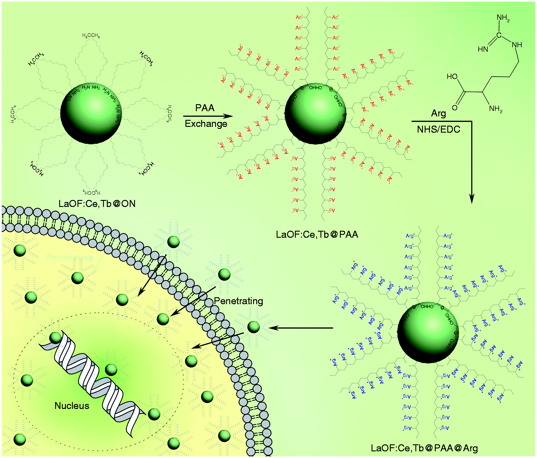
Furthermore, the morphology, size, and physical structure of as-prepared nanocrystals were further determined by transmission electron microscopy (TEM), DLS, and high-resolution transmission electron microscopy (HRTEM). TEM images showed that LaOF:Ce,Tb@PAA@Arg remained as a monodispersed nanostructure and had a uniform diameter of around 5 nm (Figure 2(a)). Due to the ultra-small thickness of functionalized shell (PAA and Arg), the hydrodynamic size of the nanocrystals was just 5.7 nm (Figure 2(a)). And the ultra-small size of functionalized nanocrystals may be beneficial for their applications in bioimaging and cell labeling. Besides, HRTEM images show that these Arg-functionalized nanocrystals retained the spherical morphology
18
(Figure 2(b)) and showed high-quality crystalline structure (Figure 2(c)). And the fast Fourier transform (FFT) patterns in Figure 2(d) confirmed that the obtained LaOF:Ce,Tb@PAA@Arg are single crystalline. All of these results demonstrated the successful synthesis of ultra-small nanocrystals with outstanding monodispersity.
Morphology and spectroscopy characterization of arginine functionalized nanocrystals. (a) TEM images and the hydrodynamic size (internal graph) of LaOF:Ce,Tb@PAA@Arg. HRTEM images (b and c) and corresponding FFT pattern (d) of LaOF:Ce,Tb@PAA@Arg. FT-IR spectroscopy (e), and UV absorption spectra (f) of LaOF:Ce,Tb@PAA@Arg and LaOF:Ce,Tb@PAA.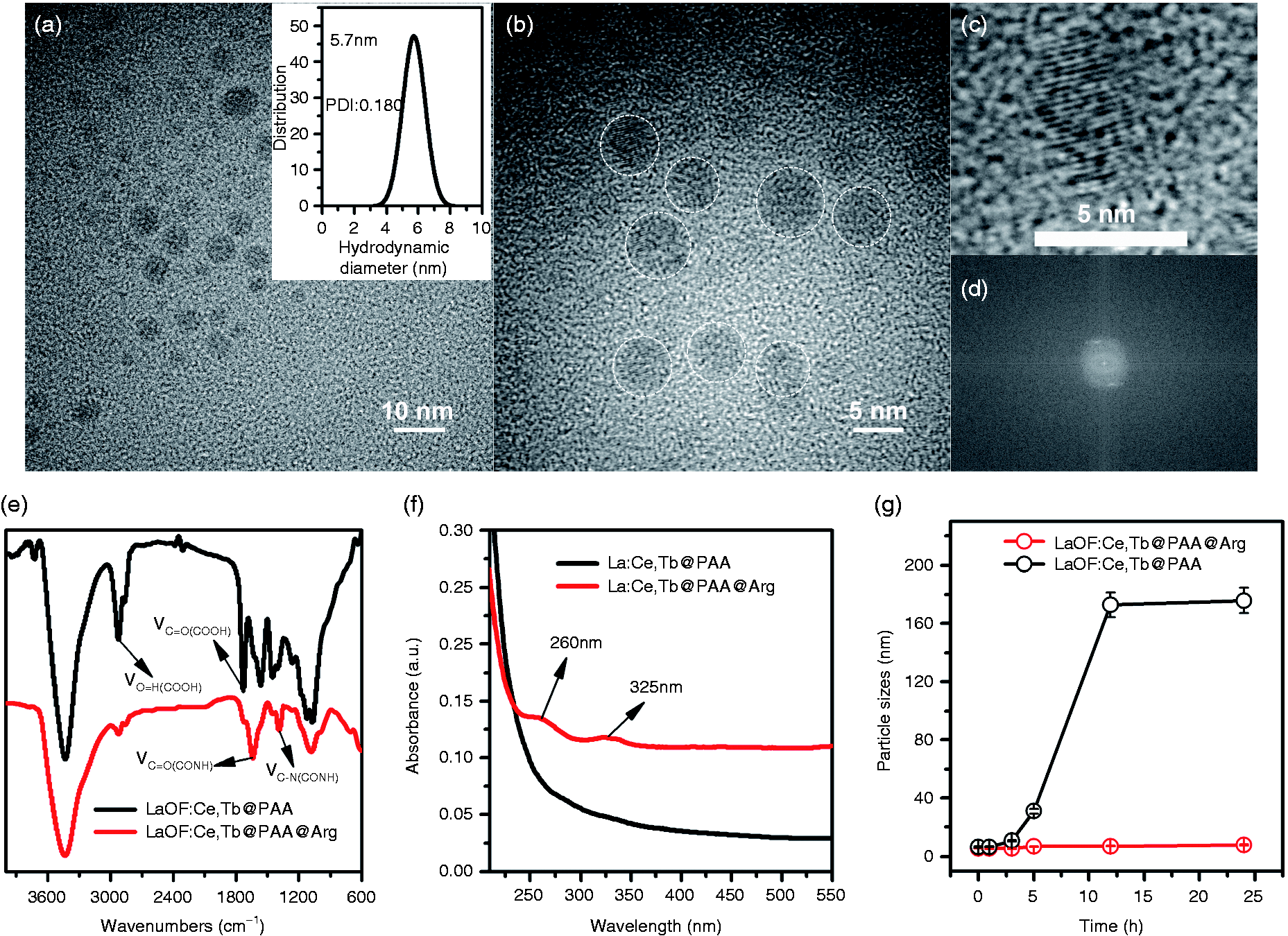
In addition, the surface functional groups of the as-grown nanocrystalline are characterized by FTIR spectroscopy (Figure 2(e)). After PAA capping, two sharp bands located at 1603 and 1715 cm−1 correlate with the stretching vibration of C−O and C=O groups, respectively, and a broad absorption in the range 2850–3485 cm−1 is attributed to the O−H vibration, suggesting that the as-grown nanocrystalline surface is covered with −COOH groups. As for the LaOF:Ce,Tb@PAA@Arg FT-IR spectroscopy, the peaks at 1640 and 1400 cm−1 assigned to CONH vibrations showed the successful Arg modification through the amido bond. And this result was verified again through the ultraviolet absorption spectrum (Figure 2(f)), by which the characteristic absorption peak at 260 and 325 nm demonstrated the amide bond formation after NHS/EDC chemical reaction. To verify the excellent temporal colloidal stability of the LaOF:Ce,Tb@PAA@Arg, the two nanocrystals were diffused in DMDM media including 10% fetal calf serum (FBS), and their particle sizes were monitored in 24 h by DLS. As expected, the sizes of LaOF:Ce,Tb@PAA@Arg keeps small during 24 h, whereas the LaOF:Ce,Tb@PAA aggregated after 12 h (Figure 2(g)). The result demonstrated that the LaOF:Ce,Tb@PAA@Arg was monodispersed during the process of living cell imaging.
The superior fluorescence property of Arg functionalized nanocrystals for cell imaging
Profitted from the suitable electronic transition levels between Ce3+ and Tb3+ ions, Ce3+ ions can absorb irradiative light and transfer the energy to Tb3+ ions, which produces green and red emission. As shown in Figure 3(a), LaOF:Ce,Tb@PAA and LaOF:Ce,Tb@PAA@Arg presented a narrow PL spectrum of dual-fluorescence: green fluorescence emission peak at 545 nm (5D4 to 7F5 transitions of Ce3+ ions), and red fluorescence emission peak at 585 nm (5D4 to 7F4) and 620 nm (5D4 to 7F3). Additionally, LaOF:Ce,Tb@PAA@Arg had higher quantum yields (QYs) of 40% than the LaOF:Ce,Tb@PAA of 16%, and it is more than likely because of the two reason: (1) Arg modification greatly enhanced the monodispersity in water and (2) the LaOF host can offer more efficient energy transfer between Ce3+ and Tb3+ ions, so the QY for the LaOF:Ce,Tb is much higher than that of the GdOF:Ce,Tb nanocrystals in our previous work.
14
The fluorescence property of arginine functionalized nanocrystals for cell imaging. (a) Fluorescence spectra of LaOF:Ce,Tb@PAA@Arg and LaOF:Ce,Tb@PAA; (b) bright pictures and fluorescence pictures of LaOF:Ce,Tb@PAA@Arg; (c) HepG2 cancer cell imaging applications of as-prepared LaOF:Ce,Tb@PAA@Arg. CLSM images of HepG2 cells after incubating with LaOF:Ce,Tb@PAA@Arg (25 mg/ml) for 1 h at 37℃. Cell nucleus was dyed in blue by Hoechst for visualization. Cell cytoplasm was stained as red and whole cell was stained as green under excitation. Cell nucleus and cytoplasm could be clearly imaged as green and yellow, respectively by merging two channels above.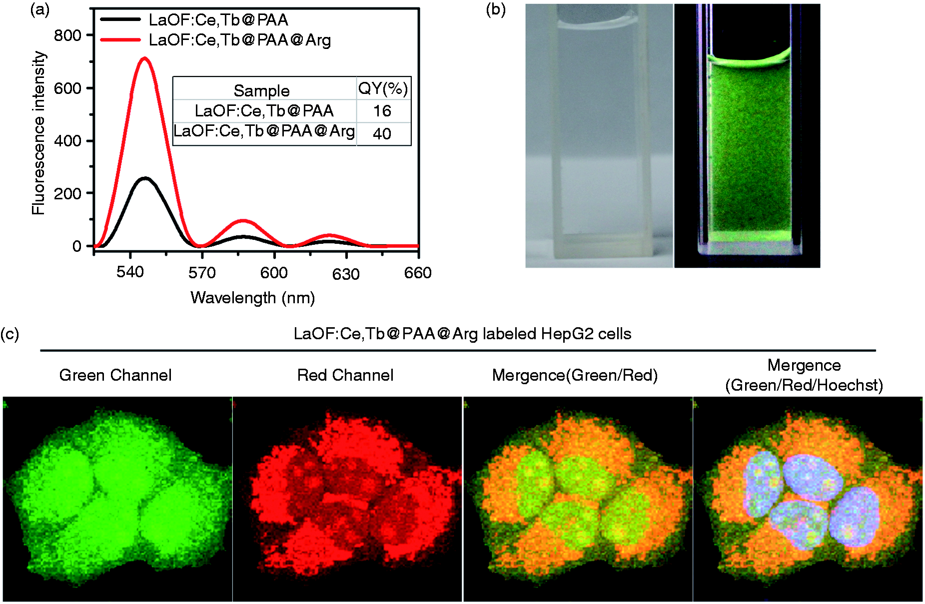
Of note, benefited from the better dispersibility from Arg and the higher electron transport from Lanthanum, the 40% QYs was far beyond our previous GdOF nanocrystalline of 16%. 14 Importantly, compared to the cell-imaging QDs, which typically possess QYs of 1–10%, 19 LaOF:Ce,Tb@PAA@Arg show tremendous performance advantages to replace the existing cell dye.
In a physiological environment mimic phosphate buffer salt (PBS) solution at pH 7.4, LaOF:Ce,Tb@PAA@Arg were well dispersed and showed a strong yellowish-green fluorescence under excitation at 254 nm (Figure 3(b)). For the cell imaging (HepG2, a human liver carcinoma), LaOF:Ce,Tb@PAA@Arg could swiftly penetrate the cellular matrix, bind to dense areas such as the nucleoli, and stain the corresponding region after 24 h incubation. Under the same excitation wavelength, two different fluorescence emissions (green and red) with different intensity could be provided by just one nanocrystals (green channel and red channel, Figure 3(c)). Merging the images from green channel and red channel, LaOF:Ce,Tb@PAA@Arg could clearly stain the nucleus in green and cytoplasm in orange (mergence (red/green), Figure 3(c)). And the nucleus position could be confirmed by the Hoechst staining (mergence (green/red/Hoechst), Figure 3(c)). This phenomenon has been reported in our previous study, 14 and the mechanism was due to the different nanocrystals concentrations in the nucleus and cytoplasm and the different fluorescence intensity between green emission and red emission.
In vitro cytotoxicity of Arg functionalized nanocrystals
Cytotoxicity is the biggest obstacle for cytochemical stain to image the living cells. Thus, the superior fluorescence performance of LaOF:Ce,Tb@PAA@Arg further compelled us to study its biocompatibility. Both of the LaOF:Ce,Tb@PAA@Arg and LaOF:Ce,Tb@PAA with different dosages were chosen to test cytotoxicity for two cell lines (HepG2 and L929) based on Alamar blue (Thermo Fisher scientific) assay. As expected, in Figure 4(a), LaOF:Ce,Tb@PAA without Arg modification induced a dose-dependent growth inhibition of HepG2 cell line. In sharp contrast, these cells lines were resistant to the treatment of LaOF:Ce,Tb@PAA@Arg (Figure 4(a)), suggesting that Arg modification significantly increased the biocompatibility of the nanocrystals. And the similar results were found in a normal somatic cell line, L929, in which LaOF:Ce,Tb@PAA@Arg showed any inhibitory at 200 µg/ml – the highest concentrations used. All of these data show satisfactory results for in vitro biocompatibility of LaOF:Ce,Tb@PAA@Arg nanocrystals, and suggested their huge potential for living cells imaging.
In vitro cytotoxicity of LaOF:Ce,Tb@PAA@Arg and LaOF:Ce,Tb@PAA to cancer and mormal cells. (a) HepG2 and (b) L929 cell viability after incubation with different dosages of LaOF:Ce,Tb@PAA@Arg and LaOF:Ce,Tb@PAA for 24 h using standard Alamar blue assay (n = 5).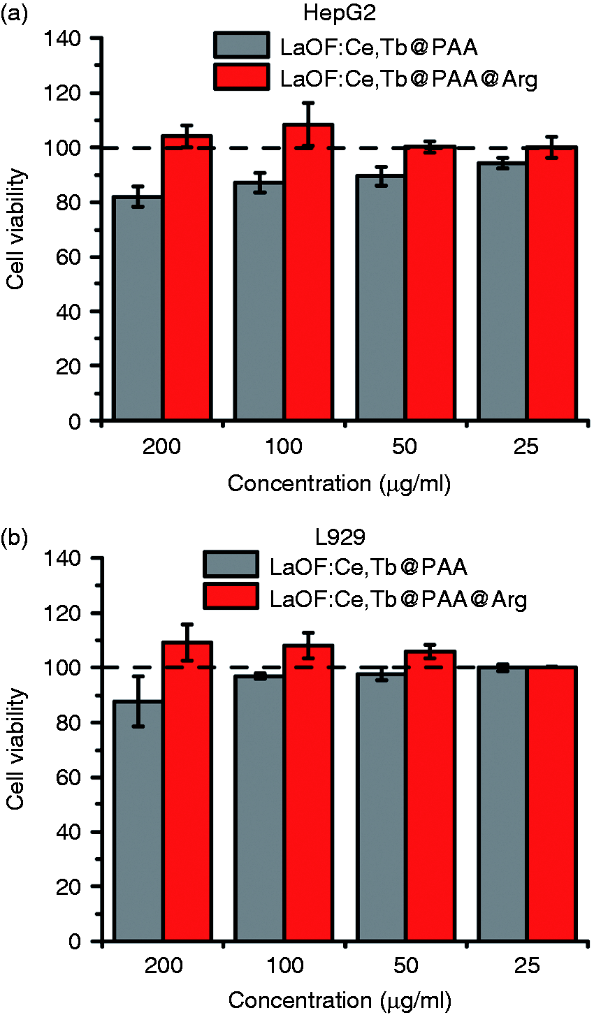
The superior cell imaging performance of Arg functionalized nanocrystals
To investigate the cell imaging performance of LaOF:Ce,Tb@PAA@Arg and LaOF:Ce,Tb@PAA, HepG2 cells was employed again to incubate with the two nanocrystal of 25 µg/ml, respectively. After the 24-h incubation, LaOF:Ce,Tb@PAA@Arg imaged the cells with bright fluorescence, whereas the imaging by LaOF:Ce,Tb@PAA were bleak nonluminous (Figure 5(a)). Quantifying the fluorescence intensity (Figure 5(b)), the LaOF:Ce,Tb@PAA@Arg exceed the LaOF:Ce,Tb@PAA by at least five times. Notably, this gap was greater than the QY of 2.5-fold, presumably due to the presence of Arg increasing the cell penetration. As our design, Arg functionalized nanocrystals will enter cells by mechanisms similar to those used by cell-penetrating peptides, specifically electrostatic binding to cell surface negative charges, induction of endocytosis and endocytic vesicle escape by osmotic and/or membrane lytic activity.
20
Meanwhile, Arg can increase the binding of our nanocrystals to the outer monolayer of cell and neutralize the surface charge of anionic lipids, leading to the development of a transmembrane electrical field that is known to result in the formation of transient pores in the lipid bilayer at cytomembrane.21,22 Thus, Arg modification would result in much higher penetration for living cells, which is conducive to the cell imaging with higher resolution and reduce the usage cost of dye.
Cancer cell imaging applications of synthesized nanocrystals with or without arginine modfication. (a) CLSM images of HepG2 after incubated with LaOF:Ce,Tb@PAA@Arg and LaOF:Ce,Tb@PAA (25 mg/ml) for 4 h at 37℃, respectively. Cell nucleus and cytoplasm could be clearly imaged as green and yellow, respectively by merging green and red fluorescence. Cell nucleus was stained as blue by Hoechst for comparison. Scale bar: 60 µm. (b) Fluorescent quantitation using Image J software.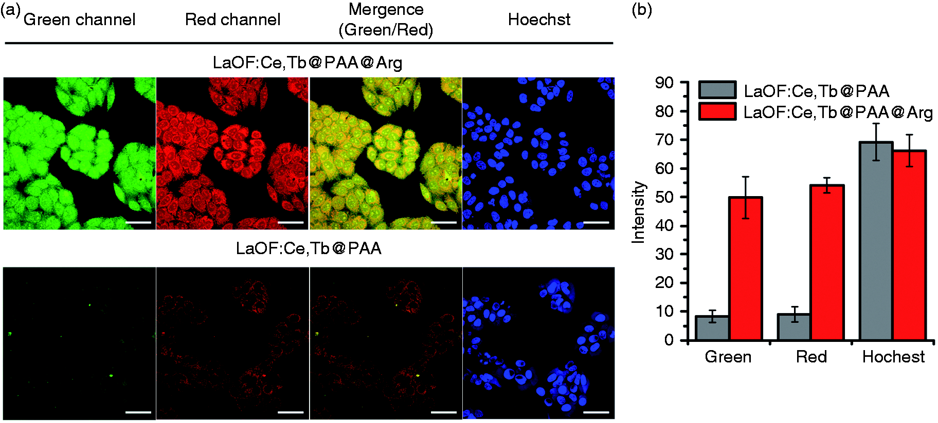
The stability of LaOF:Ce,Tb@PAA@Arg against photobleaching
Nowadays, the commercial organic dyes were widely applied in biological study and clinical diagnosis, but the notorious photobleaching hampers the applications with the need of long-term imaging. To investigate the stability of LaOF:Ce,Tb@PAA@Arg against photobleaching, Calcein Am, a commercial fluorescent dye for living cells was employed to compare the photobleach with the nanocrystal. As expected, LaOF:Ce,Tb@PAA@Arg showed much better performance to against photobleaching (Figure 6). In detail, immediately to observe after dyeing, both of Calcein Am and the nanocrystal imaged the cell morphology with bright fluorescence. After exposed to the visible light (400–800 nm) for 8 h, the green and red fluorescence signal from intracellular LaOF:Ce,Tb@PAA@Arg almost completely retained its initial fluorescence intensities (Figure 6), even in brighter fluorescence with higher resolution of nucleus and cytoplasm. In sharp contrast, the fluorescence from Calcein Am almost disappeared with the same condition. These results indicated that our functionalized nanocrystals could be employed in living cell cytoplasm and nucleus for long-term imaging.
Photostability of synthesized photoluminescent nanocrystals in cell imaging applications. CLSM images of HepG2 cells incubated with 25 mg/ml LaOF:Ce,Tb@PAA@Arg and Calcein Am before and after 8 h incandescent lamp irradiation. Scale bar: 60 µm.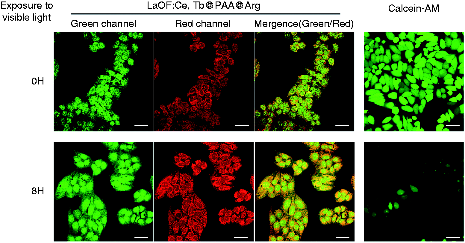
Distinguish fibration through imaging fibrosis cells by LaOF:Ce,Tb@PAA@Arg
In this work, LaOF:Ce,Tb@PAA@Arg has a superior capacity to simultaneously image the cytoplasm and nucleus of cancer cells, by which the cell cancerization can be distinguished quickly and conveniently. Except for the diagnosis of cancer, there exist another thorny problem for clinical doctors and researchers to distinguish fibrosis cells from normal tissue. To verify that LaOF:Ce,Tb@PAA@Arg has ability to solve this problem, human bronchial epithelial cells (BEAS-2B) cell line was used as a fibration modle with 10 ng/ml fibration inducer, TGF-β1. To visually observe the different morphology between common cells and fibrotic cells, Beas-2B cells were incubated with fibrosis inducer as an experimental sample and without inducer as a control. After the imaging by LaOF:Ce,Tb@PAA@Arg, the cellular morphology was unambiguously imaged. In detail, as shown in Figure 7, the shape of the control ones remain normal with the circular or elliptical profile. In sharp difference, most of the fibrotic cells was spindle shaped, but some cells were even stellate-like with thread like cytoplasm periphery. Benefitted from the superior cell imaging performance of LaOF:Ce,Tb@PAA@Arg, these morphologic changes can be clearly observed thereby bring huge convenience for fibration diagnose.
Fibrosis cell imaging applications of LaOF:Ce,Tb@PAA@Arg. Scale bar: 60 µm.
Conclusion
This paper described a facile synthesis of Arg functionalized ultra-small LaOF nanocrystals (LaOF:Ce,Tb@PAA@Arg) with a size of 5–6 nm, and presented their application for simultaneously imaging cytoplasm and nucleus with high contrast. The Arg modification for nanocrystals provided an excellent monodispersity under physiological condition and high cellular biocompatibility. These dual emission PL nanocrystals have superior cell imaging performance for imaging the shape of cancer cell and fibrosis cells without fluorescence photobleaching. In brief, the ultrafine size, strong fluorescent intensity and stability, high biocompatibility, cytoplasm and nucleus imaging, make our Arg functionalized LaOF nanocrystals particularly attractive for cellular bioimaging. And these capabilities of our engineered nanocrystals provide promise for their wide application in distinguishing cancer cells as well as fibrosis cells and determining cell types.
Footnotes
Authors' Note
Wangxiao He and Guang Yang are both the corresponding authors of the article. The contact details of Guang Yang are- No. 140, Hanzhong Road, Nanjing 210029, China. Email: Bruce.Yang@bengmedicalcenter.com.
Declaration of Conflicting Interests
The author(s) declared no potential conflicts of interest with respect to the research, authorship, and/or publication of this article.
Funding
The author(s) disclosed receipt of the following financial support for the research, authorship, and/or publication of this article: This work was supported by Project 985 of Xi’an Jiaotong University (to Wuyuan Lu) and Natural Science Foundation of Jiangsu Province (no. BK20161234).