
Abstract
The nervous system has little capacity for self-repair after injury because neurons cannot proliferate owing to lack of suitable microenvironment. Therefore, neural tissue engineering that combines neural stem, scaffolds, and growth factors may improve the chance of restoration of damaged neural tissues. A favorable niche for neural regeneration would be both fibrous and electrically conductive scaffolds. Human Wharton jelly-derived mesenchymal stem cells were seeded on wet-electrospun 3D scaffolds composed of poly lactic acid coated with natural polymers including alginate and gelatin, followed by a multi-wall carbon nanotube coating. The results show that a wet-electrospun poly lactic acid scaffold at a concentration of 15% w/v had higher porosity (above 80%) than other concentrations. Moreover, the coated scaffold supported the growth of human Wharton jelly-derived mesenchymal stem cells in 3D culture, and were incubated for 21 days with 1 mM valproic acid as the inducer resulted in improvement in human Wharton jelly-derived mesenchymal stem cells differentiation into neuron-like cells immunoreactivity to nestin, Map2, and neuron specific enolase (NSE), which were also consistent with reverse transcription polymerase chain reaction (RT-PCR) and quantitive Reverse transcription polymerase chain reaction (qRT-PCR) results. The conclusion is that the 3D composite nanofiber poly lactic acid scaffold improved the transdifferentiation of human Wharton jelly-derived mesenchymal stem cells into neuron-like cells.
Introduction
Several neurodegenerative diseases are associated with neuronal loss, such as Alzheimer's disease (AD), Huntington disease (HD), Parkinson's disease (PD), spinal muscular atrophy (SMA), and amyotrophic lateral sclerosis (ALS). 1 While the recovery is pretty difficult due to the lack of effective therapies, the cell therapy technique is an attractive approach. 2 However, the effectiveness of this method has not been yet shown because of the delivered cells need an appropriate microenvironment consisting of fibrous matrix, which can be an alternative niche for the transplants. 3 This regenerated tissue should be grown on a scaffold, which could mimic the structure and function of the natural niche. Various natural biopolymers and synthetic polymers have been used as a scaffold in tissue engineering. 4 Poly lactic acid (PLA), a well-known FDA-approved polymer for its biocompatibility and biodegradability, is one of the most widely used synthetic polymers in the biomedical field, 5 whereas gelatin (GE), as a natural hydrophilic polymer, has many functional groups (glycine, proline, glutamic acid, hydroxyproline, arginine, alanine, aspartic acid, and other amino acids). 6 The other natural biopolymer is sodium alginate (AL), which is widely applied in medicine and medical researches. 7 Whereas carbon nanotube (CNT) as a conductive material with excellent mechanical, electrical, and surface properties are extensively used in structural materials, sensors, biomedical applications, and especially for nerve tissue engineering. Chemically functionalized CNTs have been used successfully to improve neural signaling. 8 Several studies show that an increase in the efficacy of neural signal transmission may be related to the specific properties of CNT materials such as the high electrical conductivity. 9
An ideal scaffold, which mimics the natural ECM, should be biocompatible, biodegradable, and support cell proliferation, migration, and maturation. Furthermore, it should have a three-dimensional nanostructure that can mimic the environment of cells in vivo. 10
Mesenchymal stem cells (MSCs) have biological properties that make them suitable for use in regenerative medicine. Of the different sources of MSCs that have been described, one is the Wharton's jelly of the umbilical cord (WJMSCs), which exhibits unique features including primitive nature, multilineage potential immunomodulatory ability, and extensive proliferation. 11 They can easily be obtained in large numbers and could represent a valuable therapeutic tool for the treatment of various diseases or tissue damages. 12 There are different protocols for transdifferentiation of WJMSCs into neuron-like cells that have been adopted. In this study, we intended to induce human Wharton Jelly derived mesenchymal stem cells (hWJMSCs) into neuronal phenotype by valproic acid, which was seeded on porous 3D nanofibrous PLA scaffolds fabricated via the wet-electrospinning technique 13 and coated with layers of GE and AL, and subsequent coating with multi-wall carbon nanotube (MWCNT) by a self-assembly method.
Materials and methods
Materials
PLA with a density of 1.25 g/cm3 obtained from Chemiekas (Vienna, Austria) and MWCNT (Nanocyl®-7000 with an average diameter 9.5 nm; length 1.5 µm; specific surface area 250–300 m2 g−1) was procured from Nanocyl (Sambreville, Belgium).
Dulbecco's modified Eagle's medium/Nutrient F-12 Ham medium (DMEM/F12) and fetal bovine serum (FBS) were purchased from Gibco (GIBCO-BRL, Eggenstein, Germany). MTT (3-(4,5-dimethylthiazol-2-yl)-2,5-diphenyltetrazolium bromide) powder and DMSO (dimethyl sulfoxide) were purchased from Carl Roth (GmbH, Germany). All other solvents and experiment agents were obtained from Sigma-Aldrich (Hamburg, Germany).
Sample preparation via wet electrospinning
The method of scaffold preparation, coding, concentration used in the study, the porosity of the scaffold and the percentage of cellular metabolism to the scaffold.
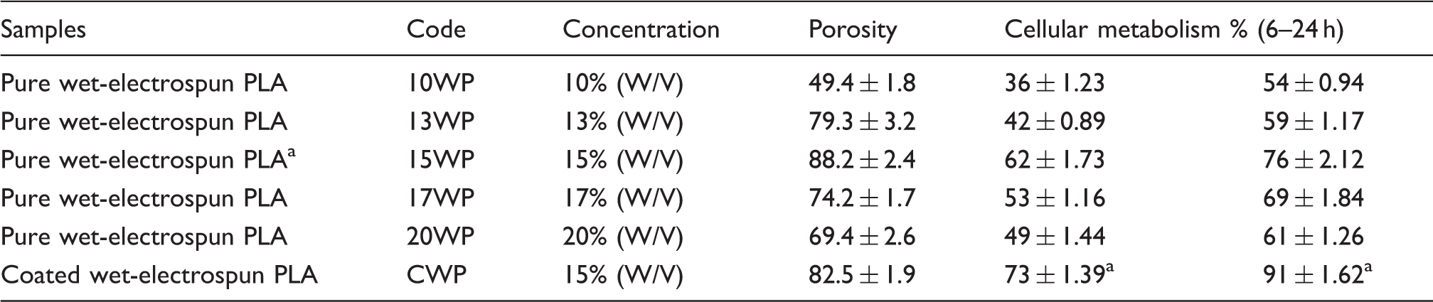
W/V indicates that preparation based on the ratio of PLA weight to the volume of the solvent. The data were presented as mean ± SE.
The corresponding concentration is significantly higher in porosity and cellular metabolism than other concentrations.
PLA: poly lactic acid; SE: standard error.
Coating of the scaffold
The best sample of pure PLA (15 WP) was chosen and functionalized by 0.25 mM NaOH/ethanol (1:1) for 30 min and, finally, coated with such natural polymers as gelatin, alginate, and MWCNT using a layer-by-layer self-assembly method. First of all, 1 mg/ml of gelatin was dissolved in phosphate-buffered saline (PBS) in pH of 3.4 at room temperature.
14
Then, 1 mg/ml aqueous solution of alginate was prepared, followed by another layer of gelatin. The next step was the preparation of 1 mg/ml of acid-treated MWCNT solution in ethanol that was sonicated for 30 min on an ultrasonic bath (SB25-12DTD, Ningbo Scientz Biotechnology Co., Ltd, Ningbo, China) to create a homogeneous solution.
15
The second gelatin layer was coated with MWCNT and followed by another gelatin layer coating. Next, the polymers' zeta potential was detected (Zetasizer Helix, Malvern Instruments Ltd, Malvern, Worcestershire, UK). These biomaterials were coated on the PLA scaffold, according to the surface charge as shown in Figure 1.
The processes of PLA scaffold coating. According to surface charge of each polymer (as characterized by zeta potential of each polymer as listed in figure) followed by nanotube coating, moreover, the self-assembly method was used in this study. Positively charged gelatin was coated on PLA (negative surface charge), then it was coated with negative charge polymer (alginate), followed by gelatin coating, then covered with negative charge MWCNTs with subsequent gelatin coating as an outer layer. PLA: poly lactic acid; MWCNT: multi-wall carbon nanotube.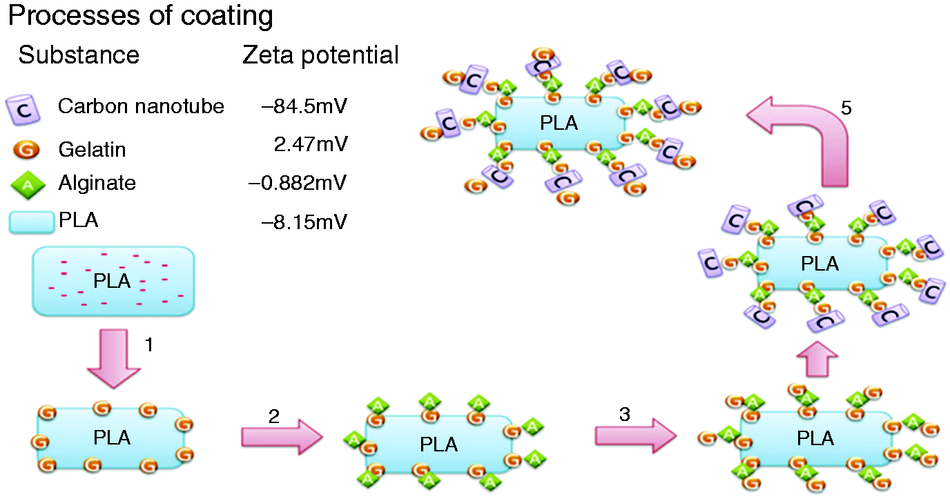
Scaffold characterization
Morphology of the scaffold
The electrospun PLA fibers and the seeded cells were morphologically characterized by scanning electron microscope (SEM, AIS2100; Seron Technology, Uiwang-si, Gyeonggi-do, South Korea). Three days after cell seeding on the scaffolds, they were prefixed by 2.5% glutaraldehyde for 1 h at room temperature post-fixed using tetroxide osmium tetroxide (1%) for 45 min and were dehydrated by an ethanol gradient, coated with gold for 180 s by a sputter Coater EMITECH model SC7620 (Quorum Technologies Ltd., East Sussex, UK) observed under SEM at an accelerating voltage of 20 kV.
Porosity of the scaffold
The porosity of the scaffolds was estimated by the liquid displacement technique, according to the formula as follows:

Tensile strength of the scaffold
The tensile properties of wet-electrospun pure and coated PLA scaffolds were determined using an Instron 5566 universal testing machine (Instron, MA, USA). Rectangular specimens of dimensions 30 mm × 10 mm were used for testing, at a crosshead speed of 10 mm/min. Three samples from each composition have been tested to get statistical data of the tensile strength for the PLA scaffolds.
Fourier transform infrared (FTIR) analysis
Initially, the zeta potential was measured and then the scaffolds of pure PLA, and PLA/GE/AL/MWCNT after cross linking and drying were analyzed by FTIR spectroscopy in the wave numbers range of 400–4000 cm−1 (FTIR Spectrometer, Bruker-EQUINOX 55, Karlsruhe, Germany). The samples were prepared by grinding particles of the scaffolds with KBr.
Hydrophilicity of the scaffolds
The hydrophilic nature of the best concentration of PLA (15%w/v) and also the coated scaffold was measured by the sessile drop water contact angle measurement using a contact angle measuring system (G10, Kruss, Germany). Distilled water was used for drop formation. The measured contact angle value reflected the hydrophilicity of the scaffolds. 17
pH monitoring of scaffold degradation
To monitor the pH alteration during the in vitro degradation, the scaffolds were immersed in normal saline (pH 6.48) at 26℃ for 3 weeks. 18 The pH was measured weekly by means of a pH meter (InoLab pH/Cond 720, WTW, Weilheim, Germany).
Weight loss during scaffold degradation
The degradation behavior of scaffolds was evaluated from its mass loss. The dried scaffold samples were weighed and equal-size samples were immersed in a falcon tube containing 10 mL of PBS (pH 7.2) and the weight loss measurement observed for maximum 3 weeks. At different time intervals (7, 14, and 21 days), triplicate specimens for each scaffold were taken out from the tubes, rinsed several times with distilled water, and lyophilized until dry. The mass of samples were then measured to determine the degree of degradation using the following equation

Mesenchymal cell isolation and characterization
The project was approved by the ethical committee in the Faculty of Medical Sciences at Tarbiat Modares University.
hWJMSCs were isolated as previously described by Yoon et al. 20 Briefly, equivalent amounts of Wharton's jelly segments were digested with 0.1% collagenase type II (GIBCO-BRL, Eggenstein, Germany) for 30 min at 37℃. The washed cells were cultured in L-DMEM (GIBCO-BRL, Eggenstein, Germany) containing 10% FBS (GIBCO-BRL, Eggenstein, Germany) and antibiotics in a 100-mm dish in a humidified 37℃, 5% CO2 incubator. The Medical Ethics Committee of Tarbiat Modares University approved the isolation of the WJMSCs from umbilical cords (UCs) for research. The isolated cells were characterized using cell surface markers by fluorescence-activated cell sorting (FACS, Sysmex Partec CyFlow® Space) analysis. They were directly labeled with different cell markers with monoclonal antibodies CD34-PE, CD45-FITC, CD29-PE, CD90-FITC, and CD73-PE (eBioscience, San Diego, USA). The leneage of the cells was confirmed by their differentiation potential. They were harvested at fourth passages, and the osteogenic and adipogenic differentiations were performed using the differentiation medium in accordance with the manufacturer's instructions in the adapted media (StemMACS™ OsteoDiff & StemMACS™ AdipoDiff Media, Miltenyi Biotec, Bergisch-Gladbach, Germany). After 21 days, the osteogenisis was demonstrated by staining with Alizarin Red, 21 while the adipogenic differentiation, with Oil O Red after 14 days. The isolated cells (hWJMSCs) were cultured in DMEM/F12 supplemented with 10% (v/v) FBS and penicillin–streptomycin–amphotericin B (100 U/ml, 100 µg/ml, and 0.25 µg/ml, respectively) in a humidified incubator at 37℃ with 5% CO2.
The scaffolds was sterilized by exposing them to UV light for 2 h and were then immersed in 70% ethanol for 1 h and dried under vacuum for 1 h. The scaffolds were washed twice with PBS and once with DMEM/F12, transferred to a 96-well plate under sterile conditions and each seeded with 5 × 10³ hWJMSCs, incubated for 2 h in a 0.1 ml cell culture medium with FBS, and the medium was changed every 2 days. 22
Live/dead cells detection
For the cell viability studies, the samples were stained using a Live/Dead staining solution freshly prepared acridine orange/ethidium bromide (AO/EB). 23 The hWJMSCs were cultured for 7 days followed by the removal of the culture medium. A live/dead assay staining solution (0.1 ml) was added to the cultured scaffolds in medium and incubated at room temperature for 5 min and, finally, examined under fluorescence microscope (Fluorescent microscope TE 2000-S, Nikon, Tokyo, Japan).
Cell metabolism studies
The MTT assay was used to quantitatively evaluate the metabolism of cells cultured onto the scaffolds. The MTT assay was performed on the first, third, and seventh days after cells seeding. At each time point, the culture medium was removed from the wells (5 × 10³ cells) and 0.2 ml of MTT (0.5 mg/ml) was added to each well, incubated at 37℃ for 3–4 h in a dark place, then the solution was removed and 0.1 ml DMSO added to each well. The formed purple formazan crystals were dissolved in DMSO. 24 The absorption was measured at a wave length of 570 nm using a microplate laser reader Anthos 2020 (Biochrom, Berlin, Germany). 25 The hWJMSCs in the same 2D culture condition were treated identically in the rest wells of the plate without scaffolds and used as a control. The mean for the triplicate wells for each sample was reported.
The cells were seeded on scaffolds and immediately transferred to an incubator and were incubated for 6 and 24 h. The samples then were washed twice with PBS for 30 s and transferred to new wells.
Differentiation of hWJMSCs into neuron-like cells
The neuronal induction was done at the forth passage; the cells were plated in 12-well plates with (3D) and without (2D) scaffolds at a density of 15 × 103 cells/cm2 and incubated in the DMEM medium supplemented by FBS10% in a humidified incubator at 37℃ with 5% CO2. After adherence of the seeded cells on scaffolds and bottom of the plate, the medium was discarded and replaced with the induction medium (DMEM/FBS10% with 1 mM valproic acid in both with scaffold (3D-VPA) and without scaffold (2D-VPA)). These cells were incubated for 21 days, half the medium was changed every 3 days, 26 and then they were processed for immunocytochemistry and polymerase chain reaction (PCR).
Neuron-like cell characterization
Immunocytochemistry
Cells were fixed with 4% (w/v) paraformaldehyde for 45 min and permeabilized with 0.3% (v/v) Triton X-100 (Sigma-Aldrich, Steinheim, Germany) in PBS for 5 min blocked in 10% (v/v) goat serum for 30 min, incubated with primary antibodies at 4℃ overnight. The immunocytochemistry was done by use of the mouse polyclonal anti-Nestin antibody (1:100), mouse polyclonal anti-Map2 (1:100), mouse polyclonal anti-NSE (1:100) (Abcam, Cambridge, UK), and then washed with PBS, labeled with FITC-conjugated secondary antibody (Abcam, Cambridge, UK), counterstained with DAPI, and finally visualized with fluorescent microscope TE 2000-S (Nikon, Tokyo, Japan).
Reverse transcription polymerase chain reaction analysis
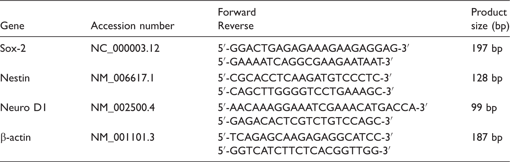
The total cellular RNA was extracted using TRIzol® reagent (Invitrogen, Karlsruhe, Germany) using 15 × 103 cells/cm2 and was carried out according to the manufacturer's protocol. Synthesis of cDNA was also done according to the manufacturer's instructions (Fermentas Inc., Hanover, MD, USA). Next, the gene expression of Sox-2, Neurod1, and Nestin were analyzed by β-actin as the housekeeping gene. PCR amplification was performed using a standard procedure with Taq DNA polymerase (Fermentas Inc., Hanover, MD, USA) with denaturation at 94℃ for 15 s, annealing at 60℃ for 30 s, and extension at 72℃ for 45 s. Depending on the abundance of the particular mRNA, the number of cycles varied between 30 and 40.
The sequence of forward and backward primers of the genes used for charactering the neuron-like cells.
Statistical analysis
The results were statistically analyzed by SPSS (IBM SPSS Statistics, V.23, Armonk, NY) software using one-way ANOVA test and the data were expressed as mean ± SE, n ≥ 3. In all evaluations, p < 0.05 was considered as statistically significant.
Results
Morphology of wet-electrospun scaffolds
SEM images show the morphology and fiber size of the scaffolds, which are characterized by a random orientation of the non-woven fibers forming interfiber porous structure. In 10WP (see Table 1), the fibers had a number of beads with a spindle-like shape (Figure 2(a)). However, with an increase in the concentration of the polymer, the mean size of fibers increased in 13WP, and the spindle-like beads were fewer with the fibers showing more regular shapes (Figure 2(b)). Moreover, in the 15WP sample similar to that of the 13WP, the shape and size of fibers were better than those at higher concentrations (Figure 2(c)). In the 17WP and 20WP samples, the diameter of the fibers in the scaffold increased with fewer cylindrical shaped fibers (Figure 2(d) and (e), respectively). The wet-electrospun method resulted in the formation of intrafiber pores and three-dimensional structure, which were scarcely noticed in the dry-electrospun (Figure 2(c1) and (c2)). The CNT in the coated scaffold by inverted microscope (see supplementary Figure 1).
Scanning electron micrographs of the PLA scaffold at different concentrations. (a) 10% concentration (W/V: weight of PLA/volume of solvent). Accordingly, (b) to (e) represent 13, 15, 17, and 20%, respectively (Scale Bar = 100 µm). (c1) and (c2) show the intrafiber pores (arrowhead). (c3) shows the wet-electrospun PLA nanofibers at 15% with the highest porosity, while C4 shows the same nanofibers coated with polymers and MWCNTs (Scale Bar = 5 µm). (f) The cellular metabolic activity, while (g) represents high-resolution image in (f) (Scale Bar: (f) = 10 µm and (g) = 5 µm, respectively). PLA: poly lactic acid; MWCNT: multi-wall carbon nanotube.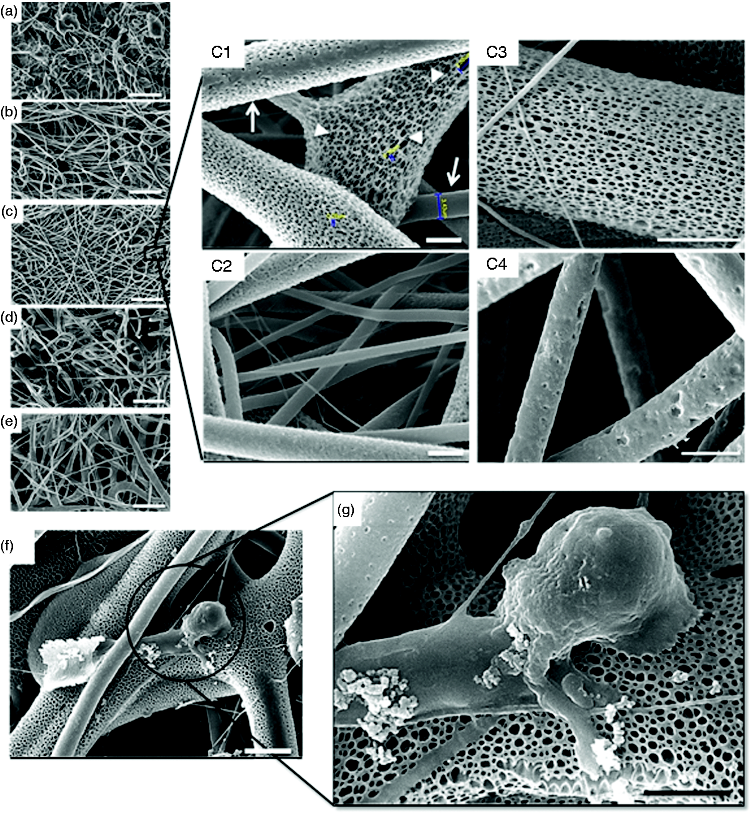
The PLA scaffold images in the concentration of 15%w/v shows many nano-size pores in the fibers but, after coating it with natural polymers, these pores were filled as displayed in Figure 2(c3) and (c4).
Cell-scaffold interaction
Figure 2(f) and (g) shows SEM images of the adherent hWJMSC cultured on the 15WP sample.
FTIR analysis
The results of zeta potential were presented in Figure 1. To confirm the coated compositions on the nanofibers, FTIR was used to examine the uncoated wet-electrospun PLA nanofibrous scaffold and the coated one (Figure 3(a) and (b), respectively). The FTIR spectrum of PLA fibers represented characteristic absorption bands at 1759 cm−1 related to the C=O stretch and at 1333 cm−1 is detected the C–O–C stretching vibration. The short bonds are also detected at 1185 and 1089 cm−1, which represent the backbone ester group of the PLA (Figure 3(a)). This assay was performed for coated scaffold as shown in Figure 3(b); it is difficult to detect the details of the entire polymer in this method, though.
The FTIR spectra of pure (a) and coated PLA scaffold (b). (a1) Macroscopic images of water drop on uncoated PLA (15% w/v) scaffold, while (b1) represents the coated one, the contact angles in (a1) and (b1) are 110 ± 3.1 and 35 ± 2.1, respectively (scale bar: 250 µm). FTIR: Fourier transform infrared; PLA: poly lactic acid.
The common bands of protein appeared at approximately 1650 cm−1 (amide I) and 1540 cm−1 (amide II), corresponding to the stretching vibrations of C=O bond, and the coupling of the bending of N–H bond and the stretching of the C–N bonds, respectively. The amide I band at 1650 cm−1 was attributable to both a random coil and α-helix conformation of gelatin (Figure 3(b)). Whereas the alginate showed a characteristic peaks at 3410 cm−1 corresponding to the hydroxyl group and 1616 cm−1 for the carboxylate group (Figure 3(b)). As the alginate concentration in the blends is low, these peaks also became progressively smaller in magnitude. Like the findings of Huang et al., 27 some of the characteristic peaks of MWNTs (–COOH) appeared at 1180 and 1350 cm−1 (C–O), 1600–1450 cm−1 (aromatic ring), 1641 cm−1 (C=C), 1721 cm−1 (C=O), and 3441 cm−1 (–OH). Moreover, the peaks appearing at around 1721 and 1180 cm−1 in Figure 3(b) were from the stretching vibration of C=O and –C–O groups in the carboxyl group (–COOH), respectively. Furthermore, Figure 3(b) illustrates the FTIR spectrum of the MWNT/PLA blend, and the result showed that strong absorption for aromatics in the region of 1600–1450 cm−1 occurred as a result of the condensation reaction between MWNT and PLA (see supplementary Figure 2).
Hydrophilicity studies
The contact angle of a water droplet with uncoated PLA scaffold was 110 ° ± 3.1, which shows the hydrophobic behavior of this polymer 28 (see Figure 3(a1)). Therefore, the surface structure of PLA scaffolds needed to be modified by adding such hydrophilic polymers as gelatin or alginate. Since the nerve is a conductive membrane, coating the scaffold with a conductive polymer like MWCNT can enhance the growth of the neural tissue. The contact angle of the coated scaffold decreased significantly by 35 ° ± 2.1 due to coating of PLA with hydrophilic biopolymers (Figure 3(b1)).
Porosity and mechanical studies
Table 1 shows the low porosity percentage of the 10WP sample (49.4 ± 1.8) rather than the other scaffolds, which could be attributed to the beaded structure of these scaffolds (P < 0.05). The porosity in 15WP was about 80% and was significantly higher than other concentrations (P < 0.05). The highest porosity percentage in the wet-electrospun scaffolds is attributed to the NaOH in the coagulation bath.
The mechanical properties of all the samples were evaluated by a tensile test and the results are shown in Figure 4(a). An increase in the porosity rate had different influences on the mechanical properties of the PLA scaffold. Although the 15WP sample had higher porosity compared with the other concentrations, it showed better mechanical properties. Moreover, the coated scaffold showed a higher mechanical behavior rather than the uncoated one (PLA: 15WP) as shown in Figure 4(a).
The histograms of the tensile strength and weight loss percentages of scaffolds. (a) The tensile strength of uncoated and coated PLA scaffolds. (α) as the lowest strength are significant (mean ± SD, P < 0.05). (b) Weight loss of pure and coated scaffolds in DMEM/FBS10% within 21 days (mean ± SE). * indicates significantly the highest degradation rate in defined time points. Different concentrations of PLA (10WP, 13WP, 15WP, 17WP, and 20WP) in preparing uncoated wet-electrospun PLA (10, 13, 15, 17, and 20%: V/W, respectively), while CWP represents the coated wet-electrospun PLA at concentration 15%. PLA: poly lactic acid; SD: standard deviation; DMEM: Dulbecco's modified Eagle's medium; FBS: fetal bovine serum; SE: standard error.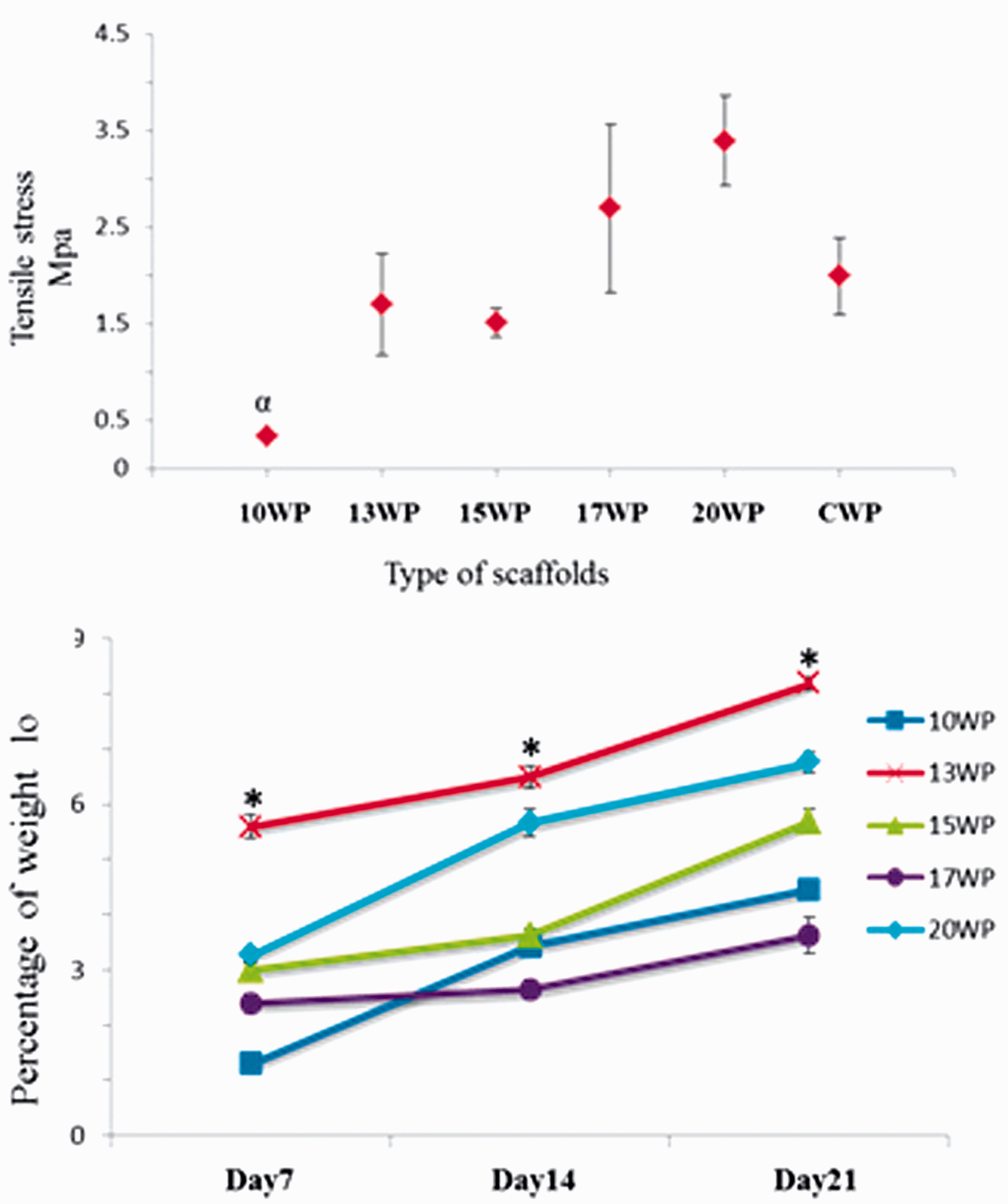
Degradation studies
The weight loss measurement was carried out for 21 days. During this time, the media were changed weekly with the samples being removed from the media, rinsed with distilled water, dried under vacuum, and their weights measured.
Weight loss in the scaffolds consisting of 10%w/v PLA was more than the other scaffolds. The high percentage of beads might have made this polymer highly degradable in the DMEM/10% FBS medium. The weight loss measurements in the 15WP sample showed that its degradation behavior was between the 10WP and 20WP samples (P < 0.05) (Figure 4(b)).
Live/dead cells evaluation
The live-and-dead test of hWJMSCs was done by green and red fluorescence via AO/EB staining (Figure 5(a)). After a 7-day incubation, only living cells in random views were observed during the first minute. Live hWJMSCs were stained green and appeared to have adhered to and attained a normal polygonal morphology. Dead cells were stained red and were not seen on surface of all the samples. There was more living hWJMSCs on CPW than all other samples.
Acridine orange/ethidium bromide staining of WJMSCs using different concentrations on day 7 post-culture (a). (b) The MTT assay histogram of uncoated and coated PLA scaffolds on 1, 3, and 7 days. * indicates significantly a good substrate for cell metabolism (P < 0.05). Different concentrations of PLA (10WP, 13WP, 15WP, 17WP, and 20WP) in preparing uncoated wet-electrospun PLA (10, 13, 15, 17, and 20%: V/W, respectively), while CWP represents the coated wet-electrospun PLA at concentration 15%. The experiments are done in triplicate for all groups (mean ± SE). WJMSC: Wharton Jelly derived mesenchymal stem cell; PLA: poly lactic acid; SE: standard error.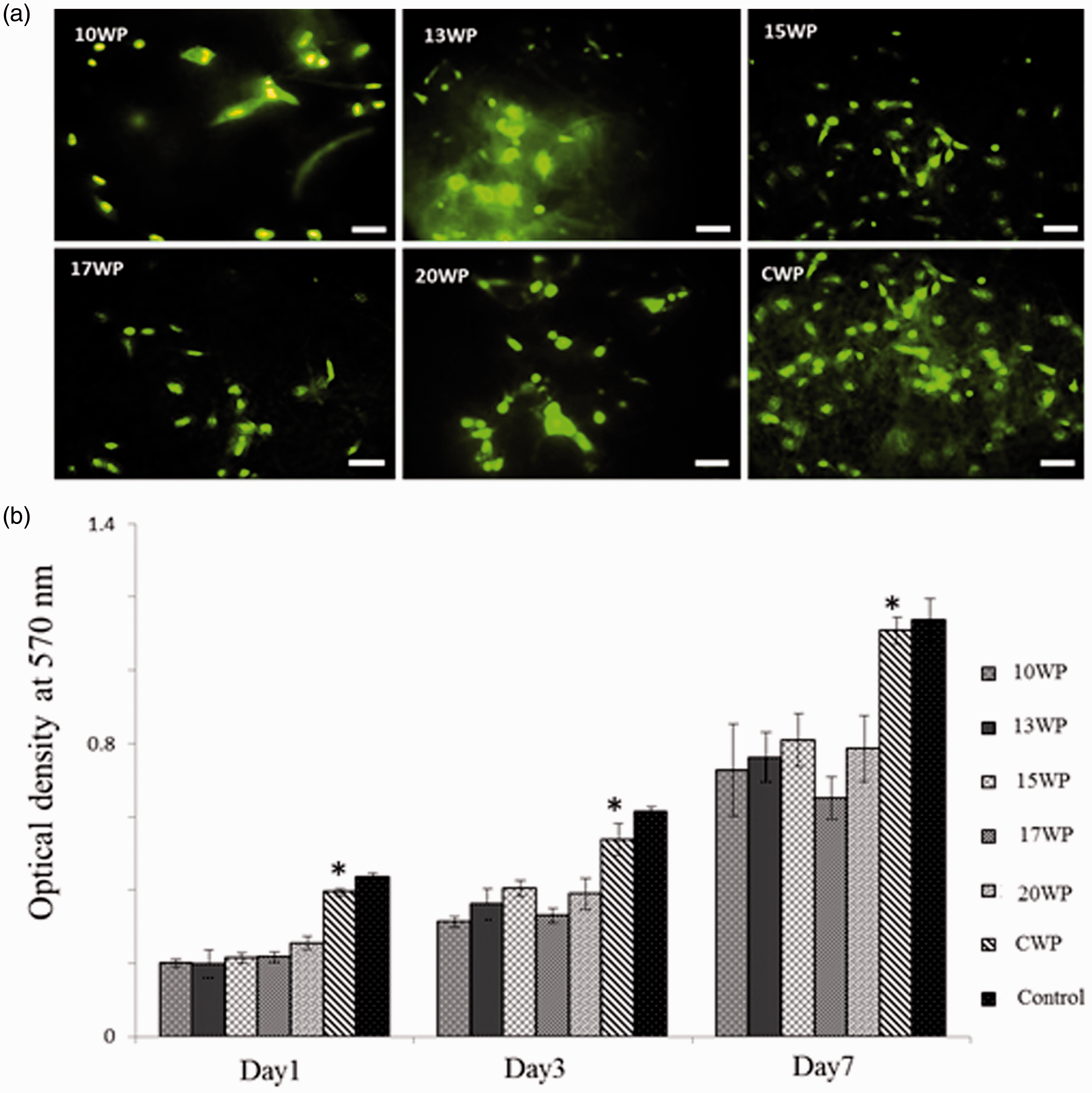
Cell metabolism studies
The MTT assay was carried out to evaluate the metabolism of hWJMSCs on uncoated and coated PLA wet-electrospun scaffolds (see Figure 5(b)). As the scaffold has a porous structure, after seeding the cells on them in a plate, some of the cells migrated from the pores and attached themselves to the bottom of the plate. However, all scaffolds provided a favorable environment in which they showed an increase in cell metabolism on the coated scaffold. The 15WP had the highest cellular metabolic activity after 6 and 24 h (62% ± 1.73 and 76% ± 2.12, respectively) compared with other concentrations scaffolds (P < 0.05). However, this scaffold (15WP), which needed surface modification for the best metabolism of cells, was coated by natural polymers. The results showed that the coated wet-electrospun PLA (CWP) was a better substrate for metabolism of MSC (73% ± 1.39 and 91% ± 1.62: 6 and 24 h, respectively; P < 0.05). The control hWJMSCs, cultured on a 96-well plate (2D group), had the highest metabolic activity (Table 1).
After 7 days of incubation, the CWP scaffolds displayed the highest absorbance compared to other samples (P < 0.05), but were the lowest compared with the control group (P > 0.05). Therefore, the coated scaffold provided a better support to the metabolic activity of hWJMSCs in this study.
pH alteration during in vitro degradation
The pH of all samples in the normal saline as a function of the degradation time, the sample codes (10WP, 13WP, 15WP, 17WP, 20WP, and CWP) are presented in Table 1.
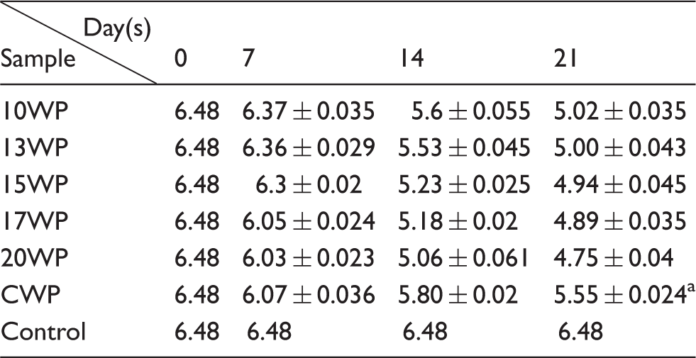
The data were presented as mean ± SE.
The coated scaffold has significantly lower pH changes during degradation than other concentration of pure PLA scaffolds.
SE: standard error.
Characterization of hWJMSCs
The phase contrast images show the hWJMSCs were flat and polygonal in shape (Figure 6(f)) and immunopositive to CD29, CD90, and CD73, while being immunonegative to CD34 and CD45 (Figure 6). Furthermore, the differentiation potential of hWJMSCs into lipogenic and osteogenic phenotypes are shown in Figure 6(g) and (h).
The characterization of hWJMSCs using flow cytometry for different markers and mesodermal lineage differentiation. (a) Flow cytometer graph of CD45 marker, whereas (b) to (e) represent the plots of CD34, CD29, CD90, and CD73 markers. (f) The phase contrast image of hWJMSCs with spindle-shaped cells. (g) and (h) represent the differentiation of hWJMSCs into osteogenic and adipogenic phenotype, respectively. Scale bar = 100 µm. hWJMSCs: human Wharton Jelly derived mesenchymal stem cells.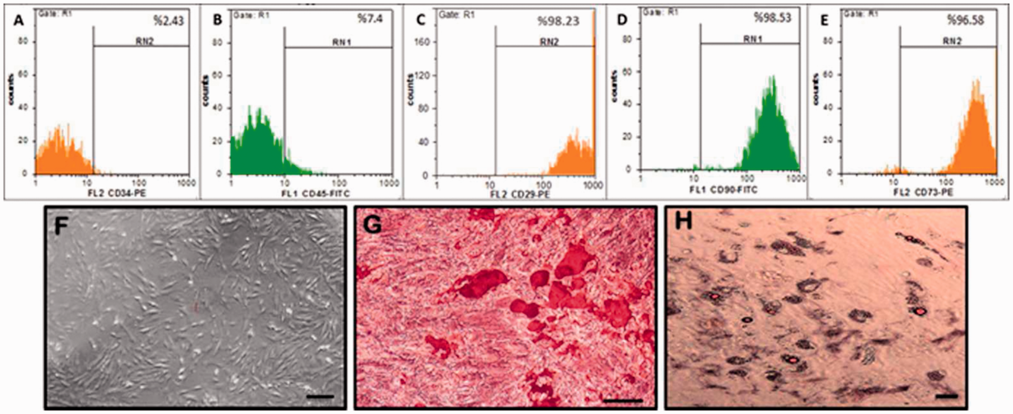
Generation and characterization of neuron-like cells
hWJMSCs were differentiated into neuron-like cells, as explained before, and characterized by immunocytochemistry and RT-PCR. The differentiated hWJMSCs into neuron-like cells were assessed by immunocytochemistry using antibodies specific to neuronal markers. The results showed the neuron-like cells expressed NSE after 21 days in both groups (2D and 3D) (Figure 7).
The immunostaining of the differentiated hWJMSCs into neuron-like cells in 2D culture (upper panel) and 3D culture (lower panel) following treatment each culture with valproic acid (21st day post-induction) using nestin (a,e), Map-2 (b,f), and NSE (c,g) (scale bar = 100 µm). hWJMSCs: human Wharton Jelly derived mesenchymal stem cells.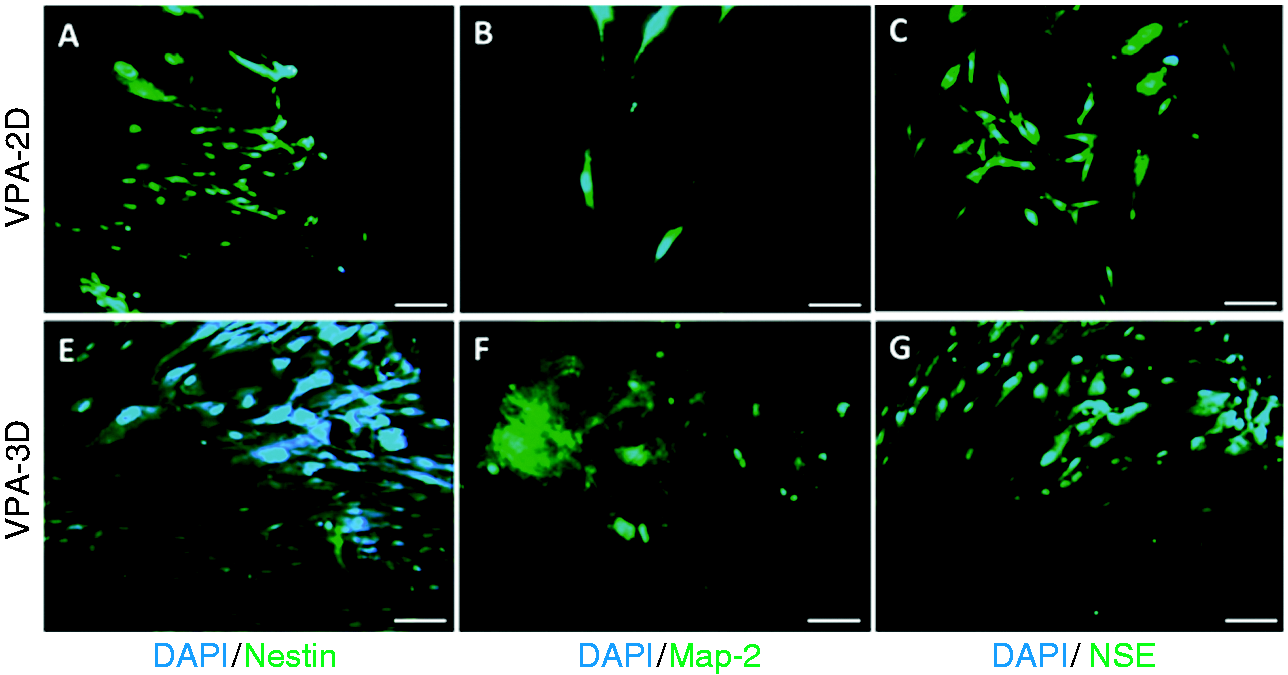
RT-PCR
To investigate the differentiation of hWJMSCs, the gene expression was analyzed using RT-PCR. The neurogenic differentiation factor, Neuro d1, was highly up-regulated in hWJMSCs cultured on the CWP scaffold compared to the hWJMSCs cultured on the 2D surface (Figure 8). Furthermore, the expression levels of stem cell marker Sox-2 was lower in the hWJMSCs on the coated scaffold than the hWJMSCs on the flat surface or 2D group.
Qualitative and quantitative evaluation of neuronal differentiation including Sox2, nestin, and neurod1 genes. (a,b) The electrophorograms of the genes in 2D and 3D cultures, respectively. The house keep gene is β-actin, NTC is non-template control. L is DNA ladder (50 bp). (c–e) The histogram of quantitative gene expression using real-time PCR of nestin, neurod1, and Sox2, respectively, in valproic acid treated 2D (gray color column) and 3D (black color column). All values are presented as mean ± SE. For nestin expression, the VPA-3D group was statistically significant (P < 0.05). For neurod1 expression, the VPA-3D group was statistically significant (P < 0.05). However, for Sox-2 expression, the VPA-2D group was statistically significant (P < 0.05). PCR: polymerase chain reaction. PCR: polymerase chain reaction; SE: standard error; VPA: valproic acid.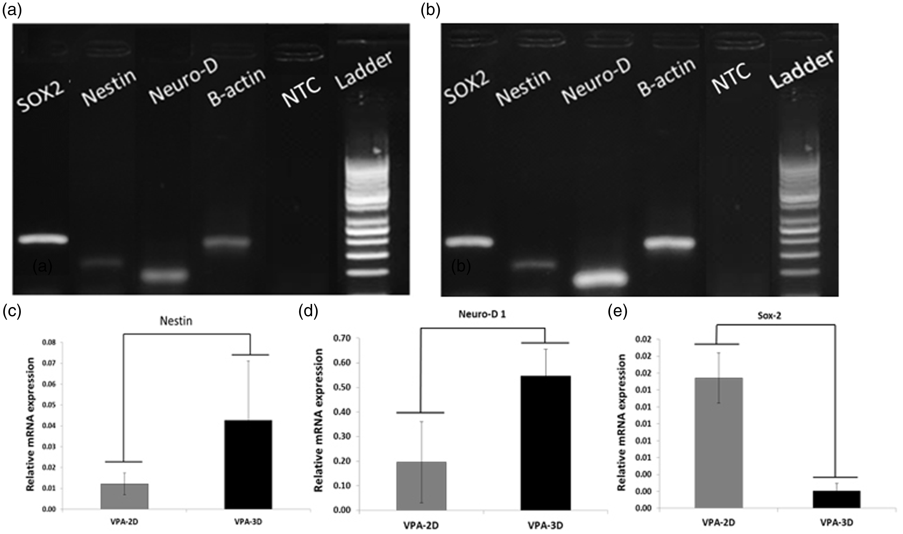
RT-qPCR
The RT-qPCR revealed the activation of neural genes in 3 weeks of differentiation. The expression of Nestin, an early neural marker, and Neuro-D1was up-regulated in VPA-3D, compared to the VPA-2D group (P < 0.05). However, the expression of Sox-2 (Figure 8), a stem cell marker, was significantly up-regulated on VPA-2D, compared with the VPA-3D group (P < 0.05).
Discussion
The purpose of fabricating this composite scaffold was to coat PLA (non-conductive) with conductive CNT. In order to detect the gelatin and alginate as well as CNTs on the PLA scaffold, we used FTIR at each step. For gelatin to be coated on PLA, the scaffold must be functionalized by NaOH. The FTIR spectrum of PLA treated with NAOH shows the functionalized PLA (FPLA) with negative charges. Then, the FPLA was coated with positively charged gelatin (GE) (see FTIR spectrum in the supplementary Figure 2). 14 This was followed by coating the PLA-GE with alginate (negatively charged) and subsequently coated with gelatin as shown in supplementary Figure 2. 29 The last gelatin layer was coated with CNT. Finally, MWCNT was coated with gelatin.
The results of this study showed that a conductive composite scaffold is a feasible option for use in regenerating nervous tissue. Neurons do not undergo mitosis and endogenous neuronal stem cells are unable to replace the quantity of neurons lost after a typical injury. The transplantation of stem cells has become an effective tool to study the mechanisms of neural tissue regeneration. However, the survival rate of transplanted cells is low. 30 One possible reason is that the injury site makes a harsh, non-permissive environment that lacks nutrients, survival factors, and, most importantly, a habitable substrate. 31 The new approach—tissue engineering—creates a new hope regarding the regeneration of defect sites in body tissues. Neural tissue engineering requires suitable precursor cells as well as a suitable microenvironment in order to replace the lost neurons. Moreover, an ideal scaffold for neural tissue engineering would be both electrically conductive and fibrous.32,33 Li et al. reported that graphene could enhance neurite numbers and average neurite length, 34 also, Lee et al., documented that graphene could induce MSC proliferation and differentiation, 35 moreover, Li et al. found that three-dimensional graphene foams could support NSC growth and active proliferation state by upregulating Ki67 expression. 36
Currently, combinations of natural and synthetic polymers are receiving attention due to additive action. 37 While the scaffold can improve the regenerating stem cells,38,39 nanotopography can have a greater influence on neural induction than the biochemical cues. 40 Therefore, the combined biochemical and physical properties could provide a synergistic inductive environment. In this study, we used wet-electrospinning to construct PLA-composite fiber scaffolds containing MWCNTs, which could provide conductivity to the scaffold and electrical stimulation enhancing nerve regeneration. 41 Moreover, PLA is a hydrophobic polymer, which has a poor cell attachment quality; so, we coated it with alginate and gelatin polymers to make the fiber surfaces hydrophilic. 42 Thus, the generation of wet-electrospun PLA composite scaffolds could mimic a three-dimensional ECM structure.
The SEM of uncoated and coated wet-electrospun PLA scaffolds displayed randomly oriented non-woven fibrous morphology, mimicking the extra cellular matrix, which consists of many nano-pores interfibers. The porosity is essential for the transport of oxygen and nutrients required for scaffolds. 43 Whereas the coating of PLA with gelatin and alginate clearly improved the hydrophilic property of the nanofibrous substrate, the contact angle supported these findings. Moreover, the coated scaffold showed a higher mechanical behavior rather than the uncoated PLA (15WP) owing to the presence of CNTs and other polymers on a scaffold that filled the fiber pores (P < 0.05), mechanical properties are critical for the scaffolds to resist the forces during a surgical operation, physiological activities, and/or tissue growth. 44 The results of cell metabolic activity demonstrated that the coated PLA scaffold was higher than the uncoated one, which was consistent with others, 37 where the modified PLA scaffold (CWP) significantly stimulated cell growth. 45
PLA coated with MWCNT could enhance the differentiation of the MSCs into neuronal phonotype 45 by improving the conductivity of PLA. 46 The in vivo studies showed that the conductive implants facilitated neural growth and reduced gliosis when implanted in the cerebral cortex, 47 and the finding of this study indicates that CNTs are biocompatible and a suitable substrate for use in neural tissue engineering. 48 The biocompatibility, low cytotoxicity, and neuron induction of these coated scaffolds make them a potential option for nervous tissue regeneration.
The degradation process of PLA is based on the hydrolytic process, as the water molecules attack the ester bonds in the polymer chains resulting in the residual oligomers, which reduce the local pH with subsequent autocatalysis of the hydrolysis. 49 Acidic environment is not suitable for the cellular growth, 50 we coated PLA by biopolymer in order to reduce the pH changes in environment.
To select a suitable stem cell source to differentiate into neuronal lineage on fabricated scaffolds, we used Wharton's jelly-derived mesenchymal cells. The extra-embryonic-tissue-derived stem, Wharton's jelly MSCs, has advantages over both adult and embryonic stem cell sources, 51 because there is a little ethical controversy with the harvest of the resident stem cells populations. In vitro, WJMSCs are capable of differentiation to multiple cell types including skeletal muscle and neurons. 52 Fu et al. also reported the generation of dopaminergic neurons from these cells. 53 WJMSCs under adequate stimulation can differentiate into neuron-like cells in vitro, exhibiting both morphological and biochemical properties of neurons and expressing typical neuronal proteins such as Nestin and Beta III Tubulin. 54 Human WJMSCs differentiation may also occur by the use of retinoic acid (RA), nerve growth factor (NGF), and fetal calf serum (FCS), but the number of yielded neuronal cells may vary widely because of the use of different culture conditions. Other strategies to improve neurons generation from MSCs include treatment with a neuronal induction medium (NIM) consisting mainly of brain-derived neurotrophic factor (BDNF) and low-serum media 55 or with a cytokine mix that include NGF, epidermal growth factor (EGF), bFGF and BDNF together with forskolin, which suggest that human UC blood 56 and Wharton's jelly 57 could be induced into neural phenotypes. Hsieh et al. reported that VPA (0.3 and 1 mM) increased neuronal differentiation up to 30–50%. 58 In their assay system, cells were isolated from adult hippocampus and passaged at least 10 times. Their studies showed that VPA up-regulates NeuroD in adult hippocampal progenitor cells. In contrast, cells in our system were isolated from human Wharton jelly and treated with VPA could promote neuronal differentiation. The neuronal marker (NeuroD) was up-regulated and significantly increased in VAP-3D than VPA-2D (P < 0.05). In contrast, the expression of Sox-2, a stem cells marker, was higher in VPA-2D than VPA-3D (P < 0.05). The induction by VPA can be attributed to VPA reprogramming property. 59
Conclusion
In this article, we prepared and modified a conductive PLA/MWCNTs nanofibrous scaffolds by the incorporation of biopolymers including gelatin and alginate via layer-by-layer self-assembly method to improve the differential potency of Wharton's jelly MSC into neuron-like cells.
We demonstrated that hMSCs isolated from the Wharton's jelly of the UC expressed markers concerning stem cells, CD29, CD90, and CD73, while were negative for hematopoietic markers, CD34 and CD45. They were seeded on PLA composite scaffold and used as a microenvironment for induction of the cells into neuron-like cells. This indicated that the composite scaffold containing conductive CNTs combined with valproic acid induction strongly influences MSCs transdifferentiation toward neuron-like cells where they expressed Nestin and Neuro-D1, neural markers.
It is needed to say that further study is required to fully understand the in vivo experiments of this scaffold.
Footnotes
Declaration of Conflicting Interests
The author(s) declared no potential conflicts of interest with respect to the research, authorship, and/or publication of this article.
Funding
The author(s) received no financial support for the research, authorship, and/or publication of this article.
Supplementary material
Supplementary material is available for this article online.
References
Supplementary Material
Please find the following supplemental material available below.
For Open Access articles published under a Creative Commons License, all supplemental material carries the same license as the article it is associated with.
For non-Open Access articles published, all supplemental material carries a non-exclusive license, and permission requests for re-use of supplemental material or any part of supplemental material shall be sent directly to the copyright owner as specified in the copyright notice associated with the article.