
Abstract
No single material can provide all requirements for wound dressings. Here, we evaluated the influence of different soy protein isolate and agar proportions (3:1, 1:1, and 1:3) in blend films on some of their physical-chemical and antibacterial properties to elucidate their potential as wound dressings. The films were synthesized by the gel casting method and ciprofloxacin hydrochloride was incorporated into the films. Films were characterized based on their surface morphology, water uptake ability, and weight loss profile. Also, the ciprofloxacin hydrochloride release kinetics was quantified spectrophotometrically. The antibacterial effect was evaluated against Staphylococcus aureus and Pseudomonas aeruginosa strains. The soy protein isolate-agar ratio affected the water uptake of the films and the release profile of ciprofloxacin hydrochloride but not the weight loss profile. The amount of drug released decreased near 80% because of the decrease in agar content in the films. The release kinetics of ciprofloxacin hydrochloride data best fitted to the Korsmeyer–Peppas model, suggesting that the mechanism of drug release was mainly of the diffusion type. All ciprofloxacin hydrochloride-releasing soy protein isolate-agar films strongly inhibited the cell viability of the bacterial strains studied. We concluded that water uptake and ciprofloxacin hydrochloride release can be controlled by changing the soy protein isolate-agar proportion. The proportions did not lead to changes in the antibacterial strength of the films.
Introduction
Wound dressing based on biopolymers is of interest due to their biocompatible, biodegradable, and non-toxic nature. 1 Several studies have shown that biopolymer matrices containing anti-microbial drugs inhibit the growth of pathogenic bacteria found in chronic wounds.2–4 Despite the various biopolymer-based products already in the market, the development or optimization of advanced biopolymer dressings still represents a very active research field.
Among biopolymers, soy protein (SP) is a potential candidate to develop wound dressing with controlled antibiotic release.5,6 SP is low-cost, is of non-animal origin, and has good water resistance and relatively long storage time and stability. 7 It has been reported that SP-based products promote tissue regeneration, such as new bone growth, and stimulate collagen deposition. 8 In vivo evaluation of a gentamicin-eluting SP dressing material in a contaminated wound in guinea pigs demonstrated its ability to accelerate epithelialization, with higher epithelial coverage (71%) than that obtained with a wound dressing available in the market (55%). 9 In addition, SP hydrogels injected subcutaneously in mice have demonstrated to be biocompatible and degraded over three weeks, indicating their potential use for wound healing. 10
In general, no single material can provide all the requirements for the wound healing process. In the case of SP, its films are very brittle and hygroscopic. 11 To overcome this limitation, SP has been blended with other proteins and polysaccharides.12–17 Some blends have demonstrated to have potential in wound healing. Chitosan/SP membranes produced by solvent casting have been shown to promote low in vitro activation of human polymorphonuclear neutrophils isolated from circulating blood, decreasing the healing time period of partial-thickness skin wounds. 18
Agar (AG) is a biodegradable polysaccharide extracted from red algae (Rhodophyceae), which shows good film-forming properties and high mechanical strength with moderate water resistance. 19 AG has been blended with different biopolymers to improve the mechanical, water resistance, and functional properties of films. 20
Hybrids based on SP and AG have demonstrated good compatibility between both.21–23 Also, the incorporation of AG decreases the solubility of films while increasing water uptake (WU), which could be useful for a controlled delivery of pharmaceutical drugs. 23 Despite many of these advantages, the functionalization of blend films with antibiotics for the development of dressings with antimicrobial activity has been little studied. Previous works have demonstrated that blends (4:1) of SP and sodium alginate (a polysaccharide) degrade more slowly in aqueous media than blends of SP and gelatin (a protein) and release the antibiotic clindamycin in a more controlled manner. Despite these differences, the antibacterial strengths of both kinds of blends against Staphylococcus aureus and Staphylococcus albus were not statistically different. 24
Ciprofloxacin hydrochloride (Cip) is a broad-spectrum antibiotic that belongs to the fluoroquinolone family and is one of the antibiotics most widely used in wound healing. 25
The aim of this work was to develop films with different proportions of SP-AG loaded with Cip and study some of their physical properties, drug release profile and antibacterial activity to elucidate their potential as biomaterials with antibacterial activity.
Materials and methods
Materials
Soy protein isolate SUPRO E (SPI) with 90% protein on fat-free dry-weight basis was donated by The Solae Company, Argentina. Agar-agar was purchased from Laboratorios Britania S.A. (Buenos Aires, Argentina) and Cip from Roemmers Laboratories SAICF (Buenos Aires, Argentina). Glutaraldehyde (GA) solution (50% in water, 5.6 M) was purchased from Merck SA Argentina. Fetal bovine serum (FBS) was purchased from Natocor SA (Córdoba, Argentina). Maximum recovery diluent was prepared according to the following formula: Peptone, 1 g (Laboratorios Britania S.A., Buenos Aires, Argentina), NaCl, 8 g (Reagents S.A., Buenos Aires, Argentina), distilled water, 1 L. Simulated wound fluid was prepared by mixing maximum recovery diluent with FBS in equal volumes. Mueller-Hinton broth was purchased from Laboratorios Britania S.A. (Buenos Aires, Argentina).
Preparation of SPI-AG blend films
SPI-AG blend films were prepared in 3:1, 1:1, and 1:3 weight ratios. In each case, the final biopolymer concentration in solution was 2% (w/v). Glycerol (4.28% w/v) was used as plasticizer. The starter materials (SPI+AG + glycerol + water) were heated at 105°C on a magnetic stirrer for 20 min and cooled to 40°C under continuous stirring. The solution pH was adjusted when necessary to pH 7.0 with NaOH (0.1 M). Then, 10 mL of Cip (20 mg mL−1) was added to the mixture under continuous stirring. Finally, 45 mL of each solution was spread in 140 mm diameter Petri dishes and dried in an oven at 37°C for 24 h to obtain the blend films. After drying, films were cross-linked for 1, 3, or 5 min in a GA solution (0.25 wt%). Blended films were named according to the SPI-AG ratio: SPI-AG 3:1, SPI-AG 1:1, and SPI-AG 1:3.
Morphological characterization
The SPI-AG films obtained were morphologically characterized by scanning electron microscopy (SEM). For this, films were fixed with GA (2.5% in 0.1 M phosphate-buffered saline solution (PBS)) overnight at 4°C. The samples were then washed with distilled water and sequentially dehydrated through a graded series of ethanol solutions. After mounting on stubs and gold sputtering, the samples were examined with SEM (JEOL JSM, 6480 LV, Japan).
WU assay
WU was determined as previously.
26
Briefly, circle-shaped films (1 cm diameter) were incubated in 15 mL distilled water. At different times, the films were removed and blotted with a filter paper. The weights were recorded and then re-incubated in distilled water at 37°C. The process was repeated until equilibrium swelling was reached. WU was calculated according to the following formula

In vitro weight loss (WL) profile
The WL was determined in PBS at pH 7.4 and 37°C for 28 days according to our previous study.
27
Briefly, the films were firstly weighed and then immersed in PBS. The samples were collected every seven days, washed and air-dried. The PBS solution was replaced twice a week. The assay was performed in triplicate and the WL was calculated as follows

In vitro Cip release study
Circle-shaped (0.5 cm diameter) SPI-AG films were immersed in closed tubes containing 10 mL of sterilized PBS at 37°C for 42 days. Each disc was weighed to normalize the data. At every data point, 1 mL was removed and used to determine the released concentration of Cip by using ultraviolet(UV)-visible spectroscopy (Bioamerican Science SP2100) at λ = 275 nm. The tubes were refilled with 1 mL of sterile PBS to restore the initial volume. The assay was performed in triplicate and the data of the amount of Cip released were presented as cumulative percentage of drug released vs. time.
Kinetics models
To gain insights into the mechanism of drug release, the in vitro drug release data were fitted to three kinetics models and studied: Zero order (cumulative % drug release vs. time), first order (log cumulative % drug remaining vs. time), and Korsmeyer–Peppas (log cumulative % drug release vs. log time). The criterion for selecting the most appropriate model was based on linear regression analysis (>r2). All models were fitted to 2–360 h, except for the Korsmeyer–Peppas model, in which case, the data were fitted only until 60%. The n value of this model is used to characterize different release mechanisms, 28 as follows: a value of n ≤ 0.5 indicates drug release through Fickian diffusion, 0.5 < n < 1 indicates that the mass transfer follows a non-Fickian model or anomalous transport, an n value of 1 is associated with Zero-order drug release or Case-II transport, and n > 1 is regarded as super Case-II transport.
Bacterial culture and preparation of the inoculum
The bacterial culture and inoculums were prepared as previously. 26 Three bacterial isolates were used: two of S. aureus and one of Pseudomonas aeruginosa. All strains were grown for 24 h in Mueller-Hinton broth at 37°C. For the assays, bacterial cell suspensions were adjusted to 6–7 log cfu mL−1. For antibacterial activity evaluation, circle-shaped (1 cm in diameter) films were obtained. Previously, film discs were UV-sterilized for 20 min on each side. The performance of samples was evaluated by the plate count method.
Viable counts
Viable counts were determined in simulated wound fluid. The films were incubated for 24 h at 37°C in 2 mL with cellular suspensions. Each cellular suspension in the absence of films served as controls. Non-Cip-releasing films were also evaluated. Cell viability was assessed after 4 and 24 h by counting in Mueller-Hinton agar plates. The assay was performed in triplicate.
Statistical analysis
Statistical analysis was performed using the SPSS 23 statistical package software (IBM, Armonk, NY, USA) with appropriate statistical tests such as one-way analysis of variance (ANOVA) with Dunnett’s and Tukey’s multiple comparison post-tests for intergroup analysis. Specifically, the data from cell suspension were used as control and compared to the Cip-releasing and non-Cip-releasing films. The level of significance was p < 0.05.
Results
Morphological properties
Figure 1(a) to (c) shows the SEM images of the peeled surfaces of the films cross-linked for 1 min. The surface morphology changed according to the SPI-AG proportion. Qualitative assessment by inspection of SEM images suggested that the surfaces became rougher and more irregular as the AG proportion increased.
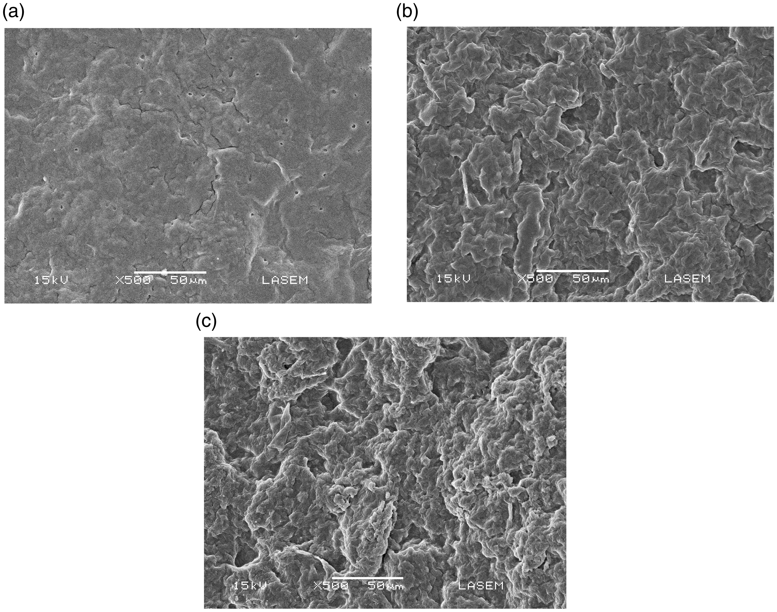
SEM images of SPI-AG films. SPI-AG 3:1 (a), SPI-AG 1:1 (b), and SPI-AG 1:3 (c).
WU ability of the films
Figure 2 shows the WU percentage of SPI-AG films cross-linked for 1 min. The WU profiles were similar for all samples. The percentage of WU increased over time. Maximum absorption was observed after 10 min. After that, the WU ability of the films reached the equilibrium. The increase in AG content increased the WU more than two fold. At the maximum peak, the percentage of WU was 152.7 ± 14.5 for SPI-AG 1:3, 121.4 ± 9.8 for SPI-AG 1:1, and 76.56 ±11.9 for SPI-AG 3:1.
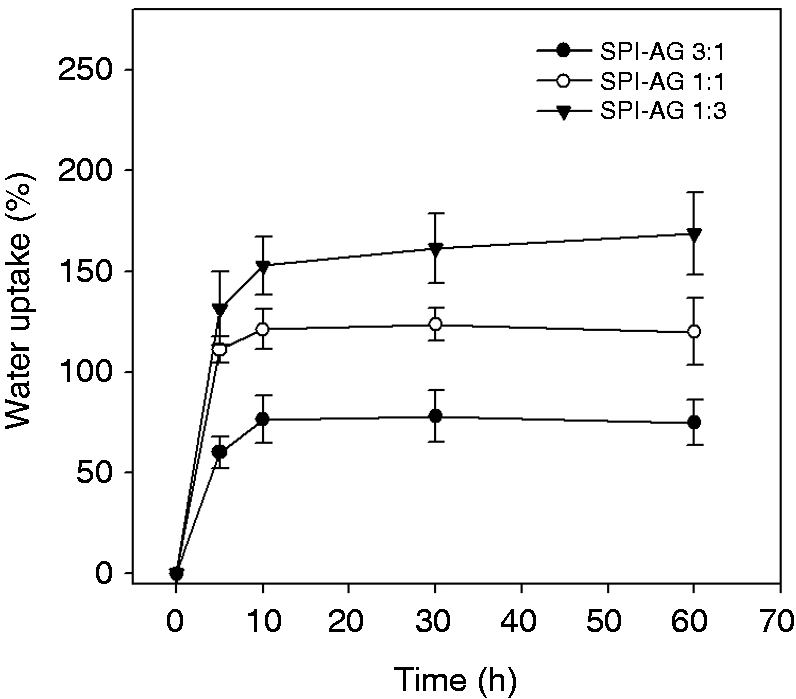
WU percentage of the SPI-AG films along the incubation time. The data are expressed as the means ± SD.
WL profile
The WL of the films was not affected by the increase in the cross-linking, so we chose 1 min of cross-linking time to work because the films maintained their structure and integrity. Figure 3 shows the WL percentage of SPI-AG films along the time. All samples lost 20–30%of their initial weight after 1 day probably due to the loss of glycerol content. After that and for 28 days, the WL of each film no longer presented significant differences and the differences between films were not significant.
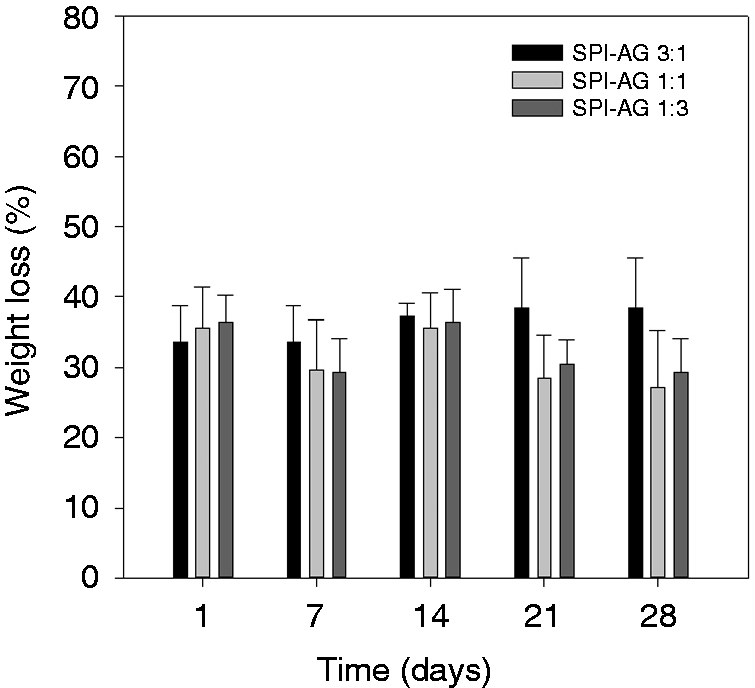
WL percentage of the SPI-AG films along the incubation time. The data are expressed as the means ± SD.
Cip release studies
Figure 4 shows the cumulative percentage of Cip released in each film. The release kinetics showed a similar behavior for the three films: a burst releasing phase during the first 2 h, followed by a slow release rate during the next two weeks, and finally a plateau phase until the end of the assay. Nevertheless, the percentage of Cip released changed considerably according to the SPI-AG ratio. For example, during the burst release phase, SPI-AG 3:1 films released 16.0 ± 0.8% of Cip, whereas SPI-AG 1:1 released 33.1 ± 1.7% and SPI-AG 1:3 47.9 ± 2.0%. Taking the time of maximum release (15 days), the decrease in AG content in the films decreased the percentage of Cip release by about 80%.
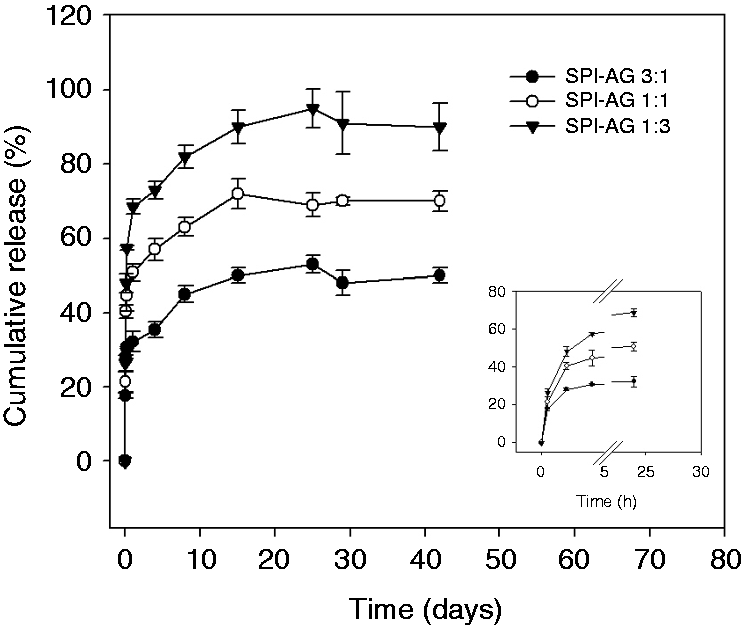
Cumulative percentage of Cip released from SPI-AG films as a function of time. The data are expressed as the means ± SD.
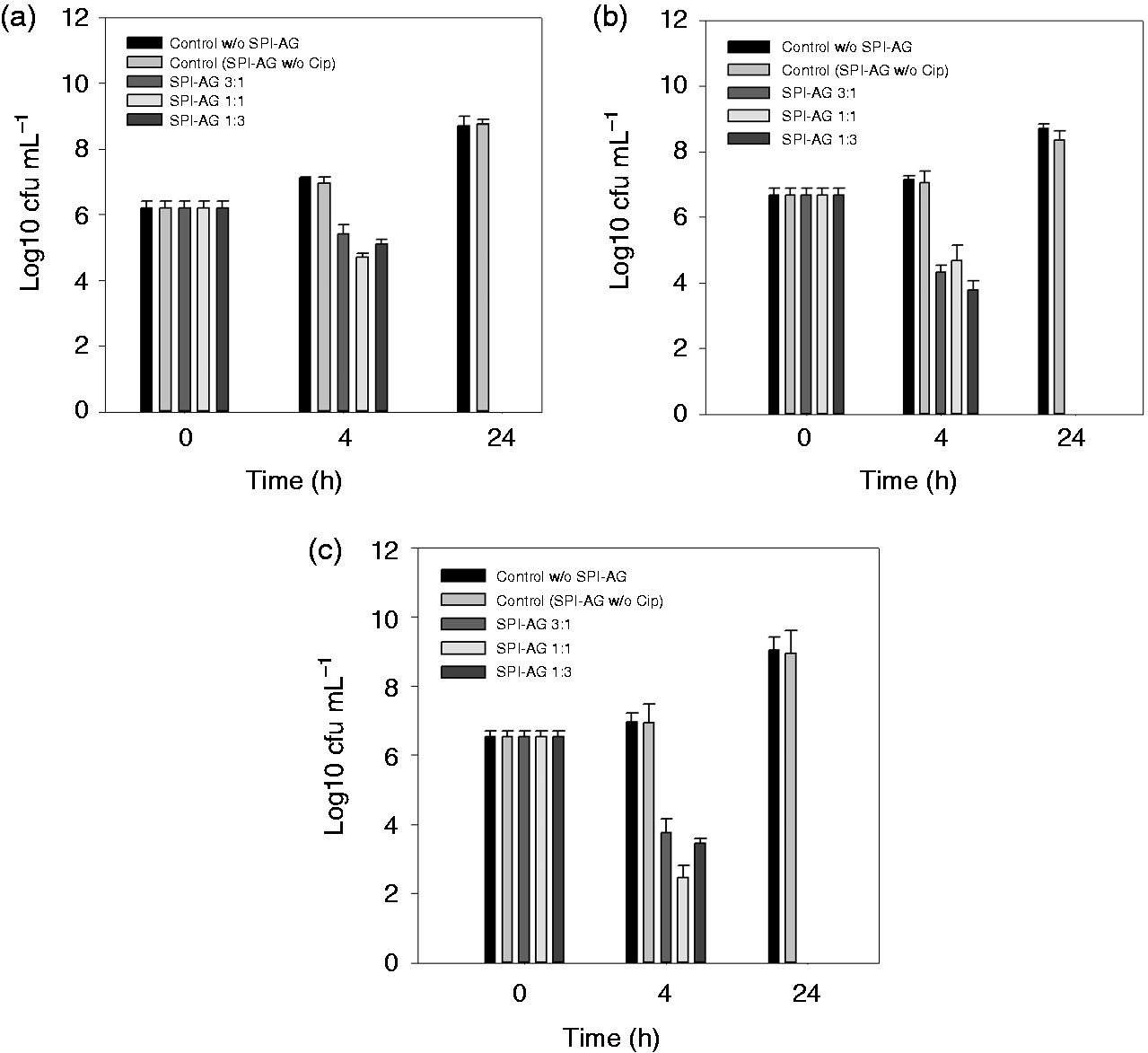
Viable counts of Staphylococcus aureus (a,b), and Pseudomonas aeruginosa (c) in the presence of films when incubated with simulated wound fluid (SWF). The data are expressed as the means ± SD.
Kinetics models
The data for the release mechanism along with the values of r2 for different samples are shown in Table 1. Acceptable correlation was achieved when the r2 values were equal to or higher than 0.970.
Kinetic data of Cip release from different blend films.
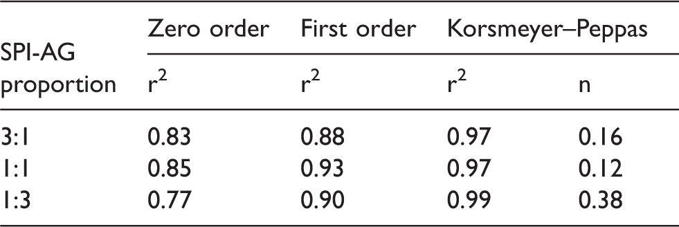
CIP: ciprofloxacin hydrochloride; SPI-AG: soy protein isolate-agar.
When data were fitted to the zero-order model, the r2 values obtained were small, suggesting that the release kinetics did not follow the zero-order. This was supported by Figure 4, which shows that the plots were curvilinear for all samples. Data fitted better first-order kinetics than zero-order kinetics but the r2 values were lower than 0.970. Finally, the data obtained from in vitro release studies were applied in Korsmeyer–Peppas equation to find out the n value, which describes the drug release mechanism. The n value of the samples was between 0.16 and 0.38, indicating the Fickian diffusion principle.
Antibacterial activity
Non-Cip-releasing films did not affect the viability of the strains (data not shown). In contrast, all Cip-releasing SPI-AG films strongly inhibited the number of bacteria studied, showing a satisfactory antibacterial activity (Figure 5). After 4 h of incubation time, the log reductions from the control were between 2.5 and 4. After 24 h, the cell viability of the three strains was below the detection limits. We found no significant antibacterial strength between the different SPI-AG films. Nevertheless, among the strains, P. aeruginosa was more susceptible to Cip than the staphylococcus strains after the 4 h incubation period.
Discussion
SPI has excellent film-forming ability and oxygen permeability. Nevertheless, SPI films have poor mechanical properties and relatively high moisture sensitivity, thus limiting their commercial application. 29 The properties of these films can be effectively improved by blending.7,11,30,31
Previously, it has been reported that blends of SPI and AG have good compatibility. 19 Tian et al. 21 revealed that hydrogen bonding interactions exist between SPI and AG and that the incorporation of AG enhances the tensile strength of casting SPI films.
Fourier-transform infrared (FTIR) spectroscopy studies showed that with the addition of AG content in blend films, characteristic amide bands of SP films (amide I at, 1656 cm−1 (C = O stretching), amide II at, 1541 cm−1 (N–H bending), and amide III at, 1240 cm−1 (C–N stretching)), shifted to higher wavenumbers while amide III remained almost invariable. This suggests hydrogen bonding interactions among hydroxyl groups of AG and protein, as well as between hydroxyl groups in AG and amino groups in protein. 19
Here, we found that the increase in AG content in the films increased their WU ability. Garrido et al. 23 suggested that water molecules may form water-polymer hydrogen bonds, thereby reducing inter-chain interactions and facilitating the WU.
In wound regeneration, some WU ability is desirable to absorb wound exudates and to provide optimal gaseous exchange, but high water penetration should be avoided because the antibiotic should be released rapidly. 5 In fact, as we discuss later, the increased WU ability of the SPI-AG 1:1 and 1:3 films over SPI-AG 3:1 correlated with a faster release of Cip.
As expected, GA was a good cross-linker for the blends. GA can react with functional groups in both proteins and carbohydrates, improving tensile properties. Also, cross-linking is essential for film integrity and stabilization during swelling, degradation, and drug release. 31 Non-crosslinked SPI-AG films disintegrated in less than 1 h (data not shown), so the cross-linking process was necessary. We found no significant WL of the films by increasing the cross-linking time duration, probably due to a plateau of cross-linking density. Since some reports have shown that GA induces cytotoxicity,32,33 we minimized the time of exposure of the films.
SPI-AG films cross-linked for 1 min lost between 20 and 30% of their initial weight during the first day. After that period, WL was not statistically significant between the samples. A similar WL profile of films based on SPI has been reported by other authors.5,31 Peles and Zilberman 5 attributed this phenomenon to the leaching out of the plasticizer and small non-crosslinked SP chains.
Many parameters, including the properties of the drug, the properties of the biopolymer, and the processing method, affect the drug release kinetics. 34 Here, the SPI-AG ratio affected the percentage of Cip released. As the content of AG in the films increased, the drug release was faster. This phenomenon correlates with a higher capacity of WU of those films. A possible explanation could be related to the interaction between SPI and AG.
Drug release from biopolymers is controlled by one or more different mechanisms, including drug diffusion, drug and/or polymer dissolution and, in some cases, polymer swelling and erosion processes. 35 It is expected that wound specific healing can be achieved by combining swelling, erosion and subsequent drug diffusion kinetics as part of the controlled drug release mechanism. The application of different mathematical models allows us to analyze the mechanism by which Cip releases from films. The data best fitted to Korsmeyer–Peppas equation, which is often used to describe the drug release behavior from polymeric systems. 28 The n values obtained suggest that the release of Cip from the films studied was mainly controlled by Fickian diffusion for all blend formulations.
Based on this, a possible mechanism of Cip release from SPI-AG films is that water penetrates into the polymeric matrix, which occurs more easily when the AG proportion is increased. Glycerol quickly leaches out of the matrix, contributing to the burst release of the drug while promoting the WL of the films. Then, Cip is released by diffusion in a more slowly manner until a plateau phase is achieved. Erosion can be overestimated.
One goal of this work was to prepare films that can release Cip to inhibit bacterial growth. For that reason, antibacterial assessment of the films prepared was crucial to determine their potential biomedical application. Here, we worked with bacterial strains most usually found in wound infections like S. aureus and P. aeruginosa. 36 We found that all blended films strongly inhibited the cell viability of the strains studied with similar strength. This means that the amount of drug released by SPI-AG 3:1 was sufficient to inhibit the bacterial population. We also observed this phenomenon in other studies with other biopolymeric systems in which an increase in the amount of drug released did not reflect an improvement in the antibacterial performance.37,38 Other authors have reported a similar pattern of inhibition. 39 The MIC for P. aeruginosa is 0.5 µg mL−1, whereas that for S. aureus is 1 µg mL−1 (www.eucast.org). After 4 h, SPI-AG 3:1 films released almost 13 µg mL−1 of Cip. So, the levels of drug released exceeded this concentration by several folds. Also, we found that P. aeruginosa was more sensitive to Cip than S. aureus strains. This difference could be attributed to the cell wall content of Gram-positive and Gram-negative bacteria.
Conclusion
Our results confirmed that some physical-chemical parameters of importance in drug release systems can be tuneable by changing the SPI and AG proportions in blend. We evaluated three proportions of SPI-AG: 3:1, 1:1, and 1:3. WL was not affected by the biopolymeric proportions, but WU ability was considerably higher as AG content increased in the films. The amount of Cip released decreased about 80% as the AG content decreased. Despite this decrease, the amount of Cip released by SPI-AG 3:1 was sufficient to inhibit the cell viability of staphylococci and P. aeruginosa. This makes the SPI-AG 1:3 film a more predictable biomaterial with an excellent antibacterial activity. Independently of the percentage of Cip released, the mechanism of drug release was the same, which, according to the Korsmeyer–Peppas model, was mainly of the diffusion type. These films could be potentially very useful as burn and ulcer dressings.
Footnotes
Declaration of Conflicting Interests
The author(s) declared no potential conflicts of interest with respect to the research, authorship, and/or publication of this article.
Funding
The author(s) disclosed receipt of the following financial support for the research, authorship, and/or publication of this article: This work was supported by the Consejo Nacional de Investigaciones Científicas y Técnicas, CONICET, Argentina (PIP0245 to AAG).